



ERα degrader 7
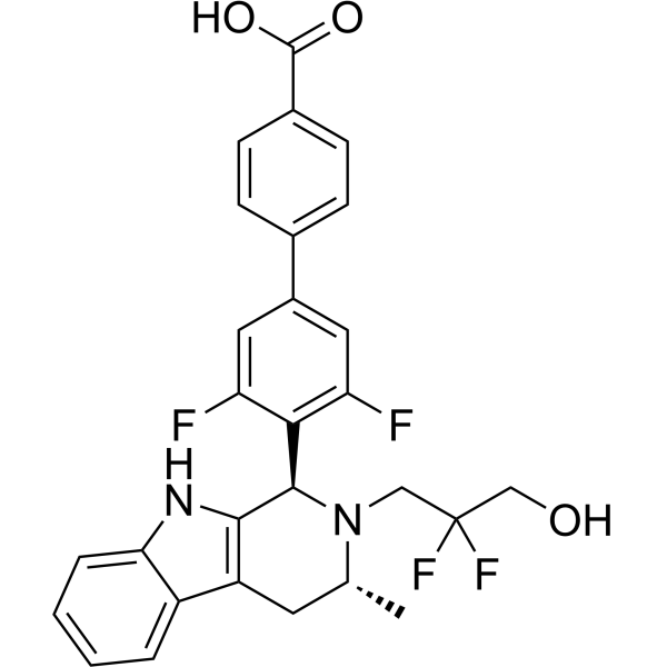
ERα degrader 7 (compound B1) is a potent ERα degrader with an IC50 of 14.6 nM and a DC50 of 9.7 nM, respectively. ERα degrader 7 shows excellent antitumor activity, indicating its potential to evolve as a promising selective estrogen-receptor degrader (SERD) for breast cancer research.
For research use only. We do not sell to patients.
- Formula: C28H24F4N2O3
- Molecular Weight:512.50
-
Storage:
Please store the product under the recommended conditions in the Certificate of Analysis.

To place orders, for customer services and technical support, please contact: MedChemExpress USA
Tel: 609-228-6898 E-mail: [email protected] [email protected]


-
 Biological Activity
-
Chemical Information
- Protocols
- Purity & Documentation
- References
-
 Help & FAQs




Biological Activity
Description
IC50 & Target
IC50: 14.6 nM (ERα)[1]
In Vitro
ERα degrader 7 (10-5-10 μM, 5 d) demonstrates antitumor activity against the breast cancer cell (IC50 = 4.21 nM) and noncytotoxicity against normal breast epithelial cell[1].
ERα degrader 7 (50-1000 nM, 5 d) shows a significant concentration-dependent degradation effect of ERα in MCF-7[1].
MedChemExpress (MCE) has not independently confirmed the accuracy of these methods. They are for reference only. Further protocols information, click here.
-
Cell Line:MCF-10 (normal breast epithelial cell)
-
Concentration:10-5-10 μM
-
Incubation Time:5 d
-
Result:Demonstrated low inhibition against MCF-10A, cell proliferation rate of each concentration are all above 80%.
-
Cell Line:MCF-7 (breast cancer cell)
-
Concentration:10-5-10 μM
-
Incubation Time:5 d
-
Result:ERα degrader 7 (10-3-10 μM) demonstrated excellent antitumor activity against the MCF-7 cell, caused an inhibition rate ranged from 60% to 75%, results were evaluated using GDC-9545 as a positive control.
-
Cell Line:MCF-7 (breast cancer cell)
-
Concentration:1, 10, 50, 100, 200, 500, 1000 nM
-
Incubation Time:5 d
-
Result:Showed a concentration-dependent degradation effect of ERα. Demonstrated a significant ERα-degradation effect (ERα expression rate < 50%) at a concentration of 50 nM, results were evaluated using GDC-9545 as a positive control.


Chemical Information
-
Molecular Weight 512.50
-
Formula C28H24F4N2O3
-
SMILES
OC(C1=CC=C(C2=CC(F)=C([C@@H]3C(NC4=CC=CC=C45)=C5C[C@@H](C)N3CC(F)(F)CO)C(F)=C2)C=C1)=O
-
Shipping
Room temperature in continental US; may vary elsewhere.
-
Storage
Please store the product under the recommended conditions in the Certificate of Analysis.

Protocols
-
Cell Cytotoxicity Assay
Cytotoxicity assays are usually based on the assessment of cell membrane damage, which can also be indirectly detected by measuring cell viability. Detection methods include MTT assay, CKK-8 assay, LDH assay and ATP assay, etc.
-
Research Protocol for Endocrine Diseases
Endocrine diseases often arise from disrupted hormone production, hormone signaling, or target-tissue responsiveness; for diabetes-focused endocrine disease models, insulin signaling regulates glucose uptake, hepatic glucose output, lipid metabolism, and β-cell compensation. Type 2 diabetes develops through interacting defects in insulin resistance, β-cell dysfunction, adipose inflammation, hepatic glucose overproduction, altered incretin signaling, and ectopic lipid metabolism. A major unresolved question is whether endocrine dysfunction is driven primarily by target-tissue insulin resistance, intrinsic β-cell failure, immune/inflammatory stress, or combined multi-organ failure that differs by disease stage.
-
Subcutaneous Cell-Line-Derived Xenograft
Subcutaneous cell-line-derived xenograft (CDX) models are established by implanting cultured human cancer cell lines into immunodeficient mice, where the injected cells form localized tumors that can be monitored in vivo as a measure of tumorigenic potential, growth kinetics, and treatment response. These models are widely used in oncology research because they allow reproducible tumor formation and enable comparative assessment of tumor growth between different cell lines or genetic manipulations in a controlled in vivo microenvironment. Subcutaneous implantation of cancer cells in immunodeficient mice is a standard approach for evaluating tumor growth behavior and therapeutic response across multiple cancer types, including prostate, esophageal, pancreatic, and colon cancer models.
-
Breast Cancer Modeling
Breast cancer is a heterogeneous cancer, and it has been distinguished into four subtypes: luminal A, luminal B, HER2-positive and basal-like. Molecular mutations, epigenetic alterations, hormone exposure and immune microenvironment are related to the progression of breast cancer.


Purity & Documentation
References

Calculators
Concentration (start) × Volume (start) = Concentration (final) × Volume (final)


ERα degrader 7 Related Classifications
HY-155492 Related Classifications
 Powered by Bioz