



ERK5-IN-2

Based on 1 Customer Validation
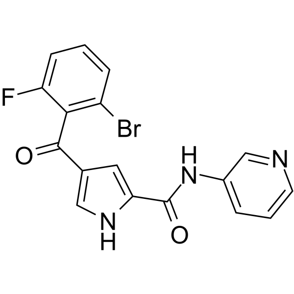
ERK5-IN-2 is an orally active, sub-micromolar, selective ERK5 inhibitor with IC50s of 0.82 μM, 3 μM for ERK5 and ERK5 MEF2D, respectively. ERK5-IN-2 does not interact with the BRD4 bromodomain. ERK5-IN-2 suppresses both tumor xenograft growth and basic fibroblast growth factor (bFGF) driven Matrigel plug angiogenesis.
For research use only. We do not sell to patients.
- Purity : 98.45%
- CAS No.: 1888305-96-1
- Formula: C17H11BrFN3O2
- Molecular Weight:388.19
-
Storage:
4°C, protect from light
* In solvent : -80°C, 6 months; -20°C, 1 month (protect from light)

To place orders, for customer services and technical support, please contact: MedChemExpress USA
Tel: 609-228-6898 E-mail: [email protected] [email protected]


-
 Biological Activity
-
Chemical Information
-
Solvent & Solubility
- Protocols
- Purity & Documentation
- References
-
 Help & FAQs


-
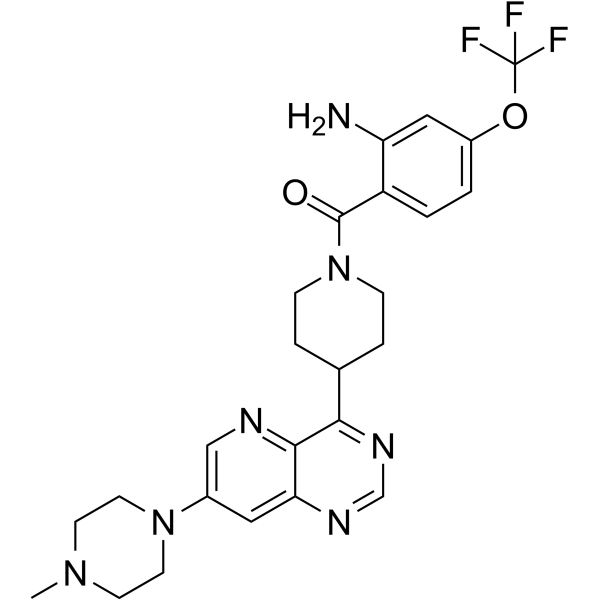
HY-112082
BAY885ERK5 Inhibitor -
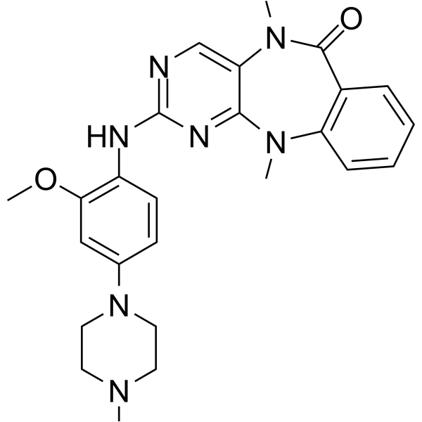
HY-14403
ERK5-IN-1ERK5 Inhibitor -
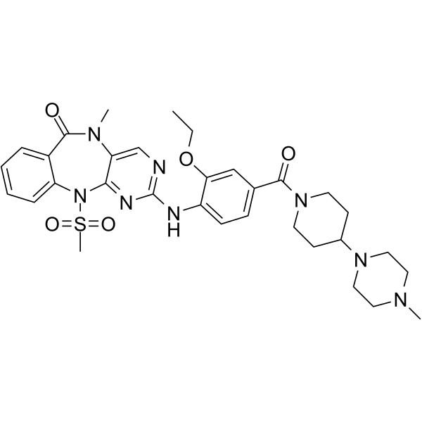
HY-101846
AX-15836ERK5 Inhibitor -
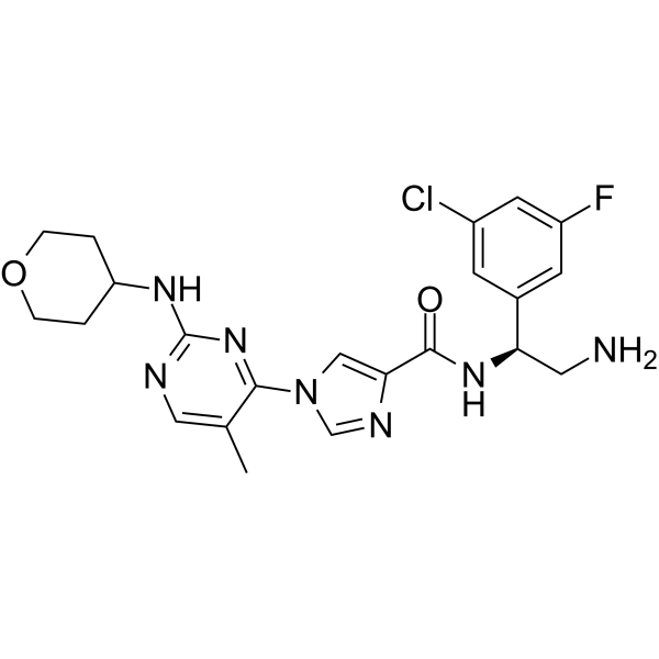
HY-136579
ASN007ERK Inhibitor -
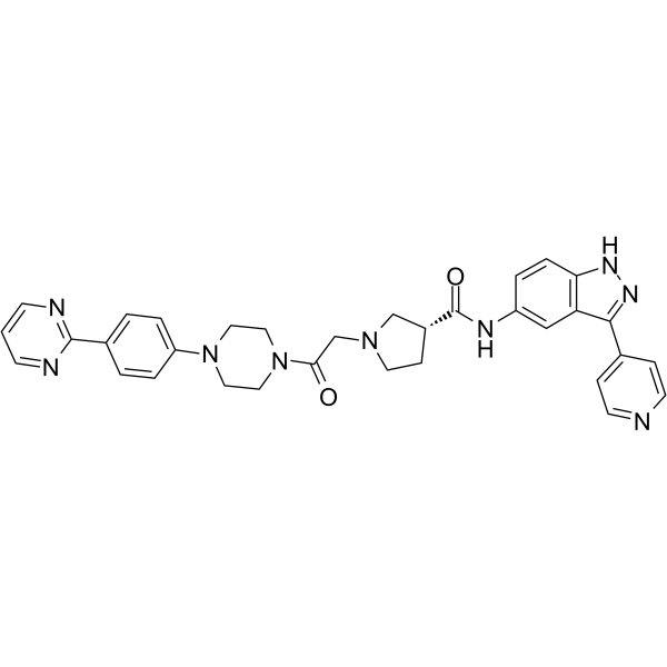
HY-50846
SCH772984ERK1/2 Inhibitor -
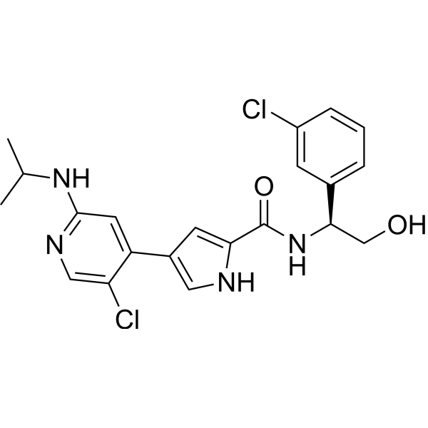
HY-15816
UlixertinibERK1/2 Inhibitor -
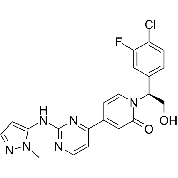
HY-15947
RavoxertinibERK1/2 Inhibitor -
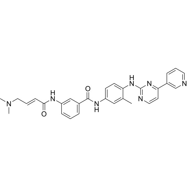
HY-13319
JNK-IN-8JNK Inhibitor -
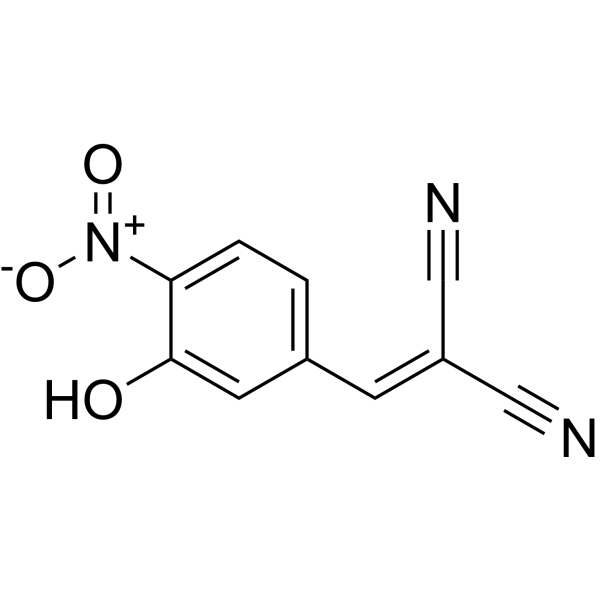
HY-108330
AG126Tyrosine Kinase Inhibitor -
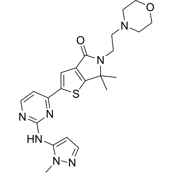
HY-101494
TemuterkibERK1/ERK2 Inhibitor -
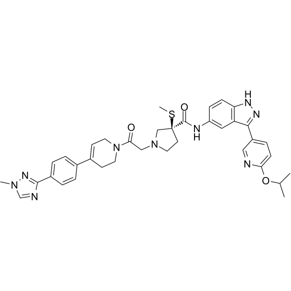
HY-111407
MK-8353ERK1/2 Inhibitor -
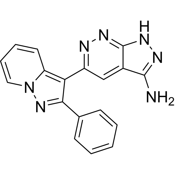
HY-12275
FR 180204ERK Inhibitor


Biological Activity
Description
IC50 & Target
[1]|
ERK5 0.82 μM (IC50) |
ERK5 MEF2D 3 μM (IC50) |
Cellular Effect
|
Cell Line

|
Type | Value | Description | References |
|---|---|---|---|---|
| HEK293 | IC50 |
3 μM
Compound: 46
|
Inhibition of HA-tagged ERK5 (unknown origin) coexpressed with mutationally activated EGFP-MEK5D and Gal4-MEF2D in HEK293 cells assessed as reduction in MEF2 transcription by measuring luciferase signal after 20 hrs by luciferase reporter gene assay
Inhibition of HA-tagged ERK5 (unknown origin) coexpressed with mutationally activated EGFP-MEK5D and Gal4-MEF2D in HEK293 cells assessed as reduction in MEF2 transcription by measuring luciferase signal after 20 hrs by luciferase reporter gene assay
|
[PMID: 31212132] |
| PC-3 | GI50 |
44 μM
Compound: 46
|
Growth inhibition of human PC3 cells
Growth inhibition of human PC3 cells
|
[PMID: 31212132] |
| Sf9 | IC50 |
0.82 μM
Compound: 46
|
Inhibition of recombinant N-terminal His-tagged human ERK5 (2 to 409 residues) co-expressed with GST-tagged human truncated MEK5DD lacking N-terminal 11 residues (13 to 448 residues) in Sf9 insect cells assessed as substrate peptide phosphorylation using
Inhibition of recombinant N-terminal His-tagged human ERK5 (2 to 409 residues) co-expressed with GST-tagged human truncated MEK5DD lacking N-terminal 11 residues (13 to 448 residues) in Sf9 insect cells assessed as substrate peptide phosphorylation using
|
[PMID: 31212132] |
In Vivo
ERK5-IN-2 (i.v. or p.o.; 10 mg/kg for 0.083-24 hours) exhibits low intrinsic clearance and has high flux and a low efflux ratio (ER) in a caco-2 cell permeability assay in both human and mouse[1].
MedChemExpress (MCE) has not independently confirmed the accuracy of these methods. They are for reference only.
-
Animal Model:Female CD1 mice (8-10 weeks old) with Matrigel inoculation and female CD1 nude (nu/nu) mice (8-10 weeks old) bearing A2780 human ovarian carcinoma xenografts[1]
-
Dosage:100 mg/kg
-
Administration:P.o.; twice-daily; CD1 mice for 7 days and CD1 nude (nu/nu) mice for 10 days
-
Result:Tumor volumes were significantly reduced.
-
Animal Model:Female CD1 mice at 8-10 weeks of age[1]
-
Dosage:10 mg/kg
-
Administration:I.v. or p.o.; 0.083-24 hours
-
Result:The terminal plasma half-life was 38 min, with a plasma clearance of 27 mL/min/kg, and oral bioavailability of 68%.


Chemical Information
-
CAS No. 1888305-96-1
-
Appearance Solid
-
Molecular Weight 388.19
-
Formula C17H11BrFN3O2
-
Color White to off-white
-
SMILES
FC1=CC=CC(Br)=C1C(C2=CNC(C(NC3=CC=CN=C3)=O)=C2)=O
-
Shipping
Room temperature in continental US; may vary elsewhere.
-
Storage
4°C, protect from light
* In solvent : -80°C, 6 months; -20°C, 1 month (protect from light)


Solvent & Solubility
In Vitro:
DMSO : 250 mg/mL (644.01 mM; Need ultrasonic; Hygroscopic DMSO has a significant impact on the solubility of product, please use newly opened DMSO)
Please refer to the solubility information to select the appropriate solvent. Once prepared, please aliquot and store the solution to prevent product inactivation from repeated freeze-thaw cycles.
Storage method and period of stock solution: -80°C, 6 months; -20°C, 1 month (protect from light). When stored at -80°C, please use it within 6 months. When stored at -20°C, please use it within 1 month.
Please refer to the solubility information to select the appropriate solvent. Once prepared, please aliquot and store the solution to prevent product inactivation from repeated freeze-thaw cycles.
Storage method and period of stock solution: -80°C, 6 months; -20°C, 1 month (protect from light). When stored at -80°C, please use it within 6 months. When stored at -20°C, please use it within 1 month.
Concentration (start) × Volume (start) = Concentration (final) × Volume (final)
In Vivo:
Select the appropriate dissolution method based on your experimental animal and administration route.
- For the following dissolution methods, please ensure to first prepare a clear stock solution using an In Vitro approach and then sequentially add co-solvents:
- To ensure reliable experimental results, the clarified stock solution can be appropriately stored based on storage conditions. As for the working solution for In Vivo experiments, it is recommended to prepare freshly and use it on the same day.
- The percentages shown for the solvents indicate their volumetric ratio in the final prepared solution. If precipitation or phase separation occurs during preparation, heat and/or sonication can be used to aid dissolution.
Add each solvent one by one: 10% DMSO 40% PEG300 5% Tween-80 45% Saline
Solubility: ≥ 2.08 mg/mL (5.36 mM); Clear solution
This protocol yields a clear solution of ≥ 2.08 mg/mL (saturation unknown).
Taking 1 mL working solution as an example, add 100 μL DMSO stock solution (20.8 mg/mL) to 400 μL PEG300, and mix evenly; then add 50 μL Tween-80 and mix evenly; then add 450 μL Saline to adjust the volume to 1 mL.
Preparation of Saline: Dissolve 0.9 g sodium chloride in ddH₂O and dilute to 100 mL to obtain a clear Saline solution.
Add each solvent one by one: 10% DMSO 90% (20% SBE-β-CD in Saline)
Solubility: ≥ 2.08 mg/mL (5.36 mM); Clear solution
This protocol yields a clear solution of ≥ 2.08 mg/mL (saturation unknown).
Taking 1 mL working solution as an example, add 100 μL DMSO stock solution (20.8 mg/mL) to 900 μL 20% SBE-β-CD in Saline, and mix evenly.
Preparation of 20% SBE-β-CD in Saline (4°C, storage for one week): 2 g SBE-β-CD powder is dissolved in 10 mL Saline, completely dissolve until clear.
In Vivo Dissolution Calculator
Please enter the basic information of animal experiments:
-
-
-
-
Recommended: Prepare an additional quantity of animals to account for potential losses during experiments.
Please enter your animal formula composition:
-
%DMSO +
Recommended: Keep the proportion of DMSO in working solution below 2% if your animal is weak.
-
%+
-
+%Tween-80 + +
-
%Saline +
The co-solvents required include: DMSO, . All of co-solvents are available by MedChemExpress (MCE). , Tween 80. All of co-solvents are available by MedChemExpress (MCE).
Working solution concentration: 0.22 mg/mL
Method for preparing stock solution: mg drug dissolved in μL DMSO. Stock solution concentration: mg/mL. * In solvent : -80°C, 6 months; -20°C, 1 month (protect from light)
1. Take μL DMSO stock solution;
2. Add μL .
μL , mix evenly;
3. Then add μL Tween 80, mix evenly;
4. Then add μL
Please ensure that the stock solution in the first step is dissolved to a clear state, and add co-solvents in sequence. You can use ultrasonic heating (ultrasonic cleaner, recommended frequency 20-40 kHz), vortexing, etc. to assist dissolution.

Protocols
-
Human pluripotent stem cell endothelial-cell differentiation
Human pluripotent stem cell endothelial differentiation is based on stepwise developmental patterning: early activation of WNT/GSK3β inhibition promotes mesodermal or vascular progenitor entry, followed by endothelial specification using VEGF-related signaling, BMP4, FGF2, Notch modulation, or cAMP depending on the published protocol. Endothelial differentiation is read out by acquisition of CD31, CD34, VE-cadherin/CD144, KDR/VEGFR2, vWF, Tie2, NOS3, acetylated LDL uptake, tube/network formation, barrier function, and in vivo vessel-forming capacity where tested.
-
Vascular/Branching Fractal Analysis
Vascular/branching fractal analysis quantifies the geometric complexity of vessel trees or vascular networks from segmented 2D images, commonly by converting vessels into binary and/or skeletonized maps and estimating fractal dimension using box-counting or related approaches. Fractal dimension is interpreted as an image-derived readout of vascular branching complexity, space filling, or density, and has been applied to retinal photographs, fluorescein angiography, OCT angiography, capillary perfusion maps, and in vitro Matrigel angiogenesis networks. The assay readout is generated from vessel-positive pixels after image preprocessing, vessel segmentation, binarization, and optional skeletonization; reported outputs include fractal dimension, vessel density, branchpoint density, endpoint density, vessel length density, tortuosity, and generation-based branching metrics when VESGEN-style analysis is used. The biological interpretation is limited to quantitative vascular patterning and s
-
Patient-Derived Xenograft (PDX)
Patient-derived xenograft (PDX) models are generated by engrafting primary human tumor tissue directly into immunodeficient mice, allowing in vivo propagation of patient tumor biology without initial in vitro adaptation. These models are used to preserve key histopathological and molecular characteristics of the original tumor and enable assessment of tumor growth dynamics and therapeutic response in a living organism. The biological readout is tumor engraftment and subsequent growth in the murine host, which reflects the ability of human tumor cells to survive, vascularize, and expand in an immunocompromised microenvironment.
-
Endothelial Tube Formation Assay
Endothelial tube formation assay evaluates the ability of endothelial cells to attach, migrate, align, and organize into capillary-like networks when cultured on gelled basement membrane extract or Matrigel; the readout is the morphology and quantity of tube-like networks, which reflects an in vitro endothelial morphogenesis step related to angiogenesis. Basement membrane extract/Matrigel provides laminin-rich extracellular matrix cues that support endothelial differentiation into capillary-like structures, but it can contain biologically active growth factors, so growth-factor-reduced matrix is preferred when testing defined angiogenic stimulators or inhibitors.
-
Splenic/Portal-Vein Liver Metastasis Xenograft
Splenic and portal-vein liver metastasis xenograft models deliver tumor cells into the portal circulation so that cells reach the liver first and form hepatic metastatic lesions; splenic injection uses the spleen as an access route to the portal system, while direct portal-vein injection introduces cells into the portal vein without requiring splenectomy. The assay detects liver colonization, intrahepatic tumor growth, tumor distribution, treatment response, survival, and liver-metastasis microenvironment changes; readouts include bioluminescence or fluorescence imaging, gross liver nodule counts, liver weight or tumor burden, histology, and survival.
-
Matrigel/ECM Transwell Invasion Assay
The Matrigel/ECM Transwell invasion assay measures the ability of cells to move toward a chemoattractant while crossing an extracellular-matrix barrier placed on a porous membrane; therefore, the readout reflects both chemotactic motility and matrix invasion rather than migration alone. Matrigel is a basement-membrane-rich matrix derived from Engelbreth-Holm-Swarm mouse sarcoma and has been used as a reconstituted basement membrane barrier in chemoinvasion assays. The assay readout is generated by quantifying cells that reach the underside of the insert membrane or lower compartment after incubation, commonly by staining and counting invaded cells or by fluorescence-based quantification.
-
Subcutaneous Cell-Line-Derived Xenograft
Subcutaneous cell-line-derived xenograft (CDX) models are established by implanting cultured human cancer cell lines into immunodeficient mice, where the injected cells form localized tumors that can be monitored in vivo as a measure of tumorigenic potential, growth kinetics, and treatment response. These models are widely used in oncology research because they allow reproducible tumor formation and enable comparative assessment of tumor growth between different cell lines or genetic manipulations in a controlled in vivo microenvironment. Subcutaneous implantation of cancer cells in immunodeficient mice is a standard approach for evaluating tumor growth behavior and therapeutic response across multiple cancer types, including prostate, esophageal, pancreatic, and colon cancer models.
-
Orthotopic Cell-Line Xenograft
Orthotopic cell-line xenograft models involve implantation of human cancer cell lines into the anatomically corresponding organ of immunodeficient mice to reproduce tumor growth within a native microenvironment, enabling more clinically relevant tumor behavior compared with subcutaneous models. These models are widely used because orthotopic placement better recapitulates tumor progression, including invasion and metastatic spread, which are often underrepresented in heterotopic implantation systems. Compared with conventional xenografts, orthotopic implantation is described as more technically complex but provides improved simulation of tumor-microenvironment interactions and metastatic behavior, making it particularly valuable for translational oncology research. Surgical orthotopic implantation approaches have been emphasized as enabling faithful reproduction of clinical cancer features, including metastasis and disease progression patterns that align with the tumor’s organ of origi
-
Endothelial Cell Migration/Angiogenic Sprouting Assay
Endothelial cell migration and angiogenic sprouting assays are in vitro (and partially ex vivo-adapted) functional models that quantify the ability of endothelial cells to undergo coordinated migration, extracellular matrix invasion, and multicellular organization into capillary-like sprouts in response to pro-angiogenic stimuli such as VEGF, bFGF, or conditioned microenvironments. These assays are used to model early angiogenic events including tip-cell formation, directional migration, and lumen-like sprout extension, which collectively reflect angiogenic activation and vascular morphogenesis processes observed in vivo.
-
Intraperitoneal/Peritoneal Dissemination Xenograft
Intraperitoneal (IP) or peritoneal dissemination xenograft models are based on the introduction of human cancer cells into the peritoneal cavity of immunodeficient mice, where they attach to peritoneal surfaces, form multicellular aggregates or spheroids, and progressively generate disseminated tumor nodules that mimic advanced peritoneal metastatic disease. These models are widely used to study ovarian cancer progression, tumor-microenvironment interactions, and intraperitoneal therapeutic responses, often incorporating bioluminescence or fluorescence imaging to longitudinally monitor tumor burden in vivo. The biological principle relies on the capacity of tumor cells such as SKOV3 or related ovarian carcinoma lines to survive in suspension, aggregate within ascites-like fluid, adhere to mesothelial surfaces, and invade peritoneal organs, thereby recapitulating human peritoneal carcinomatosis patterns observed in advanced disease.
-
Matrigel Transwell/Boyden Chamber Invasion Assay
Matrigel Transwell/Boyden chamber invasion assay measures the ability of cells to degrade or traverse an extracellular matrix-coated porous membrane and move from an upper chamber toward a chemoattractant in a lower chamber. Invasion is distinguished from migration by coating the membrane with Matrigel or basement membrane matrix; uncoated inserts measure migration, while coated inserts require cells to cross an ECM barrier before reaching the underside of the membrane.


Purity & Documentation
-
Data Sheet (279 KB)
-
SDS (394 KB)
- English - EN (394 KB)
- Français - FR (394 KB)
- Deutsch - DE (394 KB)
- Norwegian - NO (394 KB)
- Español - ES (394 KB)
- Swedish - SV (394 KB)
- Italian - IT (394 KB)
- Korean - KR (394 KB)
- Portuguese - PT (394 KB)
-
Handling Instructions (2659 KB)
References


Complete Stock Solution Preparation Table
Please refer to the solubility information to select the appropriate solvent. Once prepared, please aliquot and store the solution to prevent product inactivation from repeated freeze-thaw cycles.
Storage method and period of stock solution: -80°C, 6 months; -20°C, 1 month (protect from light). When stored at -80°C, please use it within 6 months. When stored at -20°C, please use it within 1 month.
| Optional Solvent | Concentration Solvent Mass | 1 mg | 5 mg | 10 mg | 25 mg |
|---|---|---|---|---|---|
| DMSO | 1 mM | 2.5761 mL | 12.8803 mL | 25.7606 mL | 64.4015 mL |
| 5 mM | 0.5152 mL | 2.5761 mL | 5.1521 mL | 12.8803 mL | |
| 10 mM | 0.2576 mL | 1.2880 mL | 2.5761 mL | 6.4401 mL | |
| 15 mM | 0.1717 mL | 0.8587 mL | 1.7174 mL | 4.2934 mL | |
| 20 mM | 0.1288 mL | 0.6440 mL | 1.2880 mL | 3.2201 mL | |
| 25 mM | 0.1030 mL | 0.5152 mL | 1.0304 mL | 2.5761 mL | |
| 30 mM | 0.0859 mL | 0.4293 mL | 0.8587 mL | 2.1467 mL | |
| 40 mM | 0.0644 mL | 0.3220 mL | 0.6440 mL | 1.6100 mL | |
| 50 mM | 0.0515 mL | 0.2576 mL | 0.5152 mL | 1.2880 mL | |
| 60 mM | 0.0429 mL | 0.2147 mL | 0.4293 mL | 1.0734 mL | |
| 80 mM | 0.0322 mL | 0.1610 mL | 0.3220 mL | 0.8050 mL | |
| 100 mM | 0.0258 mL | 0.1288 mL | 0.2576 mL | 0.6440 mL |


ERK5-IN-2 Related Classifications
HY-128341 Related Classifications
 Powered by Bioz