



ES 936
Based on 1 publication(s) in Google Scholar
ES 936 is a potent and specific NQO1 inhibitor. ES 936 inhibits the growth of MIA PaCa-2 and BxPC-3 cells, with IC50 values of 108 nM and 365 nM, respectively. ES936 significantly inhitbits the growth rate of MIA PaCa-2 xenograft tumors in mice. ES 936 can be used for the study of pancreatic cancer.
For research use only. We do not sell to patients.
- Purity : 98.66%
- CAS No.: 192820-78-3
- Formula: C18H16N2O6
- Molecular Weight:356.33
-
Storage:Powder -20°C, 3 years , 4°C, 2 years ; In solvent -80°C, 6 months , -20°C, 1 month


To place orders, for customer services and technical support, please contact: MedChemExpress USA
Tel: 609-228-6898 E-mail: [email protected] [email protected]


-
 Biological Activity
-
Chemical Information
-
Solvent & Solubility
- Protocols
- Purity & Documentation
- References
-
 Help & FAQs


-

HY-N0645
DicoumarolPDHK Inhibitor -

HY-13259
MG-132Proteasome Inhibitor -
HY-100229
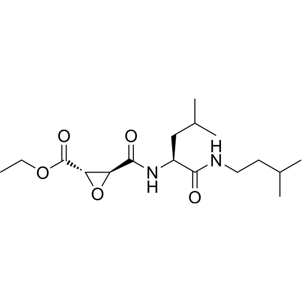
AloxistatinCysteine Protease Inhibitor -

HY-103667
2-HBANQO1 Inducer -

HY-118819
5-Bromo-3-phenyl salicylic acidAKR1C1 Inhibitor -

HY-114009
AKR1C1-IN-1AKR1C1 Inhibitor -
HY-111008A
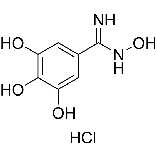
Trimidox hydrochlorideRibonucleotide Reductase Inhibitor -

HY-108534
PMX464Trx/TrxR Inhibitor
Publications Citing Use of MedChemExpress (MCE) ES 936
More

Biological Activity
Description
Cellular Effect
|
Cell Line

|
Type | Value | Description | References |
|---|---|---|---|---|
| MIA PaCa-2 | IC50 |
508 nM
Compound: 1, ES936
|
Cytotoxicity against human MIA PaCa2 cells after 72 hrs by MTT assay
Cytotoxicity against human MIA PaCa2 cells after 72 hrs by MTT assay
|
[PMID: 17944451] |
| MIA PaCa-2 | IC50 |
629 nM
Compound: 1, ES936
|
Cytotoxicity against human MIA PaCa2 cells after 4 hrs by MTT assay
Cytotoxicity against human MIA PaCa2 cells after 4 hrs by MTT assay
|
[PMID: 17944451] |
| NCI-H460 | IC50 |
2.81 μM
Compound: 5u
|
Cytotoxicity was measured on non-small cell lung cancer(NSCLC) in H460 cell lines.
Cytotoxicity was measured on non-small cell lung cancer(NSCLC) in H460 cell lines.
|
[PMID: 9822546] |
| NCI-H596 | IC50 |
2 μM
Compound: 5u
|
Cytotoxicity was measured on non-small cell lung cancer(NSCLC) in H596 cell lines.
Cytotoxicity was measured on non-small cell lung cancer(NSCLC) in H596 cell lines.
|
[PMID: 9822546] |
In Vitro
ES 936 (0-250 nM, 30-120 min) inhibits over 95% of NQO1 activity in HCT116, HT-29, MDA468 NQ16, MIA PaCa-2 and BxPC-3 cells[1][2].
ES 936 (100 nM, 30 min pretreatment) increases the IC50 of streptonigrin by ~6-fold in MDA468 NQ16 cells and ~2-fold in HCT116 cells, abrogating its toxicity[1].
ES 936 (0-500 nM, 2 h) induces a dose-dependent increase in DNA strand breaks and stimulates low levels of NAD(P)H oxidation in rat liver microsomes[1].
ES 936 (100 nM, 2 h pretreatment) significantly inhibits TNF-induced E-selectin expression and reduces TNF-induced VCAM-1 and ICAM-1 protein levels in transformed human bone marrow endothelial cells (TrHBMECs)[3].
ES936 (100 nM, 2 h pretreatment) leads to a marked reduction in TNF-induced adhesion of CD34+ KG1a cells to TrHBMECs in a parallel plate flow chamber assay[3].
MedChemExpress (MCE) has not independently confirmed the accuracy of these methods. They are for reference only. Further protocols information, click here.
-
Cell Line:Transformed human bone marrow endothelial cells (TrHBMECs)
-
Concentration:100 nM
-
Incubation Time:2 h pretreatment
-
Result:Decreased TNF-stimulated E-selectin protein levels.
Reduced TNF-induced VCAM-1 and ICAM-1 protein levels.
Resulted in lower nuclear levels of phospho-p65, phospho-c-Jun, and phospho-ATF2 in TrHBMECs.
In Vivo
MedChemExpress (MCE) has not independently confirmed the accuracy of these methods. They are for reference only.
-
Animal Model:MIA PaCa-2 cells in log-phase growth (2×10⁶ in 100 µL of 75:25 unsupplemented medium/Matrigel) were subcutaneously injected into the right flanks of 5- to 6-week-old female athymic nude mice (Ncr nu/nu)[2]
-
Dosage:5 mg/kg
-
Administration:i.p. daily for 10 days
-
Result:Achieved a maximal T/C value of 47% based on tumor volume analysis in MIA PaCa-2 xenograft tumors.
Showed no obvious toxicities and no difference in body weight between ES936-treated and control (DMSO) animals.
Significantly slowed the growth rate of MIA PaCa-2 xenograft tumors, with the inhibitory effect more significant during treatment (days 1-10) than after cessation of therapy (days 11-20).


Chemical Information
-
CAS No. 192820-78-3
-
Appearance Solid
-
Molecular Weight 356.33
-
Formula C18H16N2O6
-
Color Orange to red
-
SMILES
O=C(C(OC)=C1)C2=C(N(C)C(C)=C2COC3=CC=C([N+]([O-])=O)C=C3)C1=O
-
Shipping
Room temperature in continental US; may vary elsewhere.
-
Storage
Powder -20°C 3 years 4°C 2 years In solvent -80°C 6 months -20°C 1 month


Publications (1)
-
Journal Impact Factor
-
Most Recent
-
Cell Death Differ
A neutrophil-intrinsic CKLF1-PKM2 axis drives glycolytic flux for de novo DAG synthesis and pro-inflammatory ROS production. [Abstract]2026 May 12. PMID: 42120940


Solvent & Solubility
In Vitro:
DMSO : 2 mg/mL (5.61 mM; Need ultrasonic and warming; Hygroscopic DMSO has a significant impact on the solubility of product, please use newly opened DMSO)
Please refer to the solubility information to select the appropriate solvent. Once prepared, please aliquot and store the solution to prevent product inactivation from repeated freeze-thaw cycles.
Storage method and period of stock solution: -80°C, 6 months; -20°C, 1 month. When stored at -80°C, please use it within 6 months. When stored at -20°C, please use it within 1 month.
Please refer to the solubility information to select the appropriate solvent. Once prepared, please aliquot and store the solution to prevent product inactivation from repeated freeze-thaw cycles.
Storage method and period of stock solution: -80°C, 6 months; -20°C, 1 month. When stored at -80°C, please use it within 6 months. When stored at -20°C, please use it within 1 month.
Concentration (start) × Volume (start) = Concentration (final) × Volume (final)
In Vivo:
For the following dissolution methods, please prepare the working solution directly:
It is recommended to prepare fresh solutions and use them promptly within a short period of time.
The percentages shown for the solvents indicate their volumetric ratio in the final prepared solution. If precipitation or phase separation occurs during preparation, heat and/or sonication can be used to aid dissolution.
Add each solvent one by one: 50% PEG300 50% Saline
Solubility: 2.5 mg/mL (7.02 mM); Suspended solution; Need ultrasonic

Protocols
-
Cell Cytotoxicity Assay
Cytotoxicity assays are usually based on the assessment of cell membrane damage, which can also be indirectly detected by measuring cell viability. Detection methods include MTT assay, CKK-8 assay, LDH assay and ATP assay, etc.
-
Patient-Derived Xenograft (PDX)
Patient-derived xenograft (PDX) models are generated by engrafting primary human tumor tissue directly into immunodeficient mice, allowing in vivo propagation of patient tumor biology without initial in vitro adaptation. These models are used to preserve key histopathological and molecular characteristics of the original tumor and enable assessment of tumor growth dynamics and therapeutic response in a living organism. The biological readout is tumor engraftment and subsequent growth in the murine host, which reflects the ability of human tumor cells to survive, vascularize, and expand in an immunocompromised microenvironment.
-
Splenic/Portal-Vein Liver Metastasis Xenograft
Splenic and portal-vein liver metastasis xenograft models deliver tumor cells into the portal circulation so that cells reach the liver first and form hepatic metastatic lesions; splenic injection uses the spleen as an access route to the portal system, while direct portal-vein injection introduces cells into the portal vein without requiring splenectomy. The assay detects liver colonization, intrahepatic tumor growth, tumor distribution, treatment response, survival, and liver-metastasis microenvironment changes; readouts include bioluminescence or fluorescence imaging, gross liver nodule counts, liver weight or tumor burden, histology, and survival.
-
Subcutaneous Cell-Line-Derived Xenograft
Subcutaneous cell-line-derived xenograft (CDX) models are established by implanting cultured human cancer cell lines into immunodeficient mice, where the injected cells form localized tumors that can be monitored in vivo as a measure of tumorigenic potential, growth kinetics, and treatment response. These models are widely used in oncology research because they allow reproducible tumor formation and enable comparative assessment of tumor growth between different cell lines or genetic manipulations in a controlled in vivo microenvironment. Subcutaneous implantation of cancer cells in immunodeficient mice is a standard approach for evaluating tumor growth behavior and therapeutic response across multiple cancer types, including prostate, esophageal, pancreatic, and colon cancer models.
-
Orthotopic Cell-Line Xenograft
Orthotopic cell-line xenograft models involve implantation of human cancer cell lines into the anatomically corresponding organ of immunodeficient mice to reproduce tumor growth within a native microenvironment, enabling more clinically relevant tumor behavior compared with subcutaneous models. These models are widely used because orthotopic placement better recapitulates tumor progression, including invasion and metastatic spread, which are often underrepresented in heterotopic implantation systems. Compared with conventional xenografts, orthotopic implantation is described as more technically complex but provides improved simulation of tumor-microenvironment interactions and metastatic behavior, making it particularly valuable for translational oncology research. Surgical orthotopic implantation approaches have been emphasized as enabling faithful reproduction of clinical cancer features, including metastasis and disease progression patterns that align with the tumor’s organ of origi
-
Intraperitoneal/Peritoneal Dissemination Xenograft
Intraperitoneal (IP) or peritoneal dissemination xenograft models are based on the introduction of human cancer cells into the peritoneal cavity of immunodeficient mice, where they attach to peritoneal surfaces, form multicellular aggregates or spheroids, and progressively generate disseminated tumor nodules that mimic advanced peritoneal metastatic disease. These models are widely used to study ovarian cancer progression, tumor-microenvironment interactions, and intraperitoneal therapeutic responses, often incorporating bioluminescence or fluorescence imaging to longitudinally monitor tumor burden in vivo. The biological principle relies on the capacity of tumor cells such as SKOV3 or related ovarian carcinoma lines to survive in suspension, aggregate within ascites-like fluid, adhere to mesothelial surfaces, and invade peritoneal organs, thereby recapitulating human peritoneal carcinomatosis patterns observed in advanced disease.


Purity & Documentation
-
Data Sheet (279 KB)
-
SDS (393 KB)
- English - EN (393 KB)
- Français - FR (393 KB)
- Deutsch - DE (393 KB)
- Norwegian - NO (393 KB)
- Español - ES (393 KB)
- Swedish - SV (393 KB)
- Italian - IT (393 KB)
- Korean - KR (393 KB)
- Portuguese - PT (393 KB)
-
Handling Instructions (2659 KB)
References
[1]. Dehn DL, et al. Biochemical, cytotoxic, and genotoxic effects of ES936, a mechanism-based inhibitor of NAD(P)H:quinone oxidoreductase 1, in cellular systems. Mol Pharmacol. 2003 Sep;64(3):714-20. [Content Brief]
[2]. Dehn DL, et al. 5-Methoxy-1,2-dimethyl-3-[(4-nitrophenoxy)methyl]indole-4,7-dione, a mechanism-based inhibitor of NAD(P)H:quinone oxidoreductase 1, exhibits activity against human pancreatic cancer in vitro and in vivo. [Content Brief]
[3]. Zhou H, et al. NAD(P)H:quinone oxidoreductase 1-compromised human bone marrow endothelial cells exhibit decreased adhesion molecule expression and CD34+ hematopoietic cell adhesion. J Pharmacol Exp Ther. 2010 Jul;334(1):260-8. [Content Brief]


Complete Stock Solution Preparation Table
Please refer to the solubility information to select the appropriate solvent. Once prepared, please aliquot and store the solution to prevent product inactivation from repeated freeze-thaw cycles.
Storage method and period of stock solution: -80°C, 6 months; -20°C, 1 month. When stored at -80°C, please use it within 6 months. When stored at -20°C, please use it within 1 month.
| Optional Solvent | Concentration Solvent Mass | 1 mg | 5 mg | 10 mg | 25 mg |
|---|---|---|---|---|---|
| DMSO | 1 mM | 2.8064 mL | 14.0319 mL | 28.0639 mL | 70.1597 mL |
| 5 mM | 0.5613 mL | 2.8064 mL | 5.6128 mL | 14.0319 mL |


ES 936 Related Classifications
HY-100367 Related Classifications
 Powered by Bioz