



CPUY192018
Based on 1 publication(s) in Google Scholar
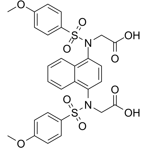
CPUY192018 is a potent inhibitor of the Keap1-Nrf2 protein-protein interaction, with an IC50 of 0.63 µM. CPUY192018 exhibits anti-inflammatory and antioxidant activities. CPUY192018 can activate the Nrf2-dependent antioxidant pathway and inhibit the NF-κB-related inflammatory response. CPUY192018 can be used in the research of inflammation-related diseases.
For research use only. We do not sell to patients.
- Purity : 98.90%
- CAS No.: 1567836-15-0
- Formula: C28H26N2O10S2
- Molecular Weight:614.64
-
Storage:Powder -20°C, 3 years , 4°C, 2 years ; In solvent -80°C, 6 months , -20°C, 1 month

To place orders, for customer services and technical support, please contact: MedChemExpress USA
Tel: 609-228-6898 E-mail: [email protected] [email protected]


-
 Biological Activity
-
Chemical Information
-
Solvent & Solubility
- Protocols
- Purity & Documentation
- References
-
 Help & FAQs


-
HY-12212
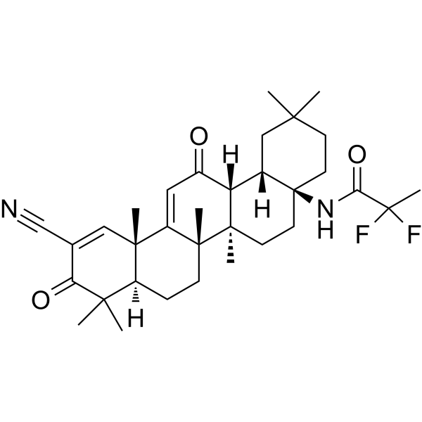
OmaveloxoloneNrf2 Activator -
HY-19543
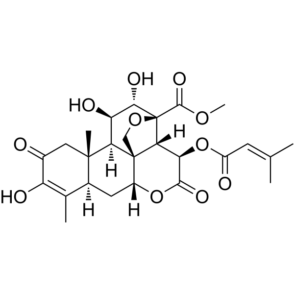
BrusatolNrf2 Inhibitor -
HY-17363
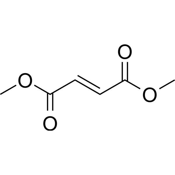
Dimethyl fumarateNrf2 Activator -
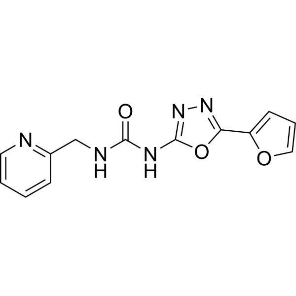
HY-19734
NK-252Nrf2 Activator -
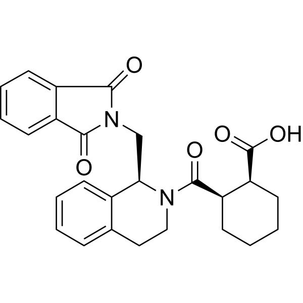
HY-110258
ML334NRF2 Activator -
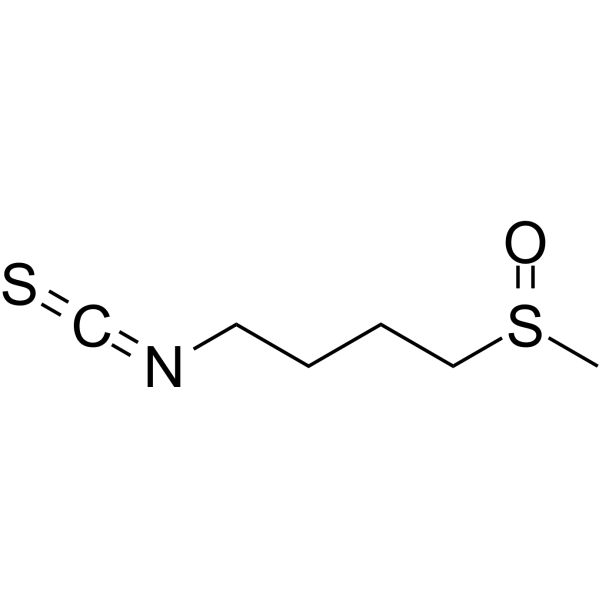
HY-13755
SulforaphaneKeap1/Nrf2/ARE Inducer -
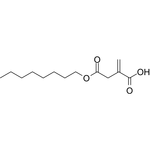
HY-112675
4-Octyl itaconateNrf2 Activator -
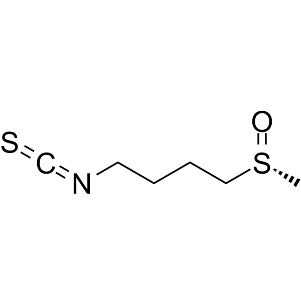
HY-13755A
(R)-SulforaphaneKeap1/Nrf2/ARE Inducer -
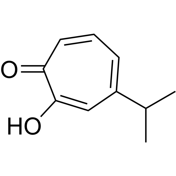
HY-B2230
HinokitiolVirus Protease Inhibitor -
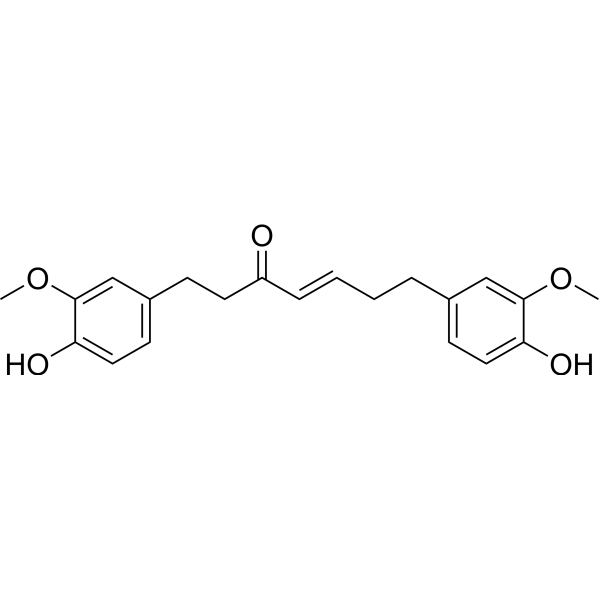
HY-120912
Gingerenone ANrf2-Gpx4 Activator -
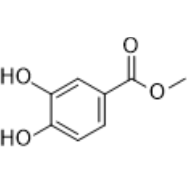
HY-Z0548
Methyl 3,4-dihydroxybenzoate -
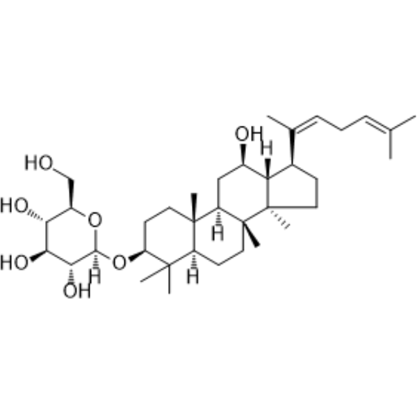
HY-N0606
Ginsenoside Rh3Nrf2 Activator
Publications Citing Use of MedChemExpress (MCE) CPUY192018
More

Biological Activity
Description
In Vitro
CPUY192018 (0-10 μM; 0-24 h) can significantly increase the nuclear translocation and level of Nrf2, as well as the expression of Nrf2 downstream proteins in HK-2 cells[1].
CPUY192018 (0-10 μM; 16-24 h) can inhibit ROS production, reduce MDA levels, increase the GSH/GSSG ratio, inhibit apoptosis, cytokine secretion, and the activation of the NF-κB signaling pathway in HK-2 cells treated with LPS (HY-D1056)[1].
MedChemExpress (MCE) has not independently confirmed the accuracy of these methods. They are for reference only.
-
Cell Line:HK-2 cells treated with LPS (HY-D1056)
-
Concentration:1 and 10μM
-
Incubation Time:Pretreated with 10 h, then co-incubation for 6 h
-
Result:Inhibited the phosphorylation levels of IKKβ, IκBα and NF-κB p65.
In Vivo
MedChemExpress (MCE) has not independently confirmed the accuracy of these methods. They are for reference only.
-
Animal Model:Female C57BL/6 mice aged 6-8 weeks old (18-20 g) treated LPS (HY-D1056)[1]
-
Dosage:5 and 20 mg/kg
-
Administration:Intraperitoneal injection; 8 weeks
-
Result:Attenuated the body weight loss in mice.
Reduced histological disease scores.
Improved glomerular pathological changes, activated the Nrf2 pathway to enhance renal antioxidant capacity and inhibited the inflammatory response.


Chemical Information
-
CAS No. 1567836-15-0
-
Appearance Solid
-
Molecular Weight 614.64
-
Formula C28H26N2O10S2
-
Color White to off-white
-
SMILES
O=C(O)CN(C1=C2C=CC=CC2=C(N(S(=O)(C3=CC=C(OC)C=C3)=O)CC(O)=O)C=C1)S(=O)(C4=CC=C(OC)C=C4)=O
-
Shipping
Room temperature in continental US; may vary elsewhere.
-
Storage
Powder -20°C 3 years 4°C 2 years In solvent -80°C 6 months -20°C 1 month


Publications (1)
-
Journal Impact Factor
-
Most Recent


Solvent & Solubility
In Vitro:
DMSO : 250 mg/mL (406.74 mM; Need ultrasonic; Hygroscopic DMSO has a significant impact on the solubility of product, please use newly opened DMSO)
Please refer to the solubility information to select the appropriate solvent. Once prepared, please aliquot and store the solution to prevent product inactivation from repeated freeze-thaw cycles.
Storage method and period of stock solution: -80°C, 6 months; -20°C, 1 month. When stored at -80°C, please use it within 6 months. When stored at -20°C, please use it within 1 month.
Please refer to the solubility information to select the appropriate solvent. Once prepared, please aliquot and store the solution to prevent product inactivation from repeated freeze-thaw cycles.
Storage method and period of stock solution: -80°C, 6 months; -20°C, 1 month. When stored at -80°C, please use it within 6 months. When stored at -20°C, please use it within 1 month.
Concentration (start) × Volume (start) = Concentration (final) × Volume (final)
In Vivo:
Select the appropriate dissolution method based on your experimental animal and administration route.
- For the following dissolution methods, please ensure to first prepare a clear stock solution using an In Vitro approach and then sequentially add co-solvents:
- To ensure reliable experimental results, the clarified stock solution can be appropriately stored based on storage conditions. As for the working solution for In Vivo experiments, it is recommended to prepare freshly and use it on the same day.
- The percentages shown for the solvents indicate their volumetric ratio in the final prepared solution. If precipitation or phase separation occurs during preparation, heat and/or sonication can be used to aid dissolution.
Add each solvent one by one: 10% DMSO 90% (20% SBE-β-CD in Saline)
Solubility: ≥ 2.08 mg/mL (3.38 mM); Clear solution
This protocol yields a clear solution of ≥ 2.08 mg/mL (saturation unknown).
Taking 1 mL working solution as an example, add 100 μL DMSO stock solution (20.8 mg/mL) to 900 μL 20% SBE-β-CD in Saline, and mix evenly.
Preparation of 20% SBE-β-CD in Saline (4°C, storage for one week): 2 g SBE-β-CD powder is dissolved in 10 mL Saline, completely dissolve until clear.
In Vivo Dissolution Calculator
Please enter the basic information of animal experiments:
-
-
-
-
Recommended: Prepare an additional quantity of animals to account for potential losses during experiments.
Please enter your animal formula composition:
-
%DMSO +
Recommended: Keep the proportion of DMSO in working solution below 2% if your animal is weak.
-
%+
-
+%Tween-80 + +
-
%Saline +
The co-solvents required include: DMSO, . All of co-solvents are available by MedChemExpress (MCE). , Tween 80. All of co-solvents are available by MedChemExpress (MCE).
Working solution concentration: 0.22 mg/mL
Method for preparing stock solution: mg drug dissolved in μL DMSO. Stock solution concentration: mg/mL.
1. Take μL DMSO stock solution;
2. Add μL .
μL , mix evenly;
3. Then add μL Tween 80, mix evenly;
4. Then add μL
Please ensure that the stock solution in the first step is dissolved to a clear state, and add co-solvents in sequence. You can use ultrasonic heating (ultrasonic cleaner, recommended frequency 20-40 kHz), vortexing, etc. to assist dissolution.


Purity & Documentation
-
Data Sheet (285 KB)
-
SDS (251 KB)
- English - EN (251 KB)
- Français - FR (251 KB)
- Deutsch - DE (251 KB)
- Norwegian - NO (251 KB)
- Español - ES (251 KB)
- Swedish - SV (251 KB)
- Italian - IT (251 KB)
- Korean - KR (251 KB)
- Portuguese - PT (251 KB)
-
Handling Instructions (2659 KB)
References
[1]. Lu MC, et al. CPUY192018, a potent inhibitor of the Keap1-Nrf2 protein-protein interaction, alleviates renal inflammation in mice by restricting oxidative stress and NF-κB activation. Redox Biol. 2019 Sep;26:101266. [Content Brief]
[2]. Lee S, et al. Structure-activity relationships of 1,4-bis(arylsulfonamido)-benzene or naphthalene-N,N'-diacetic acids with varying C2-substituents as inhibitors of Keap1-Nrf2 protein-protein interaction. Eur J Med Chem. 2022; 237:114380. [Content Brief]


Complete Stock Solution Preparation Table
Please refer to the solubility information to select the appropriate solvent. Once prepared, please aliquot and store the solution to prevent product inactivation from repeated freeze-thaw cycles.
Storage method and period of stock solution: -80°C, 6 months; -20°C, 1 month. When stored at -80°C, please use it within 6 months. When stored at -20°C, please use it within 1 month.
| Optional Solvent | Concentration Solvent Mass | 1 mg | 5 mg | 10 mg | 25 mg |
|---|---|---|---|---|---|
| DMSO | 1 mM | 1.6270 mL | 8.1348 mL | 16.2697 mL | 40.6742 mL |
| 5 mM | 0.3254 mL | 1.6270 mL | 3.2539 mL | 8.1348 mL | |
| 10 mM | 0.1627 mL | 0.8135 mL | 1.6270 mL | 4.0674 mL | |
| 15 mM | 0.1085 mL | 0.5423 mL | 1.0846 mL | 2.7116 mL | |
| 20 mM | 0.0813 mL | 0.4067 mL | 0.8135 mL | 2.0337 mL | |
| 25 mM | 0.0651 mL | 0.3254 mL | 0.6508 mL | 1.6270 mL | |
| 30 mM | 0.0542 mL | 0.2712 mL | 0.5423 mL | 1.3558 mL | |
| 40 mM | 0.0407 mL | 0.2034 mL | 0.4067 mL | 1.0169 mL | |
| 50 mM | 0.0325 mL | 0.1627 mL | 0.3254 mL | 0.8135 mL | |
| 60 mM | 0.0271 mL | 0.1356 mL | 0.2712 mL | 0.6779 mL | |
| 80 mM | 0.0203 mL | 0.1017 mL | 0.2034 mL | 0.5084 mL | |
| 100 mM | 0.0163 mL | 0.0813 mL | 0.1627 mL | 0.4067 mL |


CPUY192018 Related Classifications
HY-120371 Related Classifications
 Powered by Bioz