



Mosliciguat
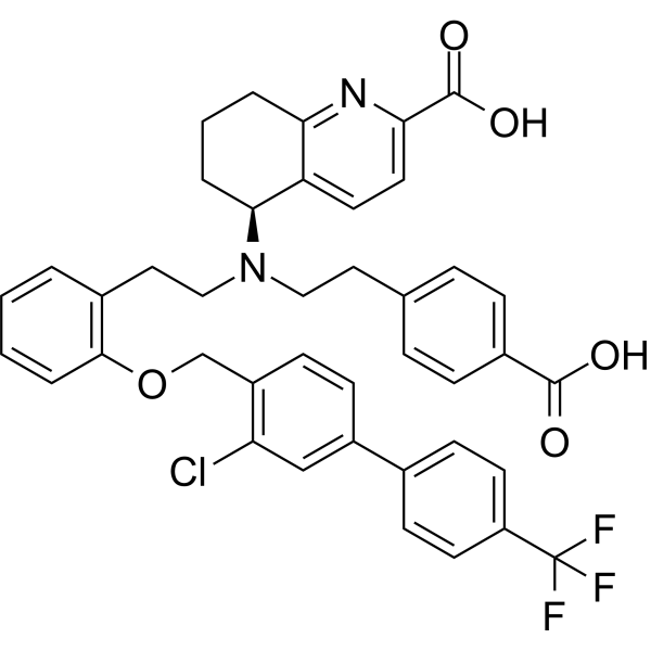
Mosliciguat (BAY 1237592) is an apo-soluble guanylate cyclase activator. Mosliciguat improves cardiopulmonary circulation, reduces pulmonary arterial pressure in animal models, and its efficacy is enhanced under oxidative stress conditions. Mosliciguat exerts a bronchodilatory effect in a rat model of acetylcholine-induced bronchoconstriction. Mosliciguat can be used for research on pulmonary arterial hypertension.
For research use only. We do not sell to patients.
- CAS No.: 2231749-54-3
- Formula: C41H36ClF3N2O5
- Molecular Weight:729.18
-
Storage:
Please store the product under the recommended conditions in the Certificate of Analysis.

To place orders, for customer services and technical support, please contact: MedChemExpress USA
Tel: 609-228-6898 E-mail: [email protected] [email protected]


-
 Biological Activity
-
Chemical Information
- Protocols
- Purity & Documentation
- References
-
 Help & FAQs


-
HY-16774
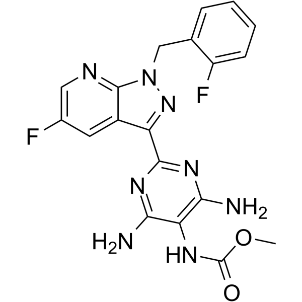
VericiguatsCG Stimulator -
HY-17584
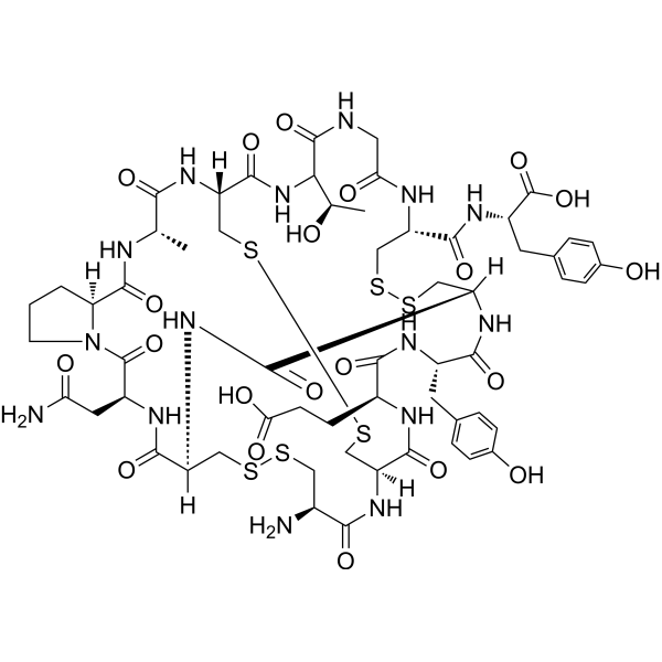
LinaclotideGuanylate Cyclase Agonist -
HY-14779
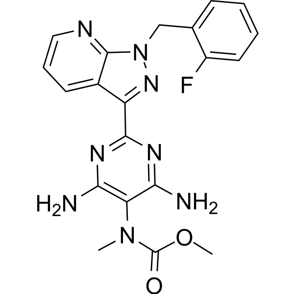
Riociguat -
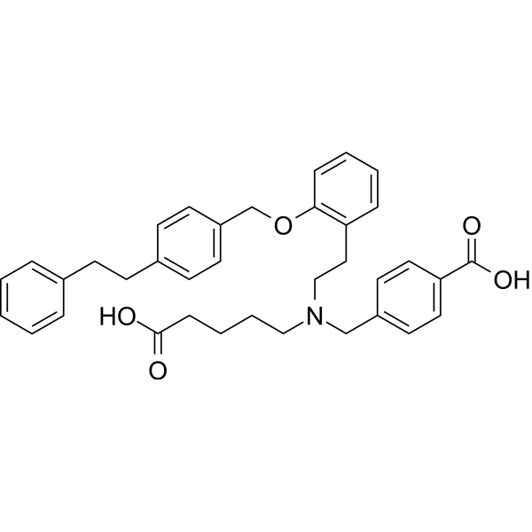
HY-14181
CinaciguatGuanylate Cyclase Activator -
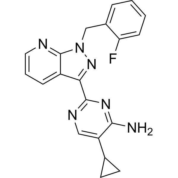
HY-12376
BAY 41-2272Guanylate Cyclase Activator -
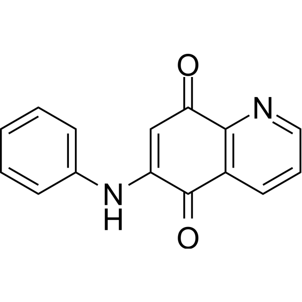
HY-W013386
LY83583sGC Inhibitor -
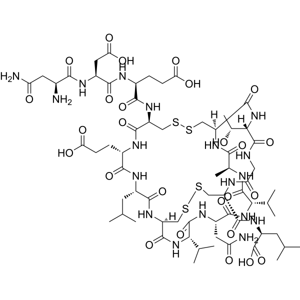
HY-108741
PlecanatideGuanylate Cyclase-C Agonist -
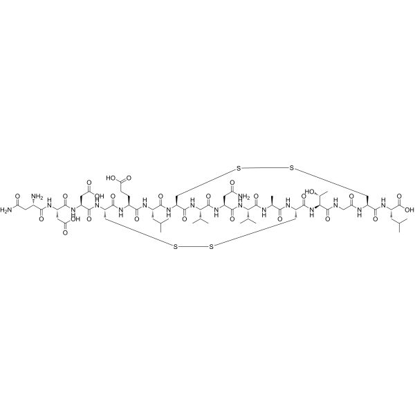
HY-P3099
Uroguanylin (human)Endogenous Peptide Hormone -
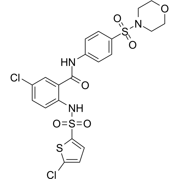
HY-17500
AtaciguatsGC Activator -
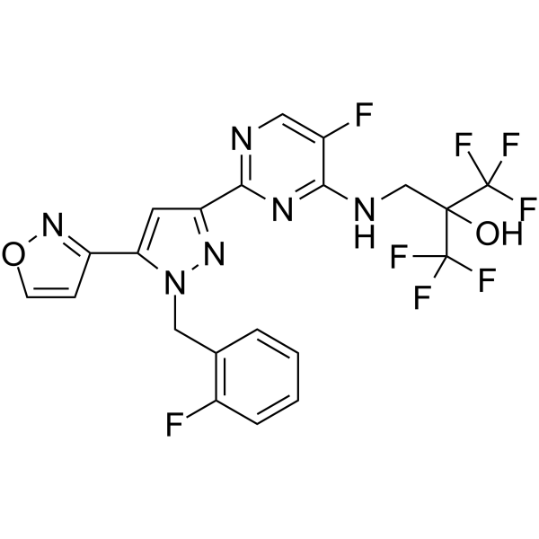
HY-109039
PraliciguatSoluble Guanylate Cyclase Stimulator -
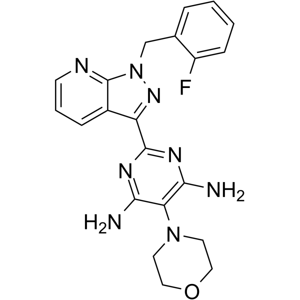
HY-W062836
BAY 41-8543sGC Stimulator -
HY-145607
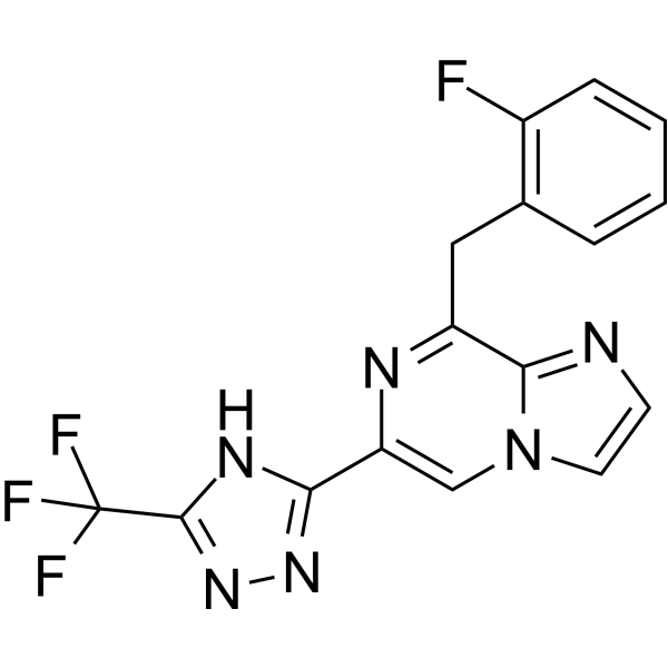
ZagociguatSoluble Guanylate Cyclase Stimulator


Biological Activity
Description
In Vitro
Mosliciguat (0.01-100 μM) potently activates both heme-containing and heme-free (apo) recombinant soluble guanylyl cyclase (sGC) in vitro. Its activity is enhanced in the presence of the sGC inhibitor ODQ (HY-101255), and its action is independent of nitric oxide[1].
MedChemExpress (MCE) has not independently confirmed the accuracy of these methods. They are for reference only. Further protocols information, click here.
In Vivo
Inhalation administration of Mosliciguat (100 µg/kg via nebulizer) produces a long-lasting (up to 17 hours) inhibitory effect on hypoxia-induced pulmonary hypertension in conscious dogs without affecting heart rate[1].
Mosliciguat (100 µg/kg; administered via inhalation; nebulized, 7 min per dose; 30-100 µg/kg; administered intravenously; cumulative bolus injection) effectively reduces pulmonary arterial pressure in a porcine model of unilateral bronchial obstruction without exacerbating ventilation-perfusion mismatch. In contrast, intravenous administration alone exacerbates desaturation and reduces systemic blood pressure[1].
Mosliciguat (1-100 µg/kg; administered via an aerosol microfeeding system) exerts a dose-dependent bronchodilatory effect in a rat model of acetylcholine-induced bronchoconstriction, with an inhibition rate of pulmonary resistance reaching 68% at the dose of 100 µg/kg[1].
MedChemExpress (MCE) has not independently confirmed the accuracy of these methods. They are for reference only.
-
Animal Model:Ellegaard Göttinger (female, 4-5 kg body weight, pulmonary hypertension induced by thromboxane A2 analog [U-46619] infusion)[1]
-
Dosage:3 µg/kg, 10 µg/kg, 30 µg/kg, 100 µg/kg, 300 µg/kg (inhaled); 1 µg/kg, 3 µg/kg, 10 µg/kg, 30 µg/kg, 100 µg/kg (intravenous)
-
Administration:inhaled; via nebulizer, 7 min/dose, cumulative doses; intravenous; cumulative infusions
-
Result:Induced a dose-dependent decrease in mean pulmonary arterial pressure (PAP) starting at 10 µg/kg, with no relevant effects on systemic mean blood pressure (BP).
Caused a >5% reduction in PAP versus vehicle at 3 µg/kg.
Achieved maximal PAP reduction 90-120 min after inhalation, maintained for 4 hours.
Did not reduce BP at inhaled 300 µg/kg, while intravenous 30 µg/kg reduced both BP and PAP.
Caused similar or greater PAP reduction than systemic endothelin receptor antagonist or phosphodiesterase type 5 inhibitor at inhaled 30 µg/kg and 100 µg/kg, without reducing BP.
Maintained PAP-lowering efficacy without additional BP effects when combined with endothelin receptor antagonist or phosphodiesterase type 5 inhibitor at inhaled 30 µg/kg.
Showed enhanced PAP reduction without BP effects under oxidative stress (ODQ or L-NAME pretreatment) at inhaled 30 µg/kg.
-
Animal Model:Beagle (male, 8-15 kg body weight, pulmonary hypertension induced by hypoxia exposure)[1]
-
Dosage:100 µg/kg
-
Administration:inhaled; via nebulizer
-
Result:Attenuated hypoxia-induced systolic pulmonary arterial pressure (sPAP) increases for 1-17 hours after administration; the effect dissipated by 24 hours.
Caused no effects on heart rate.
-
Animal Model:Ellegaard Göttinger (female, 4-5 kg body weight, ventilation/perfusion mismatch induced by unilateral broncho-occlusion)[1]
-
Dosage:100 µg/kg (inhaled); 30 µg/kg, 100 µg/kg (intravenous)
-
Administration:inhaled; via nebulizer, 7 min/dose; intravenous; cumulative bolus
-
Result:Decreased mean pulmonary arterial pressure (PAP) during unilateral ventilation cycles at inhaled 100 µg/kg, with a trend toward reduced desaturation area (no ventilation/perfusion mismatch deterioration) and no effects on systemic BP.
Decreased PAP but increased desaturation area and reduced systemic BP at intravenous 30 µg/kg and 100 µg/kg.
-
Animal Model:Brown Norway (male, 10-12 weeks old, bronchoconstriction induced by acetylcholine aerosol provocation)[1]
-
Dosage:1 µg/kg, 10 µg/kg, 100 µg/kg
-
Administration:inhaled; via aerosolized micro feeding system
-
Result:Induced a dose-dependent bronchodilatory effect, inhibiting acetylcholine-induced bronchoconstriction.
Achieved 68% inhibition of lung resistance increase and 37% improvement in dynamic compliance at 100 µg/kg, affecting both large and small airways.
Clinical Trial
| NCT Number | Sponsor | Condition | Start Date |
Phase

|
|---|---|---|---|---|
| NCT01329991 | Plexxikon| | 2011-05 | PHASE1 |


Chemical Information
-
CAS No. 2231749-54-3
-
Molecular Weight 729.18
-
Formula C41H36ClF3N2O5
-
SMILES
OC(C1=CC=C(C2=N1)[C@H](CCC2)N(CCC(C=C3)=CC=C3C(O)=O)CCC(C=CC=C4)=C4OCC(C=CC(C(C=C5)=CC=C5C(F)(F)F)=C6)=C6Cl)=O
-
Synonyms
BAY 1237592
-
Shipping
Room temperature in continental US; may vary elsewhere.
-
Storage
Please store the product under the recommended conditions in the Certificate of Analysis.

Protocols
-
ROS/oxidative-stress fluorescent staining
ROS/oxidative-stress fluorescent staining uses cell-permeant fluorogenic probes that become fluorescent after oxidation inside cells or tissues; commonly used examples include DCFH-DA/DCFDA for broad cellular oxidant detection, DHE for superoxide-related signal detection, MitoSOX for mitochondrial superoxide-related signal detection, and CellROX probes for oxidative-stress-associated fluorescence readouts. The assay detects probe oxidation rather than a single ROS species unless the probe and analysis method have been chemically validated for that species. DCFH-DA enters cells, is deacetylated by intracellular esterases to DCFH, and produces fluorescent DCF after oxidation, so the readout is used as an operational measure of total cellular oxidative stress rather than a species-specific ROS measurement. DHE and MitoSOX can report superoxide-related oxidation, but red fluorescence alone can include non-specific ethidium-like oxidation products; HPLC or optimized spectral approaches are
-
How to Select a Suitable Non-Mouse Animal Model
Selecting a suitable non-mouse animal model is a structured decision based on the research question, required anatomy or physiology, disease mechanism, endpoint feasibility, translational relevance, and ethical justification. Non-mouse models are preferred when mice cannot reproduce key human-relevant features, such as organ size, surgical anatomy, cardiovascular physiology, neuroanatomy, immune features, pharmacology, toxicology, or long-term clinical procedures. Candidate species may include rats, rabbits, guinea pigs, ferrets, zebrafish, pigs, sheep, goats, dogs, cats, horses, and non-human primates, but each species must be justified by its specific scientific advantage rather than convenience or tradition. Unresolved questions include how to quantify translational superiority across species, how to balance increased biological relevance against higher ethical burden, and when human-derived systems or new approach methodologies should replace animal use.
-
Research Protocol for Cardiovascular Diseases
Cardiovascular disease can be modeled as maladaptive cardiac remodeling, where ischemic injury or pressure overload activates inflammatory signaling, fibroblast activation, extracellular-matrix deposition, cardiomyocyte hypertrophy, vascular remodeling, and progressive ventricular dysfunction. The TGF-β/SMAD axis is a central profibrotic pathway after myocardial injury and pressure overload, while innate immune and cytokine pathways regulate leukocyte recruitment, scar formation, and adverse remodeling. Key unresolved questions include which inflammatory signals are reparative versus harmful, when fibrosis is protective versus maladaptive, and whether pathway inhibition improves function without weakening necessary infarct healing or compensatory remodeling.
-
How to Choose the Right Model Animal
Choosing the right model animal is a validity-driven decision in which the species, strain, sex, age, genetic background, disease-induction method, outcome measures, and welfare burden must match the scientific question rather than laboratory tradition or convenience. A model should be selected by judging face validity, construct validity, and predictive validity: whether it resembles the human phenotype, whether it reproduces relevant mechanisms, and whether results are likely to predict human biology or treatment response. Animal studies often fail to translate because of species differences, weak disease resemblance, poor experimental design, inadequate reporting, publication bias, and underuse of randomization, blinding, and sample-size justification. Unresolved questions include how to rank competing models objectively, how much human-disease complexity must be reproduced for a given objective, and when non-animal systems such as organoids, ex vivo tissue, or computational models


Purity & Documentation
References

Calculators
Concentration (start) × Volume (start) = Concentration (final) × Volume (final)


Mosliciguat Related Classifications
HY-137446 Related Classifications
 Powered by Bioz
Keywords
- Mosliciguat
- 2231749-54-3
- BAY 1237592
- BAY1237592
- BAY-1237592
- Guanylate Cyclase
- conscious dogs
- nitric oxide
- pulmonary hypertension
- recombinant sGC
- pulmonary vasodilator
- acetylcholine-induced bronchoconstriction rat model
- lung resistance
- ODQ
- apo-soluble guanylate cyclase
- unilateral broncho-occlusion model
- Inhibitor
- inhibitor
- inhibit