



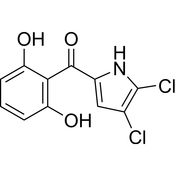
Pyoluteorin

Based on 1 Customer Validation
Pyoluteorin is an antibiotic that inhibits Oomycete fungi, including the plant pathogen Pythium ultimum, and suppresses plant diseases caused by this fungus. Pyoluteorin induces human triple-negative breast cancer MDA-MB-231 cells apoptosis in vitro. Pyoluteorin can be used for the research of human triple-negative breast cancer.
Para uso exclusivo en investigación. No vendemos a pacientes.
- Pureza : 99.90%
- No. CAS: 25683-07-2
- Fòrmula: C11H7Cl2NO3
- Peso molecular:272.08
-
Almacenamiento:
4°C, stored under nitrogen
* In solvent : -80°C, 6 months; -20°C, 1 month (stored under nitrogen)

To place orders, for customer services and technical support, please contact: MedChemExpress USA
Tel: 609-228-6898 E-mail: [email protected] [email protected]


-
 Actividad biológica
-
Chemical Information
-
Solvente y solubilidad
- Protocolo
- Pureza y Documentación
- Referencias
-
 Help & FAQs


Ver todos los productos específicos de isoformas Antibiotic
More

Actividad biológica
Descripciòn
Cellular Effect
|
Cell Line

|
Type | Value | Description | References |
|---|---|---|---|---|
| MDCK | IC50 |
4.8 μM
Compound: Pyoluteorin
|
Cytotoxicity against MDCK cells assessed as reduction in cell viability after 72 hrs by MTT assay
Cytotoxicity against MDCK cells assessed as reduction in cell viability after 72 hrs by MTT assay
|
[PMID: 30793902] |
In Vitro
Pyoluteorin is an antifungal compound composed of a bichlorinated pyrrole linked to a resorcinol moiety, were identified within a 24-kb genomic region of Pseudomonas fluorescens Pf-5[1].
Pyoluteorin has significant cytotoxicity towards MCF-7 (IC50=1.84 μM). Pyoluteorin also displays significantly selective cytotoxicity against BT474, HCC1954, MAD-MB-468, MDA-MB-231, and MCF-10A cells with IC50s of 9.75±0.16, 0.94±0.01, 3.89±0.08, 0.97±0.01, and 57.01±0.76 μM, respectively[2].
Pyoluteorin (0.1-10 μM; for 24 hours) induces change of apoptosis-related protein expressions. Pyoluteorin-induced cell apoptosis in MDA-MB-231 is related to Bcl-2 family proteins and caspase cascade[2].
Pyoluteorin (0.1-10 μM; for 24 h) induces cell cycle arrest and apoptosis in human triple-negative breast cancer cells MDA-MB-231[2].
MedChemExpress (MCE) has not independently confirmed the accuracy of these methods. They are for reference only.
-
Cell Line:Human triple-negative breast cancer cell MDA-MB-231
-
Concentration:0, 0.032, 0.16, 0.8, 4, 20, 100 μM
-
Incubation Time:24, 48, 72 hours
-
Result:Inhibited cells proliferation in a dose- and time-dependent manner.
-
Cell Line:MDA-MB-231 cells
-
Concentration:0.1, 0.3, 1, 3, 10 μM
-
Incubation Time:24 hours
-
Result:The levels of the anti-apoptotic proteins Bcl-2, Bcl-XL and PARP were obviously decreased while the pro-apoptotic proteins BAX and caspase 3 were increased in a dose-dependent manner.


Chemical Information
-
No. CAS 25683-07-2
-
Appearance Solid
-
Peso molecular 272.08
-
Fòrmula C11H7Cl2NO3
-
Color Off-white to yellow
-
SMILES
O=C(C1=C(C=CC=C1O)O)C2=CC(Cl)=C(N2)Cl
-
Structure Classification
-
Initial Source
Schroeter
-
Envío
Room temperature in continental US; may vary elsewhere.
-
Almacenamiento
4°C, stored under nitrogen
* In solvent : -80°C, 6 months; -20°C, 1 month (stored under nitrogen)


Solvente y solubilidad
In Vitro:
DMSO : 125 mg/mL (459.42 mM; Need ultrasonic; Hygroscopic DMSO has a significant impact on the solubility of product, please use newly opened DMSO)
Please refer to the solubility information to select the appropriate solvent. Once prepared, please aliquot and store the solution to prevent product inactivation from repeated freeze-thaw cycles.
Storage method and period of stock solution: -80°C, 6 months; -20°C, 1 month (stored under nitrogen). When stored at -80°C, please use it within 6 months. When stored at -20°C, please use it within 1 month.
Please refer to the solubility information to select the appropriate solvent. Once prepared, please aliquot and store the solution to prevent product inactivation from repeated freeze-thaw cycles.
Storage method and period of stock solution: -80°C, 6 months; -20°C, 1 month (stored under nitrogen). When stored at -80°C, please use it within 6 months. When stored at -20°C, please use it within 1 month.
Concentration (start) × Volume (start) = Concentration (final) × Volume (final)


Pureza y Documentación
-
Ficha de datos (319 KB)
-
SDS (480 KB)
- English - EN (480 KB)
- Français - FR (480 KB)
- Deutsch - DE (480 KB)
- Norwegian - NO (480 KB)
- Español - ES (480 KB)
- Swedish - SV (480 KB)
- Italian - IT (480 KB)
- Korean - KR (480 KB)
- Portuguese - PT (480 KB)
-
Instrucciones de manejo (2659 KB)
Referencias
[1]. B Nowak-Thompson, et al. Characterization of the pyoluteorin biosynthetic gene cluster of Pseudomonas fluorescens Pf-5. J Bacteriol. 1999 Apr;181(7):2166-74. [Content Brief]
[2]. Ting Ding, et al. Pyoluteorin induces cell cycle arrest and apoptosis in human triple-negative breast cancer cells MDA-MB-231. J Pharm Pharmacol. 2020 Jul;72(7):969-978. [Content Brief]


Complete Stock Solution Preparation Table
Please refer to the solubility information to select the appropriate solvent. Once prepared, please aliquot and store the solution to prevent product inactivation from repeated freeze-thaw cycles.
Storage method and period of stock solution: -80°C, 6 months; -20°C, 1 month (stored under nitrogen). When stored at -80°C, please use it within 6 months. When stored at -20°C, please use it within 1 month.
| Optional Solvent | Concentration Solvent Mass | 1 mg | 5 mg | 10 mg | 25 mg |
|---|---|---|---|---|---|
| DMSO | 1 mM | 3.6754 mL | 18.3769 mL | 36.7539 mL | 91.8847 mL |
| 5 mM | 0.7351 mL | 3.6754 mL | 7.3508 mL | 18.3769 mL | |
| 10 mM | 0.3675 mL | 1.8377 mL | 3.6754 mL | 9.1885 mL | |
| 15 mM | 0.2450 mL | 1.2251 mL | 2.4503 mL | 6.1256 mL | |
| 20 mM | 0.1838 mL | 0.9188 mL | 1.8377 mL | 4.5942 mL | |
| 25 mM | 0.1470 mL | 0.7351 mL | 1.4702 mL | 3.6754 mL | |
| 30 mM | 0.1225 mL | 0.6126 mL | 1.2251 mL | 3.0628 mL | |
| 40 mM | 0.0919 mL | 0.4594 mL | 0.9188 mL | 2.2971 mL | |
| 50 mM | 0.0735 mL | 0.3675 mL | 0.7351 mL | 1.8377 mL | |
| 60 mM | 0.0613 mL | 0.3063 mL | 0.6126 mL | 1.5314 mL | |
| 80 mM | 0.0459 mL | 0.2297 mL | 0.4594 mL | 1.1486 mL | |
| 100 mM | 0.0368 mL | 0.1838 mL | 0.3675 mL | 0.9188 mL |


Pyoluteorin Related Classifications
HY-114979 Related Classifications
 Powered by Bioz