



Azilsartan
Based on 6 publication(s) in Google Scholar
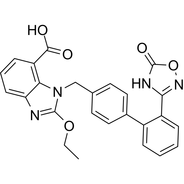
Azilsartan (TAK-536) is an orally active, potent, selective and specific angiotensin II type 1 receptor (AT1) antagonist. Azilsartan induces ROS formation and apoptosis in HepG2 cells. Azilsartan shows neuroprotective and anticancer activity. Azilsartan can be used for hypertension and stroke research.
For research use only. We do not sell to patients.
- Purity : 99.68%
- CAS No.: 147403-03-0
- Formula: C25H20N4O5
- Molecular Weight:456.45
-
Storage:Powder -20°C, 3 years , 4°C, 2 years ; In solvent -80°C, 2 years , -20°C, 1 year

To place orders, for customer services and technical support, please contact: MedChemExpress USA
Tel: 609-228-6898 E-mail: [email protected] [email protected]


-
 Biological Activity
-
Chemical Information
-
Solvent & Solubility
- Protocols
- Purity & Documentation
- References
-
 Help & FAQs


-
HY-B0205
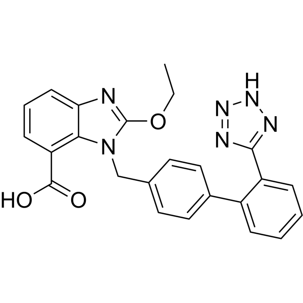
CandesartanAngiotensin II AT1-Receptor Blocker -
HY-18204
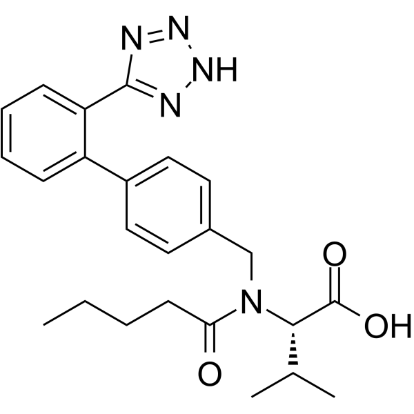
ValsartanAngiotensin II Receptor Antagonist -
HY-17512
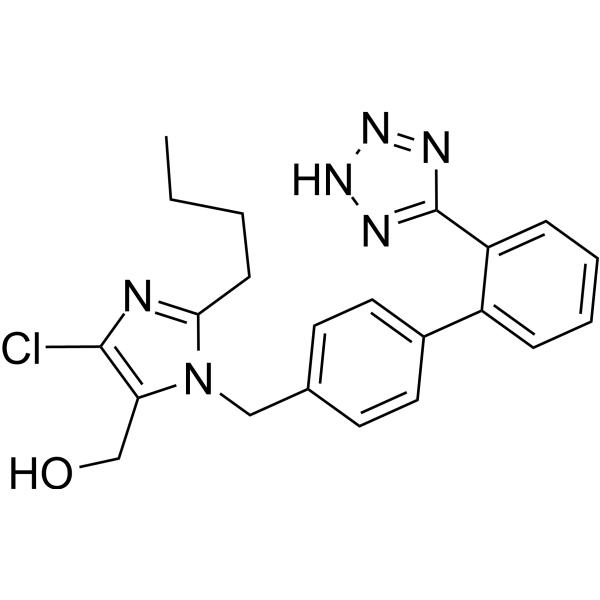
LosartanAngiotensin II Receptor Antagonist -
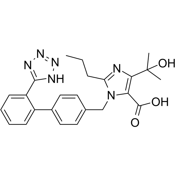
HY-17004
OlmesartanAT1R Antagonist -
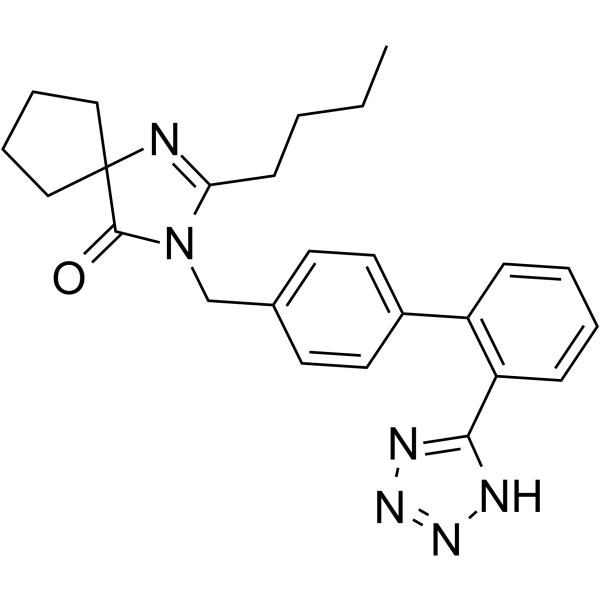
HY-B0202
IrbesartanAT1 Receptor Blocker -
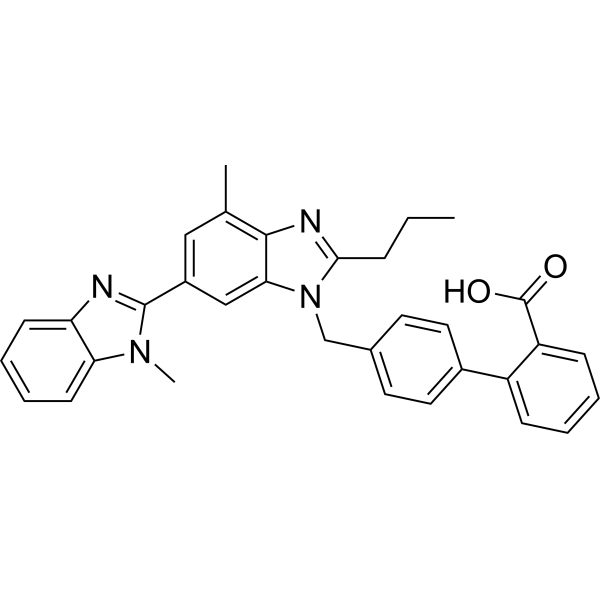
HY-13955
TelmisartanAngiotensin II Type 1 Receptor -
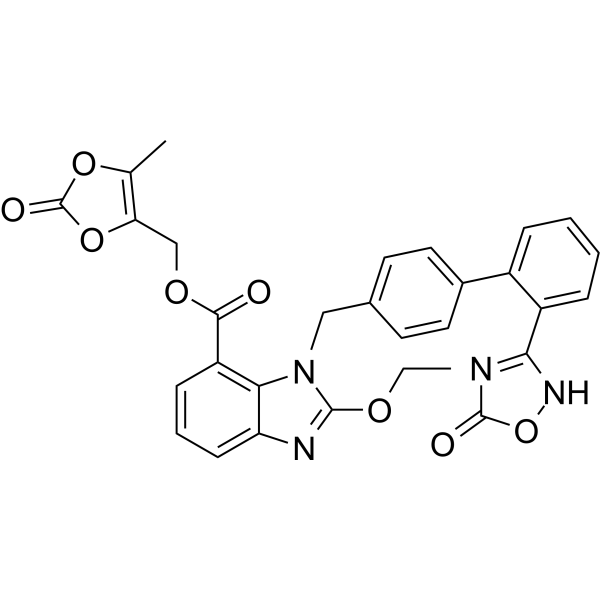
HY-14736
Azilsartan medoxomilAngiotensin Receptor Antagonist -
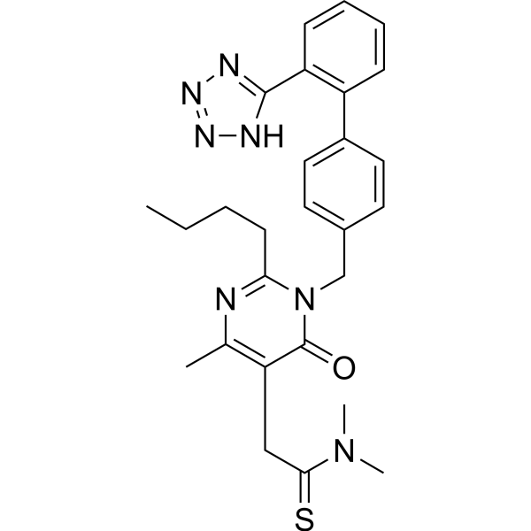
HY-B0780
FimasartanAngiotensin Receptor Antagonist -
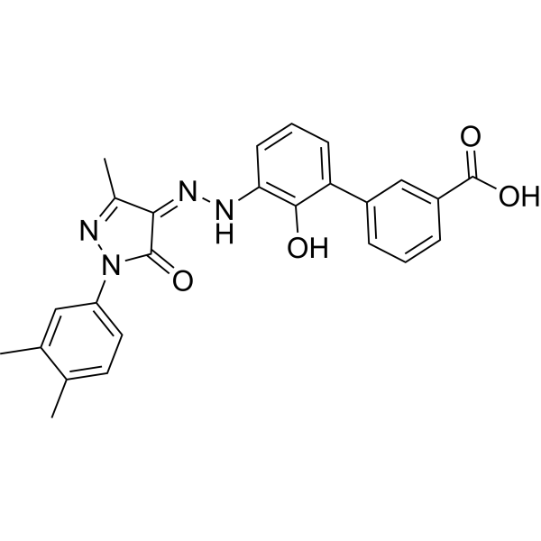
HY-15306
EltrombopagTPO Receptor Agonist -
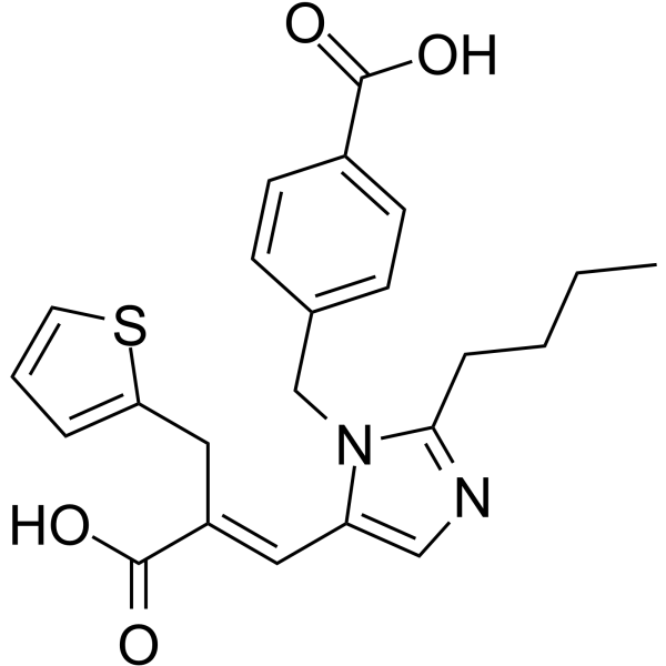
HY-117743
EprosartanAngiotensin II Receptor Antagonist -
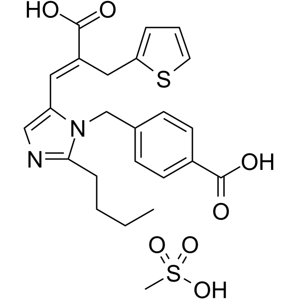
HY-15834A
Eprosartan mesylateAngiotensin II Receptor Antagonist -
HY-12765
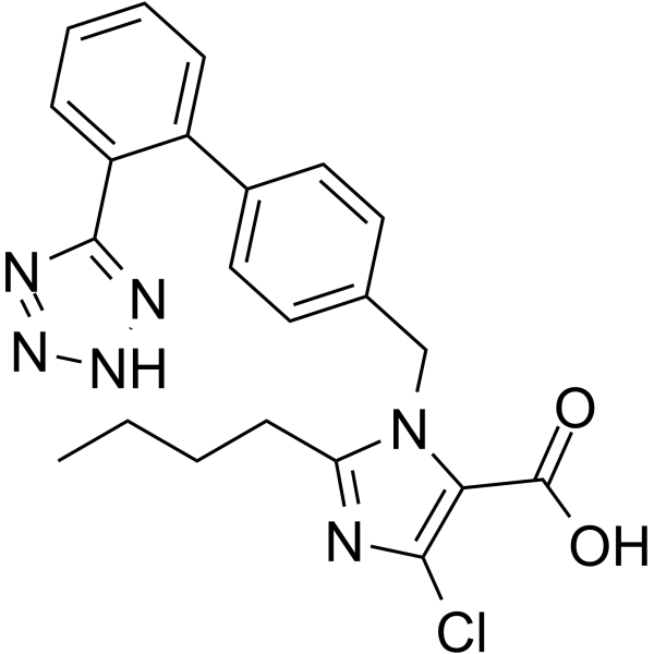
Losartan Carboxylic AcidAT1 Antagonist
Publications Citing Use of MedChemExpress (MCE) Azilsartan
More Customer Validation & Images

-
IF
All Angiotensin Receptor Isoforms
More

Biological Activity
Description
IC50 & Target
|
AT1 Receptor |
In Vitro
Azilsartan (0-200 μM, 0-72 h) decreases the viability of HepG2 cells[5].
Azilsartan (100 μM, 24 h) induces apoptosis in HepG2 cells[5].
Azilsartan inhibits the specific binding of 125I-Sar1-Ile8-AII to human angiotensin type 1 receptors with an IC50 of 2.6 nM[3].
Azilsartan potently inhibits aortic endothelial and vascular cell proliferation in the absence of exogenous Ang II supplementation[5].
Azilsartan enhances adipogenesis and exerted greater effects than Valsartan (HY-18204) on expression of genes encoding peroxisome proliferator-activated receptor-α (PPARα), PPARδ, leptin, adipsin, and adiponectin[1].
MedChemExpress (MCE) has not independently confirmed the accuracy of these methods. They are for reference only.
-
Cell Line:HepG2 and KDR cells
-
Concentration:5, 25, 50, 100 and 200 μM
-
Incubation Time:24, 48, and 72 h
-
Result:Gradually decreased the viability of HepG2 cells by increasing the incubation time and dose, the inhibitory concentration of Azilsartan (IC 50%) against HepG2 cells was 100 μM for 24 h treatment time point while in KDR epithelial normal cells no significant cytotoxic effect was observed during the similar treatment conditions.
-
Cell Line:HepG2 cells
-
Concentration:100 μM
-
Incubation Time:24 h
-
Result:Induced 57.2% early and 0.52% late apoptosis respectively after 24 h.
In Vivo
Azilsartan (0-2 mg/kg, Oral gavage, once daily for 21 days) lowers blood pressure and basal plasma insulin concentration[2].
Azilsartan (2 and 4 mg/kg; PO, daily for 9 days) offers protection against ischemia induced secondary brain injury[4].
MedChemExpress (MCE) has not independently confirmed the accuracy of these methods. They are for reference only.
-
Animal Model:Male Wistar-Kyoto (WKY) rats, obese Koletsky rats (n=6 per group)[2]
-
Dosage:0, 1, 2 and 3 mg/kg
-
Administration:Oral gavage, once daily (9:00-10:00 hours) for 5 days
-
Result:Decreased SBP (systolic blood pressure) in obese Koletsky rats to that of normal rats at 2 mg/kg, whereas the 3 mg/kg dose elicited hypotension.
-
Animal Model:Obese Koletsky rats (16, n = 8 per group)[2]
-
Dosage:0 and 2 mg/kg
-
Administration:Oral gavage, once daily (9:00-10:00 hours) for 21 days
-
Result:Lowered blood pressure, basal plasma insulin concentration and the homeostasis model assessment of insulin resistance index, and inhibited over-increase of plasma glucose and insulin concentrations during oral glucose tolerance test.
-
Animal Model:Male Wistar Rats (240–280 g)[4]
-
Dosage:0, 2, and 4 mg/kg
-
Administration:Orally, daily for 9 days, starting 7 days before the day of surgery
-
Result:Individual treatments with Azilsartan (2 & 4 mg/kg) and Coenzyme Q10 (HY-N0111) (20 & 40 mg/kg) significantly attenuated the reduction in locomotor activity. Further, combination treatment with azilsartan (2 mg/kg) and Coenzyme Q10 (20 mg/kg) significantly improved the locomotor activity of animals as compared to their effects per se in BCCAO treated animals.
Clinical Trial
| NCT Number | Sponsor | Condition | Start Date |
Phase

|
|---|---|---|---|---|
| NCT01329991 | Plexxikon| | 2011-05 | PHASE1 |


Chemical Information
-
CAS No. 147403-03-0
-
Appearance Solid
-
Molecular Weight 456.45
-
Formula C25H20N4O5
-
Color White to off-white
-
SMILES
O=C(C1=C2C(N=C(OCC)N2CC3=CC=C(C4=CC=CC=C4C(N5)=NOC5=O)C=C3)=CC=C1)O
-
Synonyms
TAK-536
-
Shipping
Room temperature in continental US; may vary elsewhere.
-
Storage
Powder -20°C 3 years 4°C 2 years In solvent -80°C 2 years -20°C 1 year


Publications (6)
-
Journal Impact Factor
-
Most Recent
-
J Immunother Cancer
Telmisartan increases olaparib efficacy in homologous recombination proficient tumors by augmenting type I interferon production. [Abstract]2026 Mar 25;14(3):e012426. PMID: 41881499 -
EMBO Rep
2022 Jun 7;23(6):e53932. PMID: 35403787 -
Int Immunopharmacol
Repurposing Azilsartan medoxomil attenuates periodontitis by targeting SERPINB9 to promote apoptosis of IL1B+ macrophages. [Abstract]2026 May 15:177:116523. PMID: 41864017 -
-
-
Oncotarget
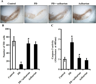
Azilsartan ameliorates apoptosis of dopaminergic neurons and rescues characteristic parkinsonian behaviors in a rat model of Parkinson's disease. [Abstract]2017 Apr 11;8(15):24099-24109. PMID: 28445961
Azilsartan purchased from MedChemExpress. Usage Cited in: Oncotarget. 2017 Apr 11;8(15):24099-24109. [Abstract]
Rats are orally administered with vehicle or Azilsartan (5 mg/kg/d) during the four-week rotenone (2.5 mg/kg/d) infusion. A. TH-immunoreactive neurons in the SN of each group. B. Quantitative analysis of TH-positive neurons in the SN of each group. Scale bar: 500 μm. C. The activity of caspase-3 in the SN is measured by a colorimetric assay kit.


Solvent & Solubility
In Vitro:
DMSO : 25 mg/mL (54.77 mM; Need ultrasonic; Hygroscopic DMSO has a significant impact on the solubility of product, please use newly opened DMSO)
Please refer to the solubility information to select the appropriate solvent. Once prepared, please aliquot and store the solution to prevent product inactivation from repeated freeze-thaw cycles.
Storage method and period of stock solution: -80°C, 2 years; -20°C, 1 year. When stored at -80°C, please use it within 2 years. When stored at -20°C, please use it within 1 year.
Please refer to the solubility information to select the appropriate solvent. Once prepared, please aliquot and store the solution to prevent product inactivation from repeated freeze-thaw cycles.
Storage method and period of stock solution: -80°C, 2 years; -20°C, 1 year. When stored at -80°C, please use it within 2 years. When stored at -20°C, please use it within 1 year.
Concentration (start) × Volume (start) = Concentration (final) × Volume (final)
In Vivo:
Select the appropriate dissolution method based on your experimental animal and administration route.
- For the following dissolution methods, please ensure to first prepare a clear stock solution using an In Vitro approach and then sequentially add co-solvents:
- To ensure reliable experimental results, the clarified stock solution can be appropriately stored based on storage conditions. As for the working solution for In Vivo experiments, it is recommended to prepare freshly and use it on the same day.
- The percentages shown for the solvents indicate their volumetric ratio in the final prepared solution. If precipitation or phase separation occurs during preparation, heat and/or sonication can be used to aid dissolution.
Add each solvent one by one: 10% DMSO 40% PEG300 5% Tween-80 45% Saline
Solubility: ≥ 2.5 mg/mL (5.48 mM); Clear solution
This protocol yields a clear solution of ≥ 2.5 mg/mL (saturation unknown).
Taking 1 mL working solution as an example, add 100 μL DMSO stock solution (25.0 mg/mL) to 400 μL PEG300, and mix evenly; then add 50 μL Tween-80 and mix evenly; then add 450 μL Saline to adjust the volume to 1 mL.
Preparation of Saline: Dissolve 0.9 g sodium chloride in ddH₂O and dilute to 100 mL to obtain a clear Saline solution.
In Vivo Dissolution Calculator
Please enter the basic information of animal experiments:
-
-
-
-
Recommended: Prepare an additional quantity of animals to account for potential losses during experiments.
Please enter your animal formula composition:
-
%DMSO +
Recommended: Keep the proportion of DMSO in working solution below 2% if your animal is weak.
-
%+
-
+%Tween-80 + +
-
%Saline +
The co-solvents required include: DMSO, . All of co-solvents are available by MedChemExpress (MCE). , Tween 80. All of co-solvents are available by MedChemExpress (MCE).
Working solution concentration: 0.22 mg/mL
Method for preparing stock solution: mg drug dissolved in μL DMSO. Stock solution concentration: mg/mL.
1. Take μL DMSO stock solution;
2. Add μL .
μL , mix evenly;
3. Then add μL Tween 80, mix evenly;
4. Then add μL
Please ensure that the stock solution in the first step is dissolved to a clear state, and add co-solvents in sequence. You can use ultrasonic heating (ultrasonic cleaner, recommended frequency 20-40 kHz), vortexing, etc. to assist dissolution.


Purity & Documentation
-
Data Sheet (291 KB)
-
SDS (393 KB)
- English - EN (393 KB)
- Français - FR (393 KB)
- Deutsch - DE (393 KB)
- Norwegian - NO (393 KB)
- Español - ES (393 KB)
- Swedish - SV (393 KB)
- Italian - IT (393 KB)
- Korean - KR (393 KB)
- Portuguese - PT (393 KB)
-
Handling Instructions (2659 KB)
References
[1]. Kajiya T, et al. Molecular and cellular effects of azilsartan: a new generation angiotensin II receptor blocker. J Hypertens. 2011 Dec;29(12):2476-83. [Content Brief]
[2]. Zhao M, et al. Azilsartan treatment improves insulin sensitivity in obese spontaneously hypertensive Koletsky rats. Diabetes Obes Metab. 2011 Dec;13(12):1123-9. [Content Brief]
[3]. Ojima M, et al. In vitro antagonistic properties of a new angiotensin type 1 receptor blocker, azilsartan, in receptor binding and function studies. J Pharmacol Exp Ther. 2011 Mar;336(3):801-8. [Content Brief]
[4]. Gupta V, et al. Neuroprotective potential of azilsartan against cerebral ischemic injury: Possible involvement of mitochondrial mechanisms. Neurochem Int. 2020 Jan;132:104604. [Content Brief]
[5]. Ahmadian E, et al. Novel angiotensin receptor blocker, azilsartan induces oxidative stress and NFkB-mediated apoptosis in hepatocellular carcinoma cell line HepG2. Biomed Pharmacother. 2018 Mar;99:939-946. [Content Brief]


Complete Stock Solution Preparation Table
Please refer to the solubility information to select the appropriate solvent. Once prepared, please aliquot and store the solution to prevent product inactivation from repeated freeze-thaw cycles.
Storage method and period of stock solution: -80°C, 2 years; -20°C, 1 year. When stored at -80°C, please use it within 2 years. When stored at -20°C, please use it within 1 year.
| Optional Solvent | Concentration Solvent Mass | 1 mg | 5 mg | 10 mg | 25 mg |
|---|---|---|---|---|---|
| DMSO | 1 mM | 2.1908 mL | 10.9541 mL | 21.9082 mL | 54.7705 mL |
| 5 mM | 0.4382 mL | 2.1908 mL | 4.3816 mL | 10.9541 mL | |
| 10 mM | 0.2191 mL | 1.0954 mL | 2.1908 mL | 5.4771 mL | |
| 15 mM | 0.1461 mL | 0.7303 mL | 1.4605 mL | 3.6514 mL | |
| 20 mM | 0.1095 mL | 0.5477 mL | 1.0954 mL | 2.7385 mL | |
| 25 mM | 0.0876 mL | 0.4382 mL | 0.8763 mL | 2.1908 mL | |
| 30 mM | 0.0730 mL | 0.3651 mL | 0.7303 mL | 1.8257 mL | |
| 40 mM | 0.0548 mL | 0.2739 mL | 0.5477 mL | 1.3693 mL | |
| 50 mM | 0.0438 mL | 0.2191 mL | 0.4382 mL | 1.0954 mL |


Azilsartan Related Classifications
HY-14914 Related Classifications
 Powered by Bioz