



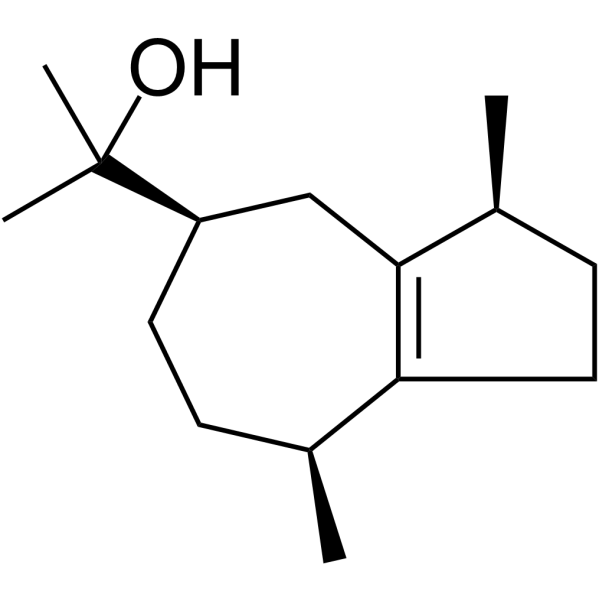
Guaiol
Based on 1 publication(s) in Google Scholar
Guaiol is a sesquiterpenoid alcohol with oral activity found in various traditional Chinese medicines, exhibiting biological activities such as anti-proliferative, autophagy-promoting, insecticidal, anti-anxiety, anti-inflammatory, diuretic, and blood pressure-lowering effects. Guaiol induces apoptosis in non-small cell lung cancer cells by regulating the stability of RAD51 through autophagy modulation. Guaiol can also act directly on parasites, inhibiting their growth by affecting the kinetoplast, mitochondrial matrix and plasma membrane of the promastigotes. Guaiol kills amastigotes at an IC50 of 0.01 µg/mL. Guaiol can be used in research related to cancer, infections, cardiovascular diseases, and inflammatory conditions[4]
For research use only. We do not sell to patients.

To place orders, for customer services and technical support, please contact: MedChemExpress USA
Tel: 609-228-6898 E-mail: [email protected] [email protected]


-
 Biological Activity
-
Chemical Information
-
Solvent & Solubility
- Protocols
- Purity & Documentation
- References
-
 Help & FAQs


Publications Citing Use of MedChemExpress (MCE) Guaiol
More

Biological Activity
Description
In Vitro
Guaiol (150 μM, 24 h) has an inhibitory effect on non-small cell lung cancer (NSCLC) cell lines (A549 cells and H1299 cells)[1]. Guaiol (0.1-50 μg/ml, 24 h) inhibits the growth of parasites in thioglycolate (HY-W115724)-stimulated macrophages from BALB/c mice[5].
MedChemExpress (MCE) has not independently confirmed the accuracy of these methods. They are for reference only.
-
Cell Line:A549 cells, H1299 cells
-
Concentration:150 μM
-
Incubation Time:24 h
-
Result:Dramatically inhibited cell growth of A549 cells and H1299 cells with IC50 values 121.7 μM and 211.5 μM.
-
Cell Line:Thioglycolate (HY-W115724)-stimulated macrophages from BALB/c mice
-
Concentration:0.1-50 μg/ml
-
Incubation Time:24 h
-
Result:Did not inhibit the macrophage phagocytic ability. Induced a significant decrease in the parasite survival index when compared with untreated control.
In Vivo
MedChemExpress (MCE) has not independently confirmed the accuracy of these methods. They are for reference only.
-
Animal Model:Non-small cell lung cancer (NSCLC) xenograft mouse model[1]
-
Dosage:8 mg/kg
-
Administration:Intraperitoneal injection (i.p.), 3 times a week for 40 days
-
Result:Reduced the tumor volume and weight in the non-small cell lung cancer (NSCLC) xenograft mouse model.
- Detection of Apoptosis
- TUNEL staining for apoptotic DNA fragmentation
- Annexin V plus membrane-impermeant dye apoptosis staining
- Autophagy
- Apoptosis Solutions
- Lysosome and acidic-vesicle live-cell staining
- Research Protocol for Inflammation-related Diseases
- Macroautophagy Solutions
- Cell Viability Determination by MTT Colorimetric Assay


Chemical Information
-
CAS No. 489-86-1
-
Appearance Solid
-
Molecular Weight 222.37
-
Formula C15H26O
-
Color White to off-white
-
SMILES
C[C@@H]1C2=C([C@H](CC[C@@H](C(C)(O)C)C2)C)CC1
-
Synonyms
Champacol; Guaiac alcohol
-
Structure Classification
-
Shipping
Room temperature in continental US; may vary elsewhere.
-
Storage
Powder -20°C 3 years In solvent -80°C 6 months -20°C 1 month


Publications (1)
-
Journal Impact Factor
-
Most Recent
-
Biol Proced Online
(-)-Guaiol Downregulates M2 Tumor-Associated Macrophage Polarization Through PPAR-γ Signaling to Suppress Lung Cancer. [Abstract]2025 Dec 5. PMID: 41350836


Solvent & Solubility
In Vitro:
DMSO : 33.33 mg/mL (149.89 mM; Need ultrasonic; Hygroscopic DMSO has a significant impact on the solubility of product, please use newly opened DMSO)
Please refer to the solubility information to select the appropriate solvent. Once prepared, please aliquot and store the solution to prevent product inactivation from repeated freeze-thaw cycles.
Storage method and period of stock solution: -80°C, 6 months; -20°C, 1 month. When stored at -80°C, please use it within 6 months. When stored at -20°C, please use it within 1 month.
Please refer to the solubility information to select the appropriate solvent. Once prepared, please aliquot and store the solution to prevent product inactivation from repeated freeze-thaw cycles.
Storage method and period of stock solution: -80°C, 6 months; -20°C, 1 month. When stored at -80°C, please use it within 6 months. When stored at -20°C, please use it within 1 month.
Concentration (start) × Volume (start) = Concentration (final) × Volume (final)
In Vivo:
Select the appropriate dissolution method based on your experimental animal and administration route.
- For the following dissolution methods, please ensure to first prepare a clear stock solution using an In Vitro approach and then sequentially add co-solvents:
- To ensure reliable experimental results, the clarified stock solution can be appropriately stored based on storage conditions. As for the working solution for In Vivo experiments, it is recommended to prepare freshly and use it on the same day.
- The percentages shown for the solvents indicate their volumetric ratio in the final prepared solution. If precipitation or phase separation occurs during preparation, heat and/or sonication can be used to aid dissolution.
For the following dissolution methods, please prepare the working solution directly:
It is recommended to prepare fresh solutions and use them promptly within a short period of time.
The percentages shown for the solvents indicate their volumetric ratio in the final prepared solution. If precipitation or phase separation occurs during preparation, heat and/or sonication can be used to aid dissolution.
Add each solvent one by one: 50% PEG300 50% Saline
Solubility: 1 mg/mL (4.50 mM); Suspended solution; Need ultrasonic
In Vivo Dissolution Calculator
Please enter the basic information of animal experiments:
-
-
-
-
Recommended: Prepare an additional quantity of animals to account for potential losses during experiments.
Please enter your animal formula composition:
-
%DMSO +
Recommended: Keep the proportion of DMSO in working solution below 2% if your animal is weak.
-
%+
-
+%Tween-80 + +
-
%Saline +
The co-solvents required include: DMSO, . All of co-solvents are available by MedChemExpress (MCE). , Tween 80. All of co-solvents are available by MedChemExpress (MCE).
Working solution concentration: 0.22 mg/mL
Method for preparing stock solution: mg drug dissolved in μL DMSO. Stock solution concentration: mg/mL.
1. Take μL DMSO stock solution;
2. Add μL .
μL , mix evenly;
3. Then add μL Tween 80, mix evenly;
4. Then add μL
Please ensure that the stock solution in the first step is dissolved to a clear state, and add co-solvents in sequence. You can use ultrasonic heating (ultrasonic cleaner, recommended frequency 20-40 kHz), vortexing, etc. to assist dissolution.


Purity & Documentation
-
Data Sheet (281 KB)
-
SDS (393 KB)
- English - EN (393 KB)
- Français - FR (393 KB)
- Deutsch - DE (393 KB)
- Norwegian - NO (393 KB)
- Español - ES (393 KB)
- Swedish - SV (393 KB)
- Italian - IT (393 KB)
- Korean - KR (393 KB)
- Portuguese - PT (393 KB)
-
Handling Instructions (2659 KB)
References
[1]. Yang, Q, et al. (−)-Guaiol regulates RAD51 stability via autophagy to induce cell apoptosis in non-small cell lung cancer Oncotarget. 2016 Sep 20;7(38):62585-62597. [Content Brief]
[2]. Ali, A., Tabanca, et al. Chemical composition and biological activity of four salvia essential oils and individual compounds against two species of mosquitoes J. Agric. Food Chem. 63(2), 447-456 (2015). [Content Brief]
[3]. Liu, T, et al. Guaiol - a naturally occurring insecticidal sesquiterpene Nat. Prod. Commun. 8(10), 1353-1354 (2013). [Content Brief]
[4]. Garcia M C F, et al. The in vitro antileishmanial activity of essential oil from Aloysia gratissima and guaiol, its major sesquiterpene against Leishmania amazonensis[J]. Parasitology, 2018, 145(9): 1219-1227. [Content Brief]
[6]. Yang M, et al. Determination and pharmacokinetic study of guaiol in rat plasma by gas chromatography–mass spectrometry with selected ion monitoring[J]. Journal of Chromatography B, 2018, 1085: 30-35. [Content Brief]


Complete Stock Solution Preparation Table
Please refer to the solubility information to select the appropriate solvent. Once prepared, please aliquot and store the solution to prevent product inactivation from repeated freeze-thaw cycles.
Storage method and period of stock solution: -80°C, 6 months; -20°C, 1 month. When stored at -80°C, please use it within 6 months. When stored at -20°C, please use it within 1 month.
| Optional Solvent | Concentration Solvent Mass | 1 mg | 5 mg | 10 mg | 25 mg |
|---|---|---|---|---|---|
| DMSO | 1 mM | 4.4970 mL | 22.4850 mL | 44.9701 mL | 112.4252 mL |
| 5 mM | 0.8994 mL | 4.4970 mL | 8.9940 mL | 22.4850 mL | |
| 10 mM | 0.4497 mL | 2.2485 mL | 4.4970 mL | 11.2425 mL | |
| 15 mM | 0.2998 mL | 1.4990 mL | 2.9980 mL | 7.4950 mL | |
| 20 mM | 0.2249 mL | 1.1243 mL | 2.2485 mL | 5.6213 mL | |
| 25 mM | 0.1799 mL | 0.8994 mL | 1.7988 mL | 4.4970 mL | |
| 30 mM | 0.1499 mL | 0.7495 mL | 1.4990 mL | 3.7475 mL | |
| 40 mM | 0.1124 mL | 0.5621 mL | 1.1243 mL | 2.8106 mL | |
| 50 mM | 0.0899 mL | 0.4497 mL | 0.8994 mL | 2.2485 mL | |
| 60 mM | 0.0750 mL | 0.3748 mL | 0.7495 mL | 1.8738 mL | |
| 80 mM | 0.0562 mL | 0.2811 mL | 0.5621 mL | 1.4053 mL | |
| 100 mM | 0.0450 mL | 0.2249 mL | 0.4497 mL | 1.1243 mL |


Guaiol Related Classifications
HY-N3980 Related Classifications
 Powered by Bioz