



Fresolimumab
Based on 2 publication(s) in Google Scholar
Fresolimumab (GC1008) is a human monoclonal antibody against TGF-β that neutralizes all mammalian active subtypes of TGF-β. The binding affinity of Fresolimumab to TGF-β2 is 1.8 nM. Fresolimumab improves Bleomycin (HY-108345)-induced acute lung injury. Fresolimumab radiolabeled with 89Zr can be used for PET analysis of TGF-β expression, antibody uptake and organ distribution. Fresolimumab can be used in the study of cancer, osteogenesis imperfecta, fibrosis and kidney disease.
For research use only. We do not sell to patients.
- Purity : 99.51%
- CAS No.: 948564-73-6
- Molecular Weight:144.4 kDa
-
Storage:
Please store the product under the recommended conditions in the Certificate of Analysis.


To place orders, for customer services and technical support, please contact: MedChemExpress USA
Tel: 609-228-6898 E-mail: [email protected] [email protected]


-
 Biological Activity
-
Chemical Information
- Protocols
- Purity & Documentation
- References
-
 Help & FAQs


-
HY-13226
GalunisertibTGFβRI Inhibitor -
HY-P99003
Human IgG4 (S228P) kappa, Isotype ControlIgG4κ Isotype Control -

HY-P99480
Bintrafusp alfaBifunctional Fusion Protein -

HY-10431
SB-431542TGF-β Receptor Kinase Inhibitor -
HY-10431G
SB-431542 (GMP)TGF-β Receptor Kinase Inhibitor -

HY-10431R
SB-431542 (Standard)TGF-β Receptor Kinase Inhibitor -
HY-12043
SB 525334ALK5 Inhibitor -
HY-50904
NintedanibPDGFR/VEGFR/FGFR Inhibitor -

HY-P99355
BimagrumabAnti-ACVR2B Antibody -
HY-10087
NavitoclaxBcl-2 Inhibitor -
HY-12815
MCC950NLRP3 Inhibitor -

HY-P9902
PembrolizumabPD-1 Inhibitor
Publications Citing Use of MedChemExpress (MCE) Fresolimumab
More Customer Validation & Images

-
Cell Imaging/Staining
-
IF


Biological Activity
Description
Isotype
Human IgG4 kappa
Recommend Isotype Controls
Species Reactivity
Human
IC50 & Target
TGFB1
In Vivo
MedChemExpress (MCE) has not independently confirmed the accuracy of these methods. They are for reference only.
Clinical Trial
| NCT Number | Sponsor | Condition | Start Date |
Phase

|
|---|---|---|---|---|
| NCT01329991 | Plexxikon| | 2011-05 | PHASE1 |
Gene ID
Accession
Conjugated
Unconjugated
Reconsititution
The product can be reconstituted/diluted with sterile PBS or saline.
Format
-
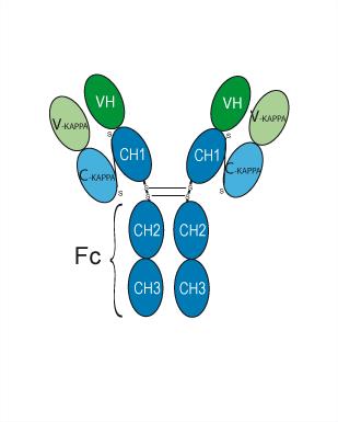
Human IgG4 kappa
Application
ELISA, FACS, Functional assay
Verified Bioactivity
-
 Immobilized TGF beta 1/TGFB1 Protein, Human can bind Fresolimumab. The EC50 for this effect is 67.28 ng/mL.
 -
 Flow Cytometry analysis of PC-3 cells labelling TGFB1 (red) with Fresolimumab (anti-TGFB1) (HY-P99020). Goat Anti-Human IgG (Alexa Fluor 488) (HY-P83776) at a dilution of 1/1000 was used as the secondary antibody. Blue-Human IgG4 kappa (HY-P99003). Black-Unlabelled control, cells without incubation with primary antibody.


Chemical Information
-
CAS No. 948564-73-6
-
Appearance Liquid
-
Molecular Weight 144.4 kDa
-
Color Colorless to light yellow
-
SMILES
[Fresolimumab]
-
Synonyms
GC1008
-
Shipping
Shipping with dry ice.
-
Formulation
Please refer to the lot-specific COA for specific buffer information.
-
Storage
Please store the product under the recommended conditions in the Certificate of Analysis.


Publications (2)
-
Journal Impact Factor
-
Most Recent
-
Cells
Investigating the Role of TGF-β Signaling Pathways in Human Corneal Endothelial Cell Primary Culture. [Abstract]2023 Jun 14;12(12):1624. PMID: 37371094
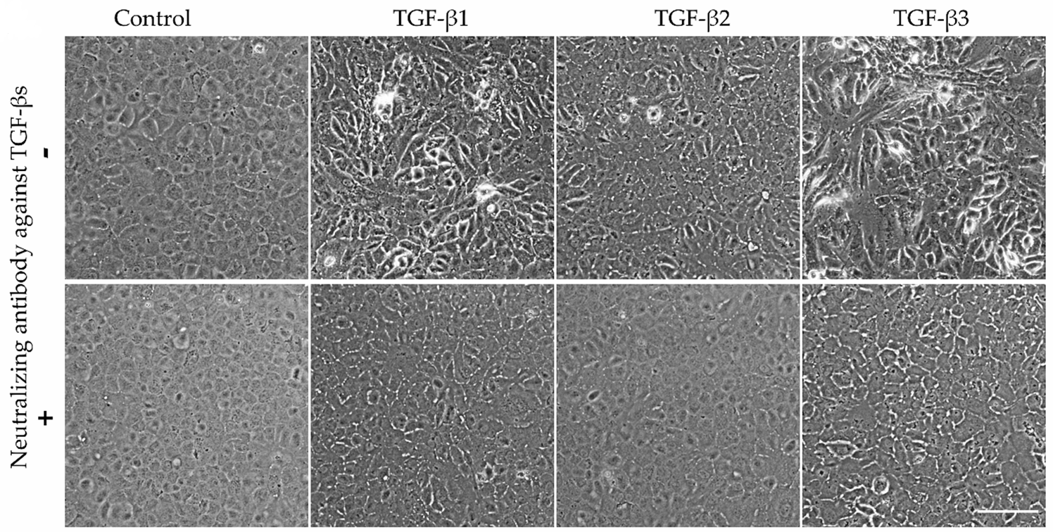
Fresolimumab purchased from MedChemExpress. Usage Cited in: Cells. 2023 Jun 14;12(12):1624. [Abstract]
Fresolimumab (Neutralizing antibody against TGF-βs) (10 μg/mL; 7 days). Illustration of cell morphology by phase-contrast images. The images were acquired using a phase-contrast microscope with X10 objective. Cropped images were used to illustrate cell morphology. Scale bar = 100 µm.
Fresolimumab purchased from MedChemExpress. Usage Cited in: Cells. 2023 Jun 14;12(12):1624. [Abstract]
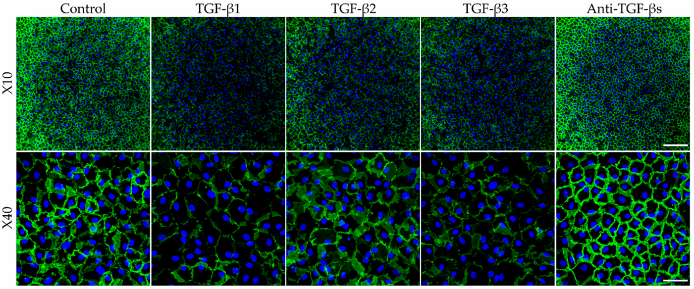
Fresolimumab (Neutralizing antibody against TGF-βs) (10 μg/mL; 4 weeks). Illustrative images. Cell lateral membranes were stained in green by NCAM and the nuclei were counter-stained in blue using DAPI. A higher magnification (X40) was added in order to show the cell morphology. The images were taken in the center of each well. Images of the same magnification were acquired with the same parameters. The scale bar was 200 µm for X10 images and 50 µm for X40 images.
-
Sci Rep
Collagen-derived biomarkers reveal distinct fibrotic responses among cancer-associated fibroblasts. [Abstract]2026 Jun 12. PMID: 42286082

Protocols
-
Fibrosis/Collagen Morphometry
Fibrosis and collagen morphometry is based on the quantitative visualization of fibrillar collagen deposition in tissue sections using histochemical stains such as Sirius Red (Picrosirius Red) or Masson's trichrome, followed by image-based or polarization-enhanced analysis to estimate collagen proportional area as a surrogate of extracellular matrix accumulation during fibrotic remodeling. Sirius Red combined with polarized light microscopy enhances detection of collagen fibers due to birefringence properties, enabling more specific visualization of collagen type I and III fibrils compared to conventional bright-field histology, while whole-section or region-restricted digital morphometry reduces field-selection bias in fibrosis assessment. Alternative quantitative approaches include second harmonic generation (SHG) and two-photon excited fluorescence microscopy, which enable label-free detection of fibrillar collagen and have been validated against histological staining and biochemica
-
RNA extraction experimental
By lysing cells, releasing RNA, and removing impurities such as proteins and DNA, high-purity RNA products are finally obtained. The commonly used traditional method is the guanidine isothiocyanate/phenol/chloroform method (Trizol), which is suitable for a variety of animal materials including animal tissues, microorganisms, cultured cells, etc., and most plant materials.
-
Collagen: Sirius Red Staining
Sirius Red or picrosirius red staining is a histochemical method for visualizing collagen-rich extracellular matrix in tissue sections, and collagen fibers are detected as red-stained structures under bright-field microscopy with enhanced birefringence under polarized light. Picrosirius red is useful for assessing total collagen organization, distribution, and fibrosis burden, but polarized color should not be interpreted as a definitive collagen type I versus type III readout because color is affected by fiber orientation, thickness, and packing.
-
Mesenchymal stromal/stem cell osteogenic differentiation
Mesenchymal stromal/stem cells can be induced toward an osteoblast-like lineage in vitro by culture in osteogenic medium containing dexamethasone, ascorbic acid or ascorbate-2-phosphate, and β-glycerophosphate; the differentiation process is commonly evaluated by alkaline phosphatase activity, osteogenic marker expression, collagenous matrix formation, and calcium-rich matrix mineralization. The main readouts are alkaline phosphatase activity as an early osteogenic marker and Alizarin Red S staining as a calcium-deposit readout for mineralized extracellular matrix; Alizarin Red S can be inspected microscopically or extracted and measured colorimetrically at 405 nm.
-
Connective Tissue: Masson's Trichrome/Collagen Trichrome Staining
Masson’s Trichrome (collagen/trichrome staining) is a histological technique that differentially stains tissue compartments using sequential acidic dyes to distinguish collagen from muscle and cytoplasmic components based on dye affinity and tissue permeability differences, enabling visualization of fibrosis and connective tissue architecture in histological sections. The classical formulation typically uses Weigert's iron hematoxylin for nuclear staining, Biebrich scarlet-acid fuchsin for cytoplasm and muscle, and aniline blue (or light green variants) for collagen, producing a characteristic blue/green collagen signal contrasted against red cytoplasm and dark nuclei. The staining principle relies on selective displacement of smaller dye molecules by larger anionic dyes in collagen-rich regions under controlled acidified conditions, which enhances collagen-specific dye retention. This property makes the method widely used for fibrosis assessment in organs such as heart, liver, lung, a
-
Inhalation Toxicity Study
Inhalation toxicity studies expose rodents to a controlled aerosol, vapor, gas, or smoke atmosphere and assess respiratory and systemic toxicity using exposure-atmosphere characterization, clinical observations, body and organ weights, bronchoalveolar lavage fluid, histopathology, blood chemistry, hematology, and, when included, molecular endpoints such as transcriptomics, proteomics, lipidomics, or tissue burden analysis. The primary biological readouts are airway irritation, pulmonary inflammation, cytotoxicity, altered surfactant or lipid homeostasis, impaired particle clearance, and tissue remodeling, reflected by BALF cell differentials, BALF protein, LDH, phosphatase activities, cytokines, lung weight, microscopic respiratory-tract lesions, and retained lung burden.


Purity & Documentation
-
Data Sheet (261 KB)
-
SDS (251 KB)
- English - EN (251 KB)
- Français - FR (251 KB)
- Deutsch - DE (251 KB)
- Norwegian - NO (251 KB)
- Español - ES (251 KB)
- Swedish - SV (251 KB)
- Italian - IT (251 KB)
- Korean - KR (251 KB)
- Portuguese - PT (251 KB)
-
Inhibitory Antibodies User Guide (603 KB)
References
[1]. Song IW, et al. Targeting TGF-β for treatment of osteogenesis imperfecta. J Clin Invest. 2022 Apr 1;132(7):e152571. [Content Brief]
[2]. Lan Y, et al. Colocalized targeting of TGF-β and PD-L1 by bintrafusp alfa elicits distinct antitumor responses. J Immunother Cancer. 2022 Jul;10(7):e004122. [Content Brief]
[3]. Oude Munnink TH, et al. PET with the 89Zr-labeled transforming growth factor-β antibody fresolimumab in tumor models. J Nucl Med. 2011 Dec;52(12):2001-8. [Content Brief]
[4]. Kadam AH, et al. Targeting caveolae to pump bispecific antibody to TGF-β into diseased lungs enables ultra-low dose therapeutic efficacy. PLoS One. 2022 Nov 22;17(11):e0276462. [Content Brief]

Calculators
Concentration (start) × Volume (start) = Concentration (final) × Volume (final)


Fresolimumab Related Classifications
HY-P99020 Related Classifications
 Powered by Bioz