



Embelin
Based on 7 publication(s) in Google Scholar
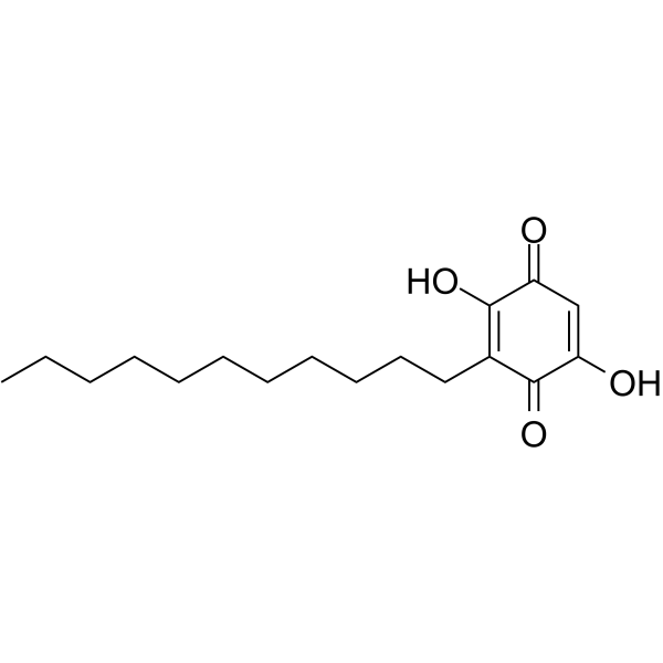
Embelin (Embelic acid), a potent, nonpeptidic XIAP inhibitor (IC50=4.1 μM), inhibits cell growth, induces apoptosis, and activates caspase-9 in prostate cancer cells with high levels of XIAP. Embelin blocks NF-kappaB signaling pathway leading to suppression of NF-kappaB-regulated antiapoptotic and metastatic gene products. Embelin also induces autophagic and apoptotic cell death in human oral squamous cell carcinoma cells.
For research use only. We do not sell to patients.
- Purity : 99.01%
- CAS No.: 550-24-3
- Formula: C17H26O4
- Molecular Weight:294.39
-
Storage:
4°C, protect from light
* In solvent : -80°C, 2 years; -20°C, 1 year (protect from light)

To place orders, for customer services and technical support, please contact: MedChemExpress USA
Tel: 609-228-6898 E-mail: [email protected] [email protected]


-
 Biological Activity
-
Chemical Information
-
Solvent & Solubility
- Protocols
- Purity & Documentation
- References
-
 Help & FAQs


-
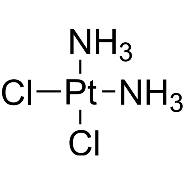
HY-17394
CisplatinDNA Alkylator -
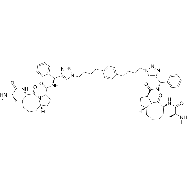
HY-15989
SM-164IAP Antagonist -
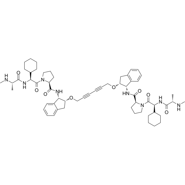
HY-12600
AZD5582 -

HY-10219
RapamycinmTOR Inhibitor -
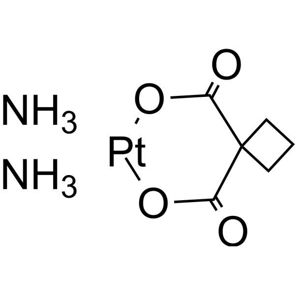
HY-17393
CarboplatinDNA Synthesis Inhibitor -
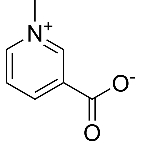
HY-N0414
TrigonellineNrf2 Inhibitor -
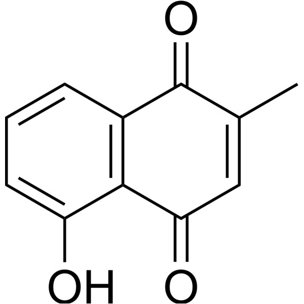
HY-N1497
PlumbaginAnticancer Agent -
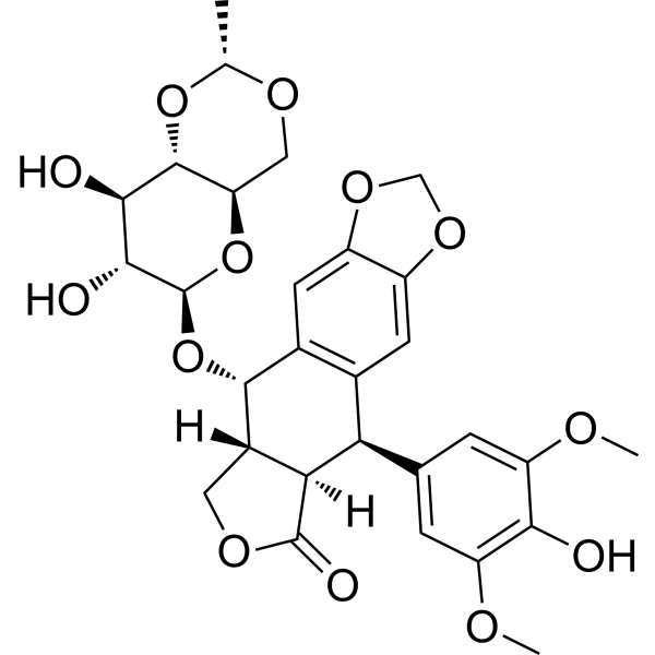
HY-13629
EtoposideTopoisomerase II Inhibitor -
HY-10358
MK-2206 dihydrochlorideAllosteric Akt Inhibitor -
HY-135303
GLPG1205GPR84 Antagonist -
HY-15893
DMOGHIF-PH Inhibitor -

HY-K1089
Cell Senescence β-Galactosidase Staining Kit
Publications Citing Use of MedChemExpress (MCE) Embelin
More- Cell Res. 2021 Mar;31(3):291-311. [Abstract]
- Autophagy. 2025 Oct 25. [Abstract]
- Sci Adv. 2025 May 2;11(18):eadu6676. [Abstract]
- Pharmacol Res. 2020 May:155:104751. [Abstract]
- Phytomedicine. 2026 May 14:157:158282. [Abstract]
- Mol Neurobiol. 2014 Apr;49(2):1087-101. [Abstract]
- Mol Pharm. 2023 Sep 4;20(9):4574-4586. [Abstract]
 Customer Validation & Images

-
WB


Biological Activity
Description
IC50 & Target
[1]|
XIAP 4.1 μM (IC50) |
Cellular Effect
|
Cell Line

|
Type | Value | Description | References |
|---|---|---|---|---|
| A2780 | IC50 |
1.79 μM
Compound: 5
|
Cytotoxicity against human A2780 cells after 96 hrs by MTT assay
Cytotoxicity against human A2780 cells after 96 hrs by MTT assay
|
[PMID: 17125236] |
| A549 | IC50 |
0.21 μM
Compound: 2
|
Inhibition of mPGES1 in IL-1beta stimulated human A549 cell microsomal membranes assessed as reduction in PGE2 synthase activity after 15 mins using PGH2 substrate by RP-HPLC method
Inhibition of mPGES1 in IL-1beta stimulated human A549 cell microsomal membranes assessed as reduction in PGE2 synthase activity after 15 mins using PGH2 substrate by RP-HPLC method
|
[PMID: 25765759] |
| A549 | IC50 |
1.47 μM
Compound: 5
|
Cytotoxicity against human A549 cells after 96 hrs by MTT assay
Cytotoxicity against human A549 cells after 96 hrs by MTT assay
|
[PMID: 17125236] |
| Bel-7402 | IC50 |
1.66 μM
Compound: 5
|
Cytotoxicity against human Bel-7402 cells after 96 hrs by MTT assay
Cytotoxicity against human Bel-7402 cells after 96 hrs by MTT assay
|
[PMID: 17125236] |
| BGC-823 | IC50 |
1.69 μM
Compound: 5
|
Cytotoxicity against human BGC-823 cells after 96 hrs by MTT assay
Cytotoxicity against human BGC-823 cells after 96 hrs by MTT assay
|
[PMID: 17125236] |
| CHO | EC50 |
0.421 μM
Compound: 2
|
Agonist activity at recombinant human GPR84 expressed in CHO cells assessed as inhibition of forskolin-induced cAMP accumulation by fluo-4AM dye based FLIPR assay
Agonist activity at recombinant human GPR84 expressed in CHO cells assessed as inhibition of forskolin-induced cAMP accumulation by fluo-4AM dye based FLIPR assay
|
[PMID: 31721581] |
| CHO | EC50 |
5 μM
Compound: 2
|
Agonist activity at recombinant human GPR84 expressed in CHO cells assessed as beta-arrestin 2 recruitment by beta-galactosidase based PathHunter assay
Agonist activity at recombinant human GPR84 expressed in CHO cells assessed as beta-arrestin 2 recruitment by beta-galactosidase based PathHunter assay
|
[PMID: 31721581] |
| HCT-8 | IC50 |
1.49 μM
Compound: 5
|
Cytotoxicity against human HCT8 cells after 96 hrs by MTT assay
Cytotoxicity against human HCT8 cells after 96 hrs by MTT assay
|
[PMID: 17125236] |
| HEK293 | EC50 |
0.089 μM
Compound: 2
|
Agonist activity at recombinant human HA-tagged GPR84 stably expressed in HEK293 cells assessed as inhibition of forskolin-induced cAMP accumulation measured after 60 mins by HTRF assay
Agonist activity at recombinant human HA-tagged GPR84 stably expressed in HEK293 cells assessed as inhibition of forskolin-induced cAMP accumulation measured after 60 mins by HTRF assay
|
[PMID: 31721581] |
| HEK293 | EC50 |
0.63 μM
Compound: 2
|
Agonist activity at recombinant human GPR84 expressed in HEK293 cells co-expressing Gqi5 assessed as inhibition of forskolin-induced cAMP accumulation by fluo-4AM dye based FLIPR assay
Agonist activity at recombinant human GPR84 expressed in HEK293 cells co-expressing Gqi5 assessed as inhibition of forskolin-induced cAMP accumulation by fluo-4AM dye based FLIPR assay
|
[PMID: 31721581] |
| Jurkat | IC50 |
20 μM
Compound: Embelin
|
Inhibitory concentration of compound against Jurkat-Vec cells
Inhibitory concentration of compound against Jurkat-Vec cells
|
[PMID: 15115387] |
| LNCaP | IC50 |
5.7 μM
Compound: Embelin
|
Compound was tested for inhibition of LnCap prostate cancer cells
Compound was tested for inhibition of LnCap prostate cancer cells
|
[PMID: 15115387] |
| LNCaP | IC50 |
5.7 μM
Compound: Embelin
|
Inhibitory concentration of compound against growth of LNCaP cells in vitro
Inhibitory concentration of compound against growth of LNCaP cells in vitro
|
[PMID: 15115387] |
| PC-3 | IC50 |
3.7 μM
Compound: Embelin
|
Compound was tested for inhibition of PC-3 prostate cancer cells
Compound was tested for inhibition of PC-3 prostate cancer cells
|
[PMID: 15115387] |
| PC-3 | IC50 |
3.7 μM
Compound: Embelin
|
Inhibitory concentration of compound against growth of PC-3 cells in vitro
Inhibitory concentration of compound against growth of PC-3 cells in vitro
|
[PMID: 15115387] |
| PrEC | IC50 |
20.1 μM
Compound: Embelin
|
Inhibitory concentration of compound against growth of PrEC cells in vitro
Inhibitory concentration of compound against growth of PrEC cells in vitro
|
[PMID: 15115387] |
| PrEC | IC50 |
20.1 μM
Compound: Embelin
|
Selectivity in normal human prostate epithelial cells (PrEC)
Selectivity in normal human prostate epithelial cells (PrEC)
|
[PMID: 15115387] |
| WI-38 | IC50 |
19.3 μM
Compound: Embelin
|
Inhibitory concentration of compound against growth of WI-38 cells in vitro
Inhibitory concentration of compound against growth of WI-38 cells in vitro
|
[PMID: 15115387] |
| WI-38 | IC50 |
19.3 μM
Compound: Embelin
|
Selectivity in normal human fibroblast cell line, WI-38 cells
Selectivity in normal human fibroblast cell line, WI-38 cells
|
[PMID: 15115387] |
In Vitro


Chemical Information
-
CAS No. 550-24-3
-
Appearance Solid
-
Molecular Weight 294.39
-
Formula C17H26O4
-
Color Yellow to orange
-
SMILES
O=C1C(O)=C(CCCCCCCCCCC)C(C(O)=C1)=O
-
Synonyms
Embelic acid; Emberine; NSC 91874
-
Structure Classification
-
Initial Source
-
Shipping
Room temperature in continental US; may vary elsewhere.
-
Storage
4°C, protect from light
* In solvent : -80°C, 2 years; -20°C, 1 year (protect from light)


Publications (7)
-
Journal Impact Factor
-
Most Recent
-
Cell Res
2021 Mar;31(3):291-311. PMID: 33299139 -
Autophagy
XIAP-ULK1-Mediated mitophagy modulates carnitine metabolism to mitigate diabetic kidney disease. [Abstract]2025 Oct 25. PMID: 41139215 -
Sci Adv
In situ protein corona-camouflaged supramolecular assemblies remodel thrombotic microenvironment for improved arterial homeostasis. [Abstract]2025 May 2;11(18):eadu6676. PMID: 40315315 -
Pharmacol Res
Cardamonin retards progression of autosomal dominant polycystic kidney disease via inhibiting renal cyst growth and interstitial fibrosis. [Abstract]2020 May:155:104751. PMID: 32151678 -
Phytomedicine
5-O-Methylembelin disrupts the ACO1-TPI1 interaction to promote ferroptosis in clear cell renal cell carcinoma. [Abstract]2026 May 14:157:158282. PMID: 42166976 -
Mol Neurobiol
Embelin suppresses dendritic cell functions and limits autoimmune encephalomyelitis through the TGF-β/β-catenin and STAT3 signaling pathways. [Abstract]2014 Apr;49(2):1087-101. PMID: 24258405
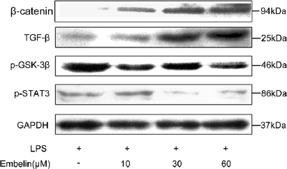
Embelin purchased from MedChemExpress. Usage Cited in: Mol Neurobiol. 2014 Apr;49(2):1087-101. [Abstract]
Embelin (EB) promotes TGF-β/β-catenin and inhibits STAT3 signaling in mDCs. DCs are treated with 10, 30, and 60 μM EB to investigate its effect on p-STAT3, TGF-β, β-catenin, and p-GSK-3β signaling (diluted 1:1,000).
-
Mol Pharm
Construction of SLC16A1/3 Targeted Gallic Acid-Iron-Embelin Nanoparticles for Regulating Glycolysis and Redox Pathways in Cervical Cancer. [Abstract]2023 Sep 4;20(9):4574-4586. PMID: 37307591


Solvent & Solubility
In Vitro:
DMSO : ≥ 50 mg/mL (169.84 mM; Hygroscopic DMSO has a significant impact on the solubility of product, please use newly opened DMSO)
* "≥" means soluble, but saturation unknown.
Please refer to the solubility information to select the appropriate solvent. Once prepared, please aliquot and store the solution to prevent product inactivation from repeated freeze-thaw cycles.
Storage method and period of stock solution: -80°C, 2 years; -20°C, 1 year (protect from light). When stored at -80°C, please use it within 2 years. When stored at -20°C, please use it within 1 year.
Please refer to the solubility information to select the appropriate solvent. Once prepared, please aliquot and store the solution to prevent product inactivation from repeated freeze-thaw cycles.
Storage method and period of stock solution: -80°C, 2 years; -20°C, 1 year (protect from light). When stored at -80°C, please use it within 2 years. When stored at -20°C, please use it within 1 year.
Concentration (start) × Volume (start) = Concentration (final) × Volume (final)
In Vivo:
Select the appropriate dissolution method based on your experimental animal and administration route.
- For the following dissolution methods, please ensure to first prepare a clear stock solution using an In Vitro approach and then sequentially add co-solvents:
- To ensure reliable experimental results, the clarified stock solution can be appropriately stored based on storage conditions. As for the working solution for In Vivo experiments, it is recommended to prepare freshly and use it on the same day.
- The percentages shown for the solvents indicate their volumetric ratio in the final prepared solution. If precipitation or phase separation occurs during preparation, heat and/or sonication can be used to aid dissolution.
Add each solvent one by one: 10% DMSO 40% PEG300 5% Tween-80 45% Saline
Solubility: 2.5 mg/mL (8.49 mM); Suspended solution; Need ultrasonic
This protocol yields a suspended solution of 2.5 mg/mL. Suspended solution can be used for oral and intraperitoneal injection.
Taking 1 mL working solution as an example, add 100 μL DMSO stock solution (25.0 mg/mL) to 400 μL PEG300, and mix evenly; then add 50 μL Tween-80 and mix evenly; then add 450 μL Saline to adjust the volume to 1 mL.
Preparation of Saline: Dissolve 0.9 g sodium chloride in ddH₂O and dilute to 100 mL to obtain a clear Saline solution.
In Vivo Dissolution Calculator
Please enter the basic information of animal experiments:
-
-
-
-
Recommended: Prepare an additional quantity of animals to account for potential losses during experiments.
Please enter your animal formula composition:
-
%DMSO +
Recommended: Keep the proportion of DMSO in working solution below 2% if your animal is weak.
-
%+
-
+%Tween-80 + +
-
%Saline +
The co-solvents required include: DMSO, . All of co-solvents are available by MedChemExpress (MCE). , Tween 80. All of co-solvents are available by MedChemExpress (MCE).
Working solution concentration: 0.22 mg/mL
Method for preparing stock solution: mg drug dissolved in μL DMSO. Stock solution concentration: mg/mL. * In solvent : -80°C, 2 years; -20°C, 1 year (protect from light)
1. Take μL DMSO stock solution;
2. Add μL .
μL , mix evenly;
3. Then add μL Tween 80, mix evenly;
4. Then add μL
Please ensure that the stock solution in the first step is dissolved to a clear state, and add co-solvents in sequence. You can use ultrasonic heating (ultrasonic cleaner, recommended frequency 20-40 kHz), vortexing, etc. to assist dissolution.

Protocols
-
Mammalian live/dead viability and cytotoxicity staining
Live/dead viability and cytotoxicity staining assays are based on the simultaneous detection of intracellular esterase activity in metabolically active (viable) cells and membrane integrity loss in non-viable cells. In commonly used dual-staining approaches, membrane-permeant fluorogenic substrates are converted by intracellular esterases into fluorescent products in live cells, while impermeant DNA-binding dyes selectively enter cells with compromised plasma membranes and label nucleic acids in dead or dying cells, enabling discrimination between viable and non-viable populations by fluorescence microscopy or flow cytometry.
-
Annexin V plus membrane-impermeant dye apoptosis staining
Annexin V-based apoptosis assays rely on the detection of phosphatidylserine (PS) externalization from the inner leaflet of the plasma membrane to the outer leaflet, an early biochemical hallmark of apoptosis. Fluorescently labeled Annexin V binds PS in a calcium-dependent manner, enabling identification of early apoptotic cells by flow cytometry or fluorescence microscopy. When combined with a membrane-impermeant DNA-binding dye (e. g. , propidium iodide), this approach allows discrimination between viable (Annexin V−/dye−), early apoptotic (Annexin V+/dye−), and late apoptotic or necrotic (Annexin V+/dye+) cell populations by assessing membrane integrity and PS exposure.
-
Somatic Cell Culture
A method of simulating the in vivo environment in vitro to maintain the cell growth, differentation and main functions.
-
CCK-8/WST-8 Cell Proliferation Assay
The CCK-8/WST-8 assay is based on the reduction of the water-soluble tetrazolium salt WST-8 to a water-soluble formazan product by cellular dehydrogenases in metabolically active cells, where the generated formazan amount is proportional to the number of living cells and is quantified by measuring absorbance in the visible range, providing a colorimetric readout for cell viability and proliferation assessment. This class of tetrazolium-based assays improves upon earlier MTT-based systems by producing a water-soluble formazan, eliminating the need for organic solubilization steps and enabling direct spectrophotometric measurement in culture medium.
-
Cell Counting-Based Growth Curve Assay
Cell counting-based growth curve assays quantify cell proliferation by directly measuring changes in viable cell number over time using manual or automated counting methods such as hemocytometer-based counting or instrument-assisted cell enumeration, enabling construction of growth curves that reflect population expansion dynamics in response to culture conditions. A widely used approach is trypan blue exclusion with hemocytometer counting, where membrane-compromised (non-viable) cells take up the dye, allowing discrimination between viable and non-viable cells while simultaneously enabling total cell number quantification. Repeated sampling across time points allows estimation of proliferation rate, growth phases, and comparative growth kinetics between experimental conditions.
-
Cell Viability Determination by MTT Colorimetric Assay
The following protocol uses the MTT colorimetric assay as a classic literature-established method for assessing cell viability/metabolic activity in cultured mammalian cells. MTT[3-(4,5-dimethylthiazol-2-yl)-2,5-diphenyltetrazolium bromide] is reduced by metabolically active cells to a colored formazan product; the amount of formazan is quantified spectrophotometrically and provides an indirect measure of metabolically active viable cells. Importantly, MTT reduction reflects cellular oxidoreductase/metabolic activity rather than an absolute direct count of living cells, so changes in cellular metabolism can alter the signal independently of cell number.
-
Apoptosis
Apoptosis, also called programmed cell death, is generally characterized by distinct morphological characteristics.
-
TUNEL staining for apoptotic DNA fragmentation
TUNEL staining detects DNA strand breaks by using terminal deoxynucleotidyl transferase to add labeled nucleotides to exposed 3′-OH DNA termini, generating either microscopic staining in fixed cells or tissue sections, or fluorescence/cytometric signal in cell suspensions. TUNEL positivity reflects DNA fragmentation but should not be interpreted alone as definitive apoptosis, because TUNEL can also label necrotic, autolytic, mechanically damaged, or DNA-repair-associated DNA breaks.
-
Cell Cytotoxicity Assay
Cytotoxicity assays are usually based on the assessment of cell membrane damage, which can also be indirectly detected by measuring cell viability. Detection methods include MTT assay, CKK-8 assay, LDH assay and ATP assay, etc.
-
Protocol for Cell Counting and Cell Density Analysis
Cell counting and cell-density analysis estimate the number of cells in a known volume or field area. Manual hemocytometer counting uses a chamber of defined geometry to convert counted cells into cells/mL, while automated counters and image-analysis workflows detect cell objects from optical, brightfield, fluorescence, impedance, or digital-image features. Trypan blue viability counting is based on dye exclusion: viable cells with intact membranes exclude dye, while non-viable cells with compromised membranes stain blue. The readout is total cell density, viable-cell density, dead-cell density, and percent viability. Cell density can also be estimated from microscopy images by counting objects per image area, from flow cytometry using calibrated volume or reference particles, or from in situ microscopy in bioreactors after calibration against reference methods such as hemocytometer or flow cytometry.
-
Autophagy
Autophagy is a process in which eukaryotic cells use lysosomes to degrade their own cytoplasmic proteins and damaged organelles under the regulation of autophagy related gene (Atg). Microtubule-associated proteins light chain 3 (LC3) is recognized as autophagy marker, which transfers from cytoplasmic LC3 (LC3-I) to membrane type (LC3-II). LC3-II/I ratio could be detected by Western Blot and fluorescence microscopy.
-
Apoptosis Solutions
Apoptosis is a regulated, generally non-lytic cell-death pathway that removes unwanted, damaged, infected, or abnormal cells through coordinated morphological changes, caspase activation, DNA fragmentation, and membrane remodeling. The intrinsic apoptosis pathway is controlled mainly by mitochondrial outer membrane permeabilization, BCL-2 family proteins, cytochrome c release, apoptosome formation, caspase-9 activation, and downstream executioner caspase-3/7 activation. The extrinsic apoptosis pathway is initiated by death receptors such as Fas, TNFR, and TRAIL receptors, which recruit adaptor proteins and activate caspase-8 before engaging executioner caspases or mitochondrial amplification through BID cleavage. Apoptosis is linked to many phenotypes, including cancer cell killing, tissue homeostasis, immune regulation, neurodegeneration, infection response, and treatment-induced cytotoxicity; unresolved questions include how apoptosis interacts with necroptosis, pyroptosis, ferroptos
-
MTT Cell Proliferation Assay
The MTT assay is a colorimetric endpoint assay for estimating viable cell number, cell growth, cytotoxicity, or cell activation in cultured mammalian cells. Living cells reduce the yellow tetrazolium salt MTT into purple/blue formazan, while dead cells do not generate the same signal; the resulting color can be quantified with a multiwell spectrophotometer. MTT reduction is commonly interpreted as a readout of metabolic activity that often correlates with viable cell number, but it should not be treated as a direct cell-counting method unless the assay is optimized for the cell type and experimental condition. Studies show that MTT reduction can involve mitochondrial and non-mitochondrial reducing systems, and formazan may accumulate in intracellular lipid droplets rather than simply marking mitochondria.


Purity & Documentation
-
Data Sheet (270 KB)
-
SDS (396 KB)
- English - EN (396 KB)
- Français - FR (396 KB)
- Deutsch - DE (396 KB)
- Norwegian - NO (396 KB)
- Español - ES (396 KB)
- Swedish - SV (396 KB)
- Italian - IT (396 KB)
- Korean - KR (396 KB)
- Portuguese - PT (396 KB)
-
Handling Instructions (2659 KB)
References
[1]. Nikolovska-Coleska Z, et al. Discovery of embelin as a cell-permeable, small-molecular weight inhibitor of XIAP through structure-based computational screening of a traditional herbal medicine three-dimensional structure database. J Med Chem. 2004;47(10):2430-2440. [Content Brief]
[2]. Ahn KS, et al. Embelin, an inhibitor of X chromosome-linked inhibitor-of-apoptosis protein, blocks nuclear factor-kappaB (NF-kappaB) signaling pathway leading to suppression of NF-kappaB-regulated antiapoptotic and metastatic gene products. Mol Pharmacol. 2007;71(1):209-219. [Content Brief]
[3]. Lee YJ, et al. XIAP inhibitor embelin induces autophagic and apoptotic cell death in human oral squamous cell carcinoma cells. Environ Toxicol. 2017;32(11):2371-2378. [Content Brief]


Complete Stock Solution Preparation Table
Please refer to the solubility information to select the appropriate solvent. Once prepared, please aliquot and store the solution to prevent product inactivation from repeated freeze-thaw cycles.
Storage method and period of stock solution: -80°C, 2 years; -20°C, 1 year (protect from light). When stored at -80°C, please use it within 2 years. When stored at -20°C, please use it within 1 year.
| Optional Solvent | Concentration Solvent Mass | 1 mg | 5 mg | 10 mg | 25 mg |
|---|---|---|---|---|---|
| DMSO | 1 mM | 3.3969 mL | 16.9843 mL | 33.9685 mL | 84.9214 mL |
| 5 mM | 0.6794 mL | 3.3969 mL | 6.7937 mL | 16.9843 mL | |
| 10 mM | 0.3397 mL | 1.6984 mL | 3.3969 mL | 8.4921 mL | |
| 15 mM | 0.2265 mL | 1.1323 mL | 2.2646 mL | 5.6614 mL | |
| 20 mM | 0.1698 mL | 0.8492 mL | 1.6984 mL | 4.2461 mL | |
| 25 mM | 0.1359 mL | 0.6794 mL | 1.3587 mL | 3.3969 mL | |
| 30 mM | 0.1132 mL | 0.5661 mL | 1.1323 mL | 2.8307 mL | |
| 40 mM | 0.0849 mL | 0.4246 mL | 0.8492 mL | 2.1230 mL | |
| 50 mM | 0.0679 mL | 0.3397 mL | 0.6794 mL | 1.6984 mL | |
| 60 mM | 0.0566 mL | 0.2831 mL | 0.5661 mL | 1.4154 mL | |
| 80 mM | 0.0425 mL | 0.2123 mL | 0.4246 mL | 1.0615 mL | |
| 100 mM | 0.0340 mL | 0.1698 mL | 0.3397 mL | 0.8492 mL |


Embelin Related Classifications
HY-17473 Related Classifications
 Powered by Bioz