



IC 100
IC 100 is a humanized IgG4κ monoclonal antibody targeting apoptosis-associated speck-like protein (ASC) with blood-brain barrier permeability. IC 100 specifically inhibits ASC after being endocytosed via its Fc segment, blocks ASC polymerization and inflammasome activation, suppresses IL-1β release, forms complexes with ASC and TRIM21, and evades TRIM21-mediated proteasomal degradation. IC 100 alleviates symptoms associated with autoimmune encephalomyelitis, reduces immune cell infiltration and microglial activation in the mouse EAE model. IC 100 is suitable for research on neuroinflammatory and inflammasome-related diseases such as multiple sclerosis. Isotype comparison: HY-P99003.
For research use only. We do not sell to patients.
-
Storage:
Please store the product under the recommended conditions in the Certificate of Analysis.


To place orders, for customer services and technical support, please contact: MedChemExpress USA
Tel: 609-228-6898 E-mail: [email protected] [email protected]


-
 Biological Activity
-
Chemical Information
- Protocols
- Purity & Documentation
- References
-
 Help & FAQs




Biological Activity
Description
Isotype
Human IgG4 kappa
Recommend Isotype Controls
Species Reactivity
Human
IC50 & Target
[2]|
IL-1β |
In Vitro
IC 100 (0.001-1 μg/mL) dose-dependently inhibits IL-1β release in LPS/ATP-stimulated whole human blood, with significant inhibition observed at 1.0 and 0.1 μg/mL[2].
IC 100 (1 μg/mL) is rapidly internalized into iBMDM, remains detectable intracellularly for at least 2 hours, and is partially recycled and secreted back into the extracellular medium[2].
IC 100 (5 μg/mL) can bind to intracellular ASC and TRIM21 in THP-1 cells and iBMDM, directly interact with purified TRIM21 in a cell-free system, evade degradation through the TRIM21-mediated ADIN pathway, and remain detectable in A549 WT and TRIM21 KO cells for at least 3 days after cellular uptake[2].
IC 100 (3 μM; 1 hour pre-incubation prior to ASC PYD filament formation; 1 hour incubation with pre-formed filaments for immunogold labeling) specifically binds to ASC PYD filaments and alters their architecture, without inhibiting TEV protease activity or binding to non-ASC control filaments[2].
MedChemExpress (MCE) has not independently confirmed the accuracy of these methods. They are for reference only.
In Vivo
IC 100 (100 μg per animal; i.v.; single dose) exhibits broad tissue distribution in healthy mice, crossing the blood-brain barrier to penetrate brain and spinal cord parenchyma, with significant accumulation in multiple peripheral tissues (excluding thyroid and pancreas) at 48 hours post-administration[2].
MedChemExpress (MCE) has not independently confirmed the accuracy of these methods. They are for reference only.
-
Animal Model:C57BL/6 (2-month-old female; experimental autoimmune encephalomyelitis induced via intraperitoneal pertussis toxin injection on day 0, subcutaneous myelin oligodendrocyte glycoprotein peptide 35-55 injection on day 1, and second intraperitoneal pertussis toxin injection on day 2)[1]
-
Dosage:10 mg/kg; 30 mg/kg; 45 mg/kg
-
Administration:i.p.; every 4 days; 27 days
-
Result:Significantly reduced daily clinical EAE scores throughout the experiment at 30 mg/kg and 45 mg/kg, with overall disease curves significantly different from vehicle controls.
Reduced average peak clinical score, cut cumulative disease index by half, significantly decreased spinal cord-infiltrating CD4+ T cells and CD8+ T cells, significantly reduced total spinal cord microglia (CD45lowCD11b+ population) and MHCII+ activated microglia, and reduced spinal cord infiltration of CD11b+ MHCII+ activated myeloid cells at 30 mg/kg.
Reduced average peak clinical score and decreased cumulative disease index.
Penetrated the brain, spinal cord, liver, and spleen at all tested doses, with maximal levels in the brain and spinal cord observed at 30 mg/kg and 45 mg/kg.
-
Animal Model:C57BL/6 (B6N-Tyrc-Brd/BrdCrCrl) (female, 8 weeks old, 18.1-23.1 g)[2]
-
Dosage:100 μg per animal
-
Administration:i.v.; single dose
-
Result:Showed broad tissue distribution, including penetration into the central nervous system (brain and spinal cord parenchyma).
Detected distribution in brain, spinal cord, heart, lungs, kidneys, liver, eyes, thyroid, stomach, pancreas, small intestine, large intestine, bladder, and ovaries at 48 hours post-injection.
Demonstrated statistically significant accumulation compared to untreated controls in brain, spinal cord, heart, lungs, kidneys, liver, eyes, stomach, small intestine, large intestine, bladder, and ovaries at 48 hours post-injection.
- Multiplex immunofluorescence IHC
- Detection of Apoptosis
- TUNEL staining for apoptotic DNA fragmentation
- Annexin V plus membrane-impermeant dye apoptosis staining
- Multiplex immunohistochemistry
- Apoptosis Solutions
- Transepithelial/transendothelial electrical resistance assay
- Research Protocol for Cancer Immunology
- Pyroptosis Solutions
Gene ID
Accession
Q9ULZ3
Target
PYCARD
Conjugated
Unconjugated
Reconsititution
The product can be reconstituted/diluted with sterile PBS or saline.
Format
-
Product Image
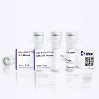
Application
ELISA, FACS, Functional assay


Chemical Information
-
Formulation
Please refer to the lot-specific COA for specific buffer information.
-
Storage
Please store the product under the recommended conditions in the Certificate of Analysis.


Purity & Documentation
References
[1]. Desu HL, et al. IC100: a novel anti-ASC monoclonal antibody improves functional outcomes in an animal model of multiple sclerosis. J Neuroinflammation. 2020;17(1):143. Published 2020 May 4. [Content Brief]
[2]. de Rivero Vaccari JP, et al. Mechanism of action of IC 100, a humanized IgG4 monoclonal antibody targeting apoptosis-associated speck-like protein containing a caspase recruitment domain (ASC). Transl Res. 2023;251:27-40. [Content Brief]

Calculators
Concentration (start) × Volume (start) = Concentration (final) × Volume (final)


IC 100 Related Classifications
HY-P992382 Related Classifications
 Powered by Bioz