



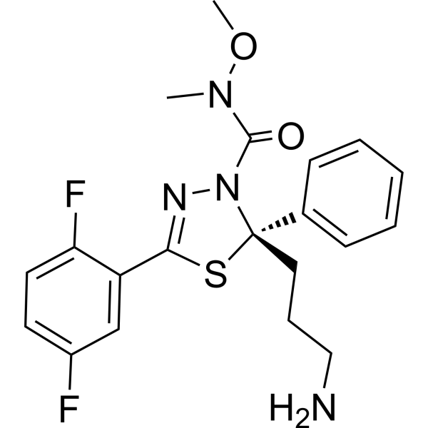
Filanesib
Based on 7 publication(s) in Google Scholar
Filanesib (ARRY-520) is a selective and noncompetitive kinesin spindle protein (KSP) inhibitor, with an IC50 of 6 nM for human KSP. Filanesib induces cell death by apoptosis in vitro. Filanesib has potent anti-proliferative activity.
Para uso exclusivo en investigación. No vendemos a pacientes.
- Pureza : 99.59%
- No. CAS: 885060-09-3
- Fòrmula: C20H22F2N4O2S
- Peso molecular:420.48
-
Almacenamiento:Powder -20°C, 3 years , 4°C, 2 years ; In solvent -80°C, 2 years , -20°C, 1 year

To place orders, for customer services and technical support, please contact: MedChemExpress USA
Tel: 609-228-6898 E-mail: [email protected] [email protected]


-
 Actividad biológica
-
Chemical Information
-
Solvente y solubilidad
- Protocolo
- Pureza y Documentación
- Referencias
-
 Help & FAQs


-
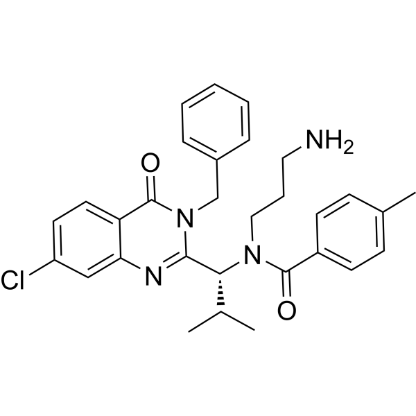
HY-50759
IspinesibKSP Inhibitor -
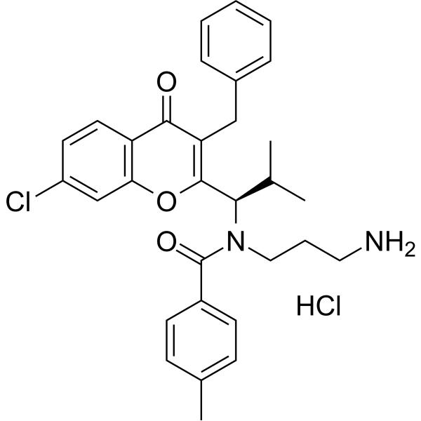
HY-12069
SB-743921 hydrochlorideEg5 Inhibitor -
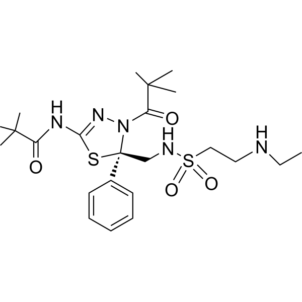
HY-14846
LitronesibKinesin Inhibitor -
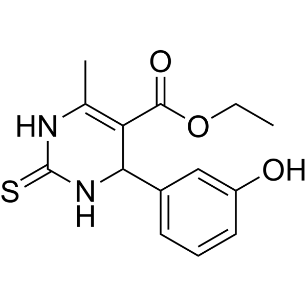
HY-101071A
MonastrolMitotic Kinesin Eg5 Inhibitor -
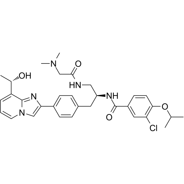
HY-10299
GSK-923295CENP-E Inhibitor -
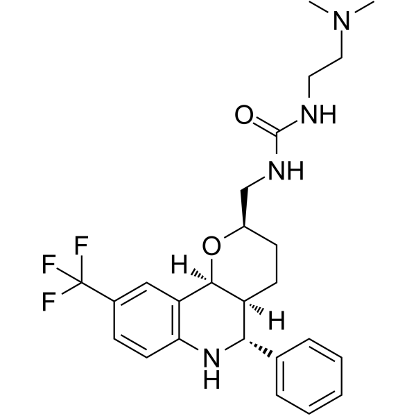
HY-15000
EMD534085Kinesin Inhibitor -
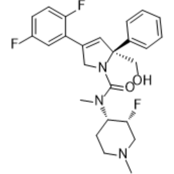
HY-50672
MK-0731KSP Inhibitor -
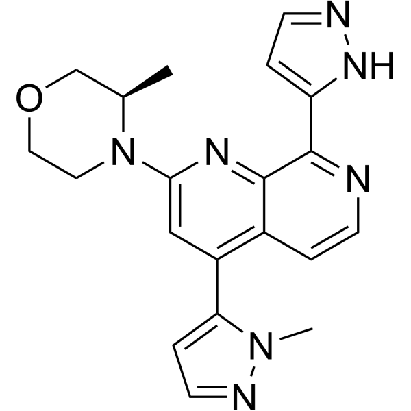
HY-101566
ElimusertibATR Inhibitor -
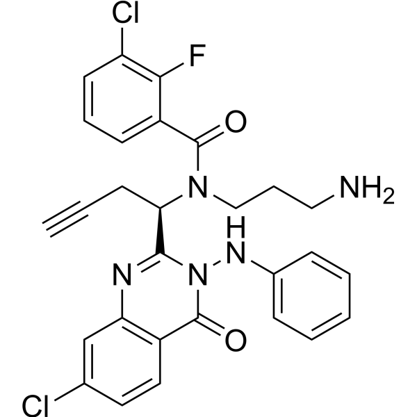
HY-16062
ARQ 621Eg5 Inhibitor -
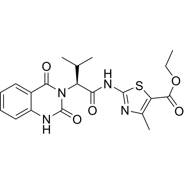
HY-15948
Kif15-IN-1Kinesin Inhibitor -
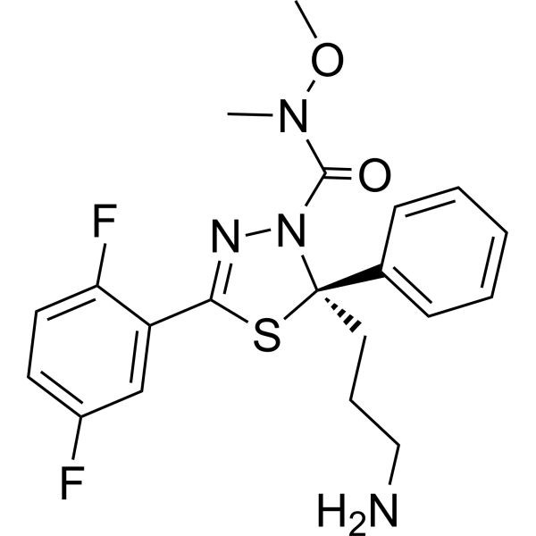
HY-15187A
(R)-FilanesibKSP Inhibitor -

HY-10227
BortezomibProteasome Inhibitor
Publications Citing Use of MedChemExpress (MCE) Filanesib
More Customer Validation & Images

-
Cell Proliferation/Viability Assay
-
Cell Proliferation/Viability Assay

-
Flow Cytometry
-
Apoptosis Analysis
-
In Vivo Efficacy Study
Ver todos los productos específicos de isoformas Kinesin
More

Actividad biológica
Descripciòn
IC50 & Target
[1]|
KSP 6 nM (IC50) |
Cellular Effect
|
Cell Line

|
Type | Value | Description | References |
|---|---|---|---|---|
| HCT-116 | IC50 |
0.7 nM
Compound: 2; ARRY-520
|
Growth inhibition of human HCT116 cells after 72 hrs by MTS assay
Growth inhibition of human HCT116 cells after 72 hrs by MTS assay
|
[PMID: 26396688] |
In Vitro
Filanesib induces mitotic arrest in multiple cell lines[1].
Filanesib exhibits anti-proliferative against a broad range of human and rodent tumor cell lines, including a variety of leukemias and solid tumors, with EC50s between 0.4 nM and 14.4 nM[1].
Filanesib (0.001-0.1 nM; 36 hours) induces apoptosis in a dose-dependent manner in HeLa cells[1].
Filanesib (3.13-6.25 nM; 44 hours) causes accumulation of cells in the G2/M phase of the cell cycle in a dose-dependent manner in HeLa cells[1].
Filanesib potently induces cell cycle block and subsequent death in leukemic cells via the mitochondrial pathway and has potential to eradicate AML progenitor cells[2].
Filanesib (3 μM; 6-24 hours) is able to induce caspase-2 activation[3].
Filanesib (0.003-3 μM; 24-48 hours) is cytotoxic in Type II EOC cells[3].
MedChemExpress (MCE) has not independently confirmed the accuracy of these methods. They are for reference only.
-
Cell Line:Hela cells
-
Concentration:0.01-0.1 nM
-
Incubation Time:36 hours
-
Result:Induced the formation of nucleosomes and activation of caspases-3 and 7.
-
Cell Line:HeLa cells
-
Concentration:0.78 nM, 1.56 nM, 3.13 nM, 6.25 nM
-
Incubation Time:44 hours
-
Result:Resulted in G2/M arrest.
-
Cell Line:Type II EOC cells
-
Concentration:3 μM
-
Incubation Time:6 hours, 12 hours, 24 hours
-
Result:Induced caspase-2 activation in a time-dependent manner.
-
Cell Line:Type II EOC cell lines (A2780, CP70, 01-28)
-
Concentration:0.003 μM, 0.03 μM, 0.3μM, 3 μM
-
Incubation Time:24 hours , 48 hours
-
Result:Effectively decreased cell viability in a time-dependent manner in the Type II EOC cell lines.
In Vivo
MedChemExpress (MCE) has not independently confirmed the accuracy of these methods. They are for reference only.
-
Animal Model:Female nude mice, EOC mice xenograft model[3]
-
Dosage:20 mg/kg, 30 mg/kg
-
Administration:Intraperitoneal injection, q4dx3
-
Result:Induced a decrease in tumor kinetics in a dose-dependent manner.
Ensayo clínico
| NCT Number | Sponsor | Condition | Start Date |
Phase
|
|---|---|---|---|---|
| NCT01329991 | 2011-05 | PHASE1 |


Chemical Information
-
No. CAS 885060-09-3
-
Appearance Solid
-
Peso molecular 420.48
-
Fòrmula C20H22F2N4O2S
-
Color White to off-white
-
SMILES
O=C(N1[C@@](C2=CC=CC=C2)(CCCN)SC(C3=CC(F)=CC=C3F)=N1)N(OC)C
-
Synonyms
ARRY-520
-
Envío
Room temperature in continental US; may vary elsewhere.
-
Almacenamiento
Powder -20°C 3 years 4°C 2 years In solvent -80°C 2 years -20°C 1 year


Publications (7)
-
Journal Impact Factor
-
Most Recent
-
Cell Discov
Single-cell profiling reveals molecular basis of malignant phenotypes and tumor microenvironments in small bowel adenocarcinomas. [Abstract]2022 Sep 14;8(1):92. PMID: 36104333
Filanesib purchased from MedChemExpress. Usage Cited in: Cell Discov. 2022 Sep 14;8(1):92. [Abstract]
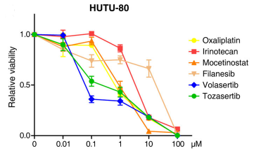
Filanesib (0-100 μM; 36 h) inhibited HUTU-80 cell survival in a dose-dependent manner.
-
Cancer Lett
2021 May 28:506:1-10. PMID: 33652084
Filanesib purchased from MedChemExpress. Usage Cited in: Cancer Lett. 2021 May 28:506:1-10. [Abstract]
Filanesib (0-10 nM; 48 h) showed antiproliferative effects in the benign meningioma cell line Ben-Men-1 and the anaplastic cell lines NCH93, IOMM-Lee, and KT21-MG cells.
Filanesib purchased from MedChemExpress. Usage Cited in: Cancer Lett. 2021 May 28:506:1-10. [Abstract]
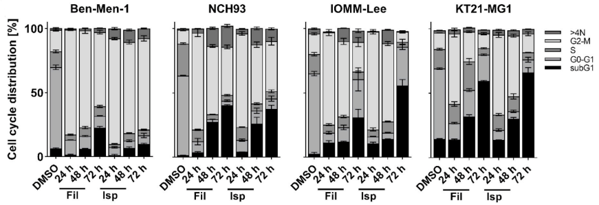
Filanesib (10 nM; 24, 48, 72 h) induced cell cycle arrest at G2/M-phase in Ben-Men-1, NCH93, IOMM-Lee, and KT21-MG cells.
Filanesib purchased from MedChemExpress. Usage Cited in: Cancer Lett. 2021 May 28:506:1-10. [Abstract]
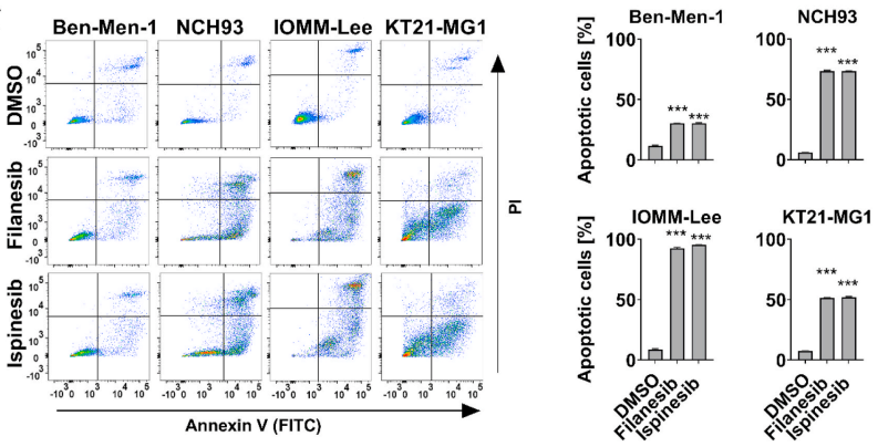
Filanesib (10 nM; 72 h) induced cell apoptosis in Ben-Men-1, NCH93, IOMM-Lee, and KT21-MG cells.
Filanesib purchased from MedChemExpress. Usage Cited in: Cancer Lett. 2021 May 28:506:1-10. [Abstract]
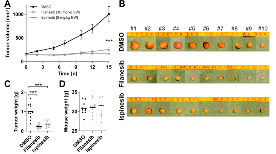
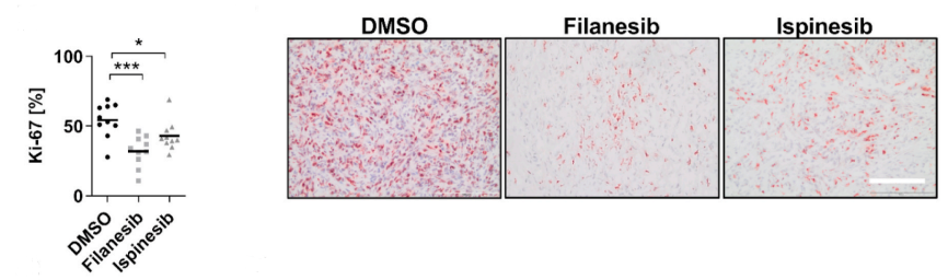
Filanesib (10 mg/kg; i.p.; once every three days on five occasions) inhibited NCH93 tumor growth in NCH93 tumor-bearing mice.
Filanesib purchased from MedChemExpress. Usage Cited in: Cancer Lett. 2021 May 28:506:1-10. [Abstract]
Filanesib (10 mg/kg; i.p.; once every three days on five occasions) showed a significant reduction of Ki-67-positive cells in tumor sections.
-
NPJ Precis Oncol
Functional screening identifies kinesin spindle protein inhibitor filanesib as a potential treatment option for hepatoblastoma. [Abstract]2025 Apr 25;9(1):122. PMID: 40281281 -
Gene
2025 Jul 5:955:149458. PMID: 40187619 -
-
-
Methods Mol Biol
2018:1711:351-398. PMID: 29344898


Solvente y solubilidad
In Vitro:
DMSO : ≥ 100 mg/mL (237.82 mM; Hygroscopic DMSO has a significant impact on the solubility of product, please use newly opened DMSO)
* "≥" means soluble, but saturation unknown.
Please refer to the solubility information to select the appropriate solvent. Once prepared, please aliquot and store the solution to prevent product inactivation from repeated freeze-thaw cycles.
Storage method and period of stock solution: -80°C, 2 years; -20°C, 1 year. When stored at -80°C, please use it within 2 years. When stored at -20°C, please use it within 1 year.
Please refer to the solubility information to select the appropriate solvent. Once prepared, please aliquot and store the solution to prevent product inactivation from repeated freeze-thaw cycles.
Storage method and period of stock solution: -80°C, 2 years; -20°C, 1 year. When stored at -80°C, please use it within 2 years. When stored at -20°C, please use it within 1 year.
Concentration (start) × Volume (start) = Concentration (final) × Volume (final)
In Vivo:
Select the appropriate dissolution method based on your experimental animal and administration route.
- For the following dissolution methods, please ensure to first prepare a clear stock solution using an In Vitro approach and then sequentially add co-solvents:
- To ensure reliable experimental results, the clarified stock solution can be appropriately stored based on storage conditions. As for the working solution for In Vivo experiments, it is recommended to prepare freshly and use it on the same day.
- The percentages shown for the solvents indicate their volumetric ratio in the final prepared solution. If precipitation or phase separation occurs during preparation, heat and/or sonication can be used to aid dissolution.
Add each solvent one by one: 10% DMSO 40% PEG300 5% Tween-80 45% Saline
Solubility: ≥ 2.5 mg/mL (5.95 mM); Clear solution
This protocol yields a clear solution of ≥ 2.5 mg/mL (saturation unknown).
Taking 1 mL working solution as an example, add 100 μL DMSO stock solution (25.0 mg/mL) to 400 μL PEG300, and mix evenly; then add 50 μL Tween-80 and mix evenly; then add 450 μL Saline to adjust the volume to 1 mL.
Preparation of Saline: Dissolve 0.9 g sodium chloride in ddH₂O and dilute to 100 mL to obtain a clear Saline solution.
Add each solvent one by one: 10% DMSO 90% (20% SBE-β-CD in Saline)
Solubility: ≥ 2.5 mg/mL (5.95 mM); Clear solution
This protocol yields a clear solution of ≥ 2.5 mg/mL (saturation unknown).
Taking 1 mL working solution as an example, add 100 μL DMSO stock solution (25.0 mg/mL) to 900 μL 20% SBE-β-CD in Saline, and mix evenly.
Preparation of 20% SBE-β-CD in Saline (4°C, storage for one week): 2 g SBE-β-CD powder is dissolved in 10 mL Saline, completely dissolve until clear.
In Vivo Dissolution Calculator
Please enter the basic information of animal experiments:
-
-
-
-
Recommended: Prepare an additional quantity of animals to account for potential losses during experiments.
Please enter your animal formula composition:
-
%DMSO +
Recommended: Keep the proportion of DMSO in working solution below 2% if your animal is weak.
-
%+
-
+%Tween-80 + +
-
%Saline +
The co-solvents required include: DMSO, . All of co-solvents are available by MedChemExpress (MCE). , Tween 80. All of co-solvents are available by MedChemExpress (MCE).
Working solution concentration: 0.22 mg/mL
Method for preparing stock solution: mg drug dissolved in μL DMSO. Stock solution concentration: mg/mL.
1. Take μL DMSO stock solution;
2. Add μL .
μL , mix evenly;
3. Then add μL Tween 80, mix evenly;
4. Then add μL
Please ensure that the stock solution in the first step is dissolved to a clear state, and add co-solvents in sequence. You can use ultrasonic heating (ultrasonic cleaner, recommended frequency 20-40 kHz), vortexing, etc. to assist dissolution.


Pureza y Documentación
-
Ficha de datos (303 KB)
-
SDS (393 KB)
- English - EN (393 KB)
- Français - FR (393 KB)
- Deutsch - DE (393 KB)
- Norwegian - NO (393 KB)
- Español - ES (393 KB)
- Swedish - SV (393 KB)
- Italian - IT (393 KB)
- Korean - KR (393 KB)
- Portuguese - PT (393 KB)
-
Instrucciones de manejo (2659 KB)
Referencias
[2]. BZ Carter, et al. Inhibition of KSP by ARRY-520 Induces Cell Cycle Block and Cell Death via the Mitochondrial Pathway in AML Cells. [Content Brief]
[3]. Ki Hyung Kim, et al. KSP inhibitor ARRY-520 as a substitute for Paclitaxel in Type I ovarian cancer cells. J Transl Med. 2009; 7: 63. [Content Brief]


Complete Stock Solution Preparation Table
Please refer to the solubility information to select the appropriate solvent. Once prepared, please aliquot and store the solution to prevent product inactivation from repeated freeze-thaw cycles.
Storage method and period of stock solution: -80°C, 2 years; -20°C, 1 year. When stored at -80°C, please use it within 2 years. When stored at -20°C, please use it within 1 year.
| Optional Solvent | Concentration Solvent Mass | 1 mg | 5 mg | 10 mg | 25 mg |
|---|---|---|---|---|---|
| DMSO | 1 mM | 2.3782 mL | 11.8912 mL | 23.7823 mL | 59.4559 mL |
| 5 mM | 0.4756 mL | 2.3782 mL | 4.7565 mL | 11.8912 mL | |
| 10 mM | 0.2378 mL | 1.1891 mL | 2.3782 mL | 5.9456 mL | |
| 15 mM | 0.1585 mL | 0.7927 mL | 1.5855 mL | 3.9637 mL | |
| 20 mM | 0.1189 mL | 0.5946 mL | 1.1891 mL | 2.9728 mL | |
| 25 mM | 0.0951 mL | 0.4756 mL | 0.9513 mL | 2.3782 mL | |
| 30 mM | 0.0793 mL | 0.3964 mL | 0.7927 mL | 1.9819 mL | |
| 40 mM | 0.0595 mL | 0.2973 mL | 0.5946 mL | 1.4864 mL | |
| 50 mM | 0.0476 mL | 0.2378 mL | 0.4756 mL | 1.1891 mL | |
| 60 mM | 0.0396 mL | 0.1982 mL | 0.3964 mL | 0.9909 mL | |
| 80 mM | 0.0297 mL | 0.1486 mL | 0.2973 mL | 0.7432 mL | |
| 100 mM | 0.0238 mL | 0.1189 mL | 0.2378 mL | 0.5946 mL |


Filanesib Related Classifications
HY-15187 Related Classifications
 Powered by Bioz