



Filanesib TFA
Based on 7 publication(s) in Google Scholar
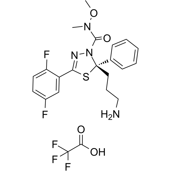
Filanesib TFA (ARRY-520 TFA) is a selective and noncompetitive kinesin spindle protein (KSP) inhibitor, with an IC50 of 6 nM for human KSP. Filanesib TFA induces cell death by apoptosis in vitro. Filanesib TFA has potent anti-proliferative activity.
For research use only. We do not sell to patients.
- CAS No.: 1781834-99-8
- Formula: C22H23F5N4O4S
- Molecular Weight:534.50
-
Storage:
Please store the product under the recommended conditions in the Certificate of Analysis.

To place orders, for customer services and technical support, please contact: MedChemExpress USA
Tel: 609-228-6898 E-mail: [email protected] [email protected]


-
 Biological Activity
-
Chemical Information
- Protocols
- Purity & Documentation
- References
-
 Help & FAQs


-
HY-50759
IspinesibKSP Inhibitor -
HY-12069
SB-743921 hydrochlorideEg5 Inhibitor -
HY-15187
FilanesibKSP Inhibitor -
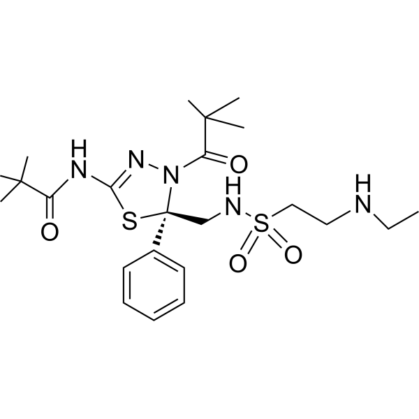
HY-14846
LitronesibKinesin Inhibitor -
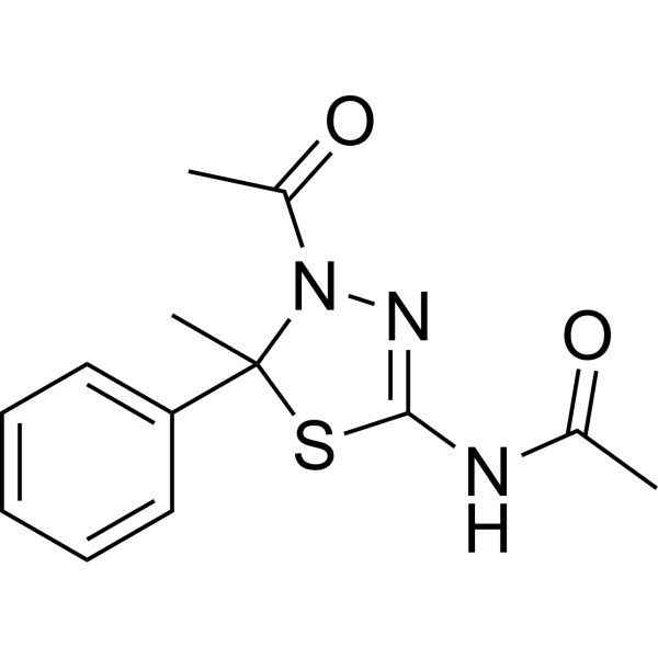
HY-19966
K858 (Racemic)Apoptosis Inducer -
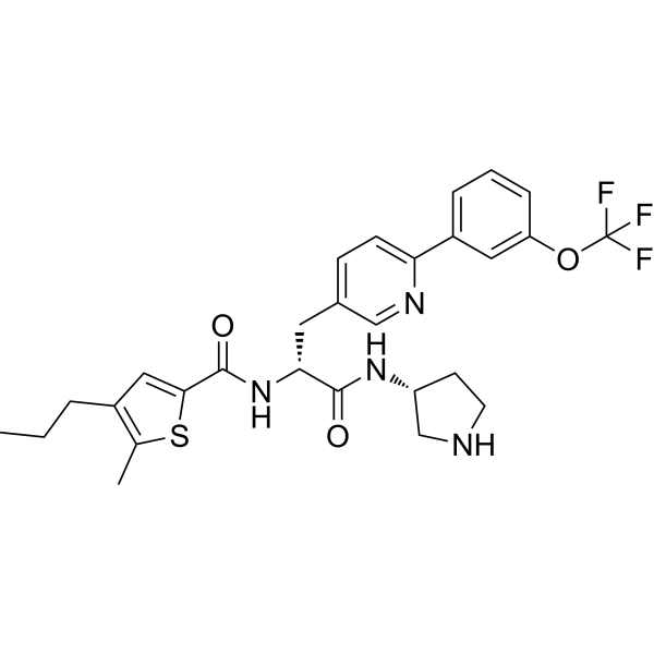
HY-12241
AZ82KIFC1 Inhibitor -
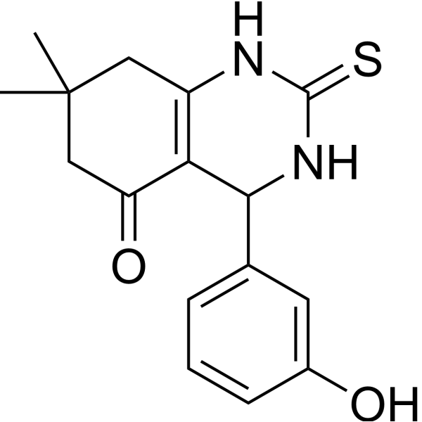
HY-19944
DimethylenastronKinesin Inhibitor -
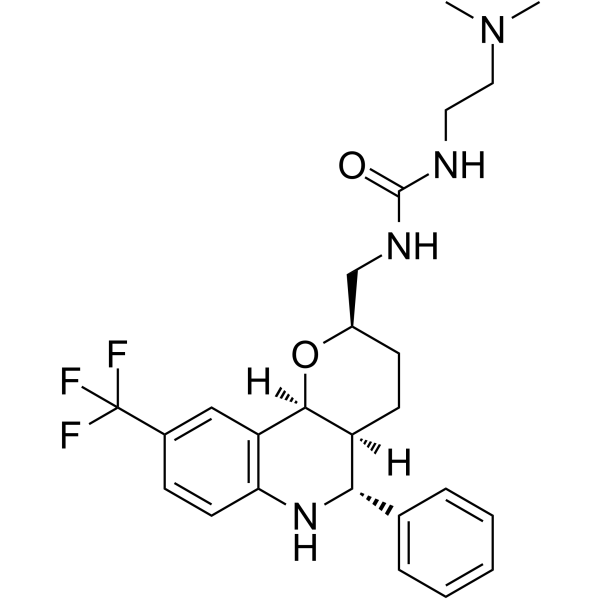
HY-15000
EMD534085Kinesin Inhibitor -
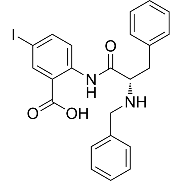
HY-15857
CW-069HSET Inhibitor -
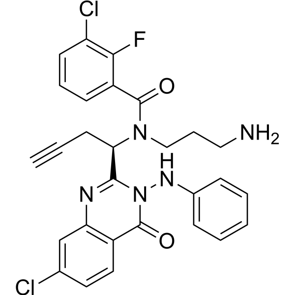
HY-16062
ARQ 621Eg5 Inhibitor -
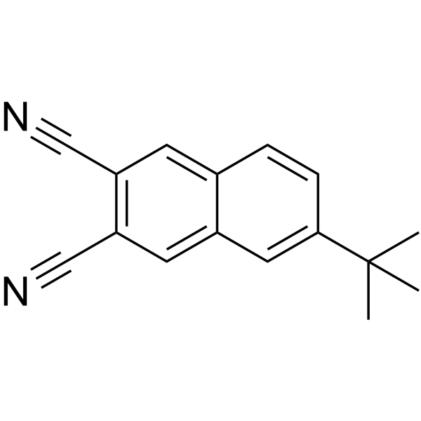
HY-110208
BRD9876 -
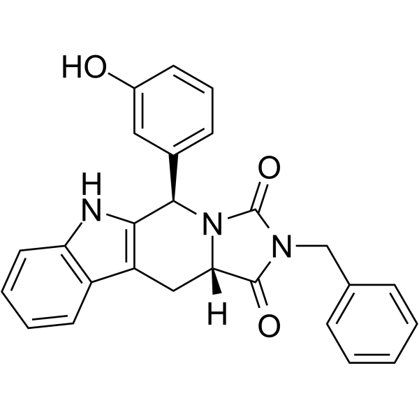
HY-112915
Eg5 Inhibitor V, trans-24Kinesin Inhibitor
Publications Citing Use of MedChemExpress (MCE) Filanesib TFA
More Customer Validation & Images

-
Cell Proliferation/Viability Assay
-
Cell Proliferation/Viability Assay

-
Flow Cytometry
-
Apoptosis Analysis
-
In Vivo Efficacy Study
All Kinesin Isoforms
More

Biological Activity
Description
IC50 & Target
IC50: 6 nM (KSP)[1]
In Vitro
Filanesib TFA induces mitotic arrest in multiple cell lines[1].
Filanesib TFA exhibits anti-proliferative against a broad range of human and rodent tumor cell lines, including a variety of leukemias and solid tumors, with EC50s between 0.4 nM and 14.4 nM[1].
Filanesib (0.001-0.1 nM; 36 hours) TFA induces apoptosis in a dose-dependent manner in HeLa cells[1].
Filanesib (3.13-6.25 nM; 44 hours) TFA causes accumulation of cells in the G2/M phase of the cell cycle in a dose-dependent manner in HeLa cells[1].
Filanesib TFA potently induces cell cycle block and subsequent death in leukemic cells via the mitochondrial pathway and has potential to eradicate AML progenitor cells[2].
Filanesib (3 μM; 6-24 hours) TFA is able to induce caspase-2 activation[3].
Filanesib (0.003-3 μM; 24-48 hours) TFA is cytotoxic in Type II EOC cells[3].
MedChemExpress (MCE) has not independently confirmed the accuracy of these methods. They are for reference only.
In Vivo
MedChemExpress (MCE) has not independently confirmed the accuracy of these methods. They are for reference only.


Chemical Information
-
CAS No. 1781834-99-8
-
Molecular Weight 534.50
-
Formula C22H23F5N4O4S
-
SMILES
O=C(N1[C@@](C2=CC=CC=C2)(CCCN)SC(C3=CC(F)=CC=C3F)=N1)N(OC)C.OC(C(F)(F)F)=O
-
Synonyms
ARRY-520 TFA
-
Shipping
Room temperature in continental US; may vary elsewhere.
-
Storage
Please store the product under the recommended conditions in the Certificate of Analysis.


Publications (7)
-
Journal Impact Factor
-
Most Recent
-
Cell Discov
Single-cell profiling reveals molecular basis of malignant phenotypes and tumor microenvironments in small bowel adenocarcinomas. [Abstract]2022 Sep 14;8(1):92. PMID: 36104333
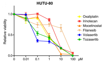
Filanesib TFA purchased from MedChemExpress. Usage Cited in: Cell Discov. 2022 Sep 14;8(1):92. [Abstract]
Filanesib (0-100 μM; 36 h) inhibited HUTU-80 cell survival in a dose-dependent manner.
-
Cancer Lett
2021 May 28:506:1-10. PMID: 33652084
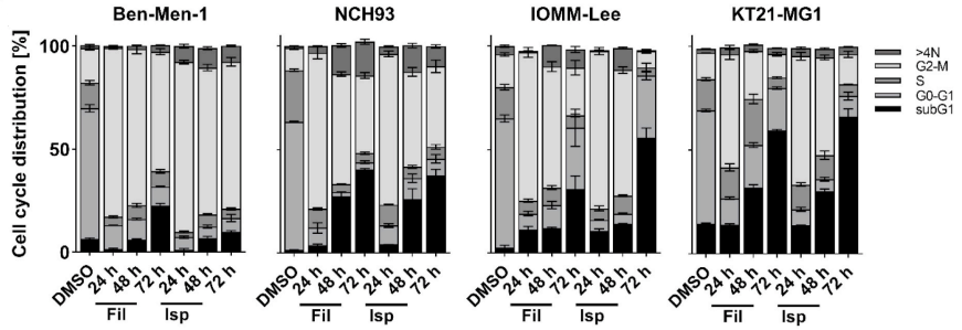
Filanesib TFA purchased from MedChemExpress. Usage Cited in: Cancer Lett. 2021 May 28:506:1-10. [Abstract]
Filanesib (0-10 nM; 48 h) showed antiproliferative effects in the benign meningioma cell line Ben-Men-1 and the anaplastic cell lines NCH93, IOMM-Lee, and KT21-MG cells.
Filanesib TFA purchased from MedChemExpress. Usage Cited in: Cancer Lett. 2021 May 28:506:1-10. [Abstract]
Filanesib (10 nM; 24, 48, 72 h) induced cell cycle arrest at G2/M-phase in Ben-Men-1, NCH93, IOMM-Lee, and KT21-MG cells.
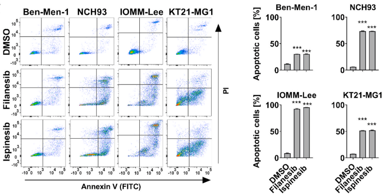
Filanesib TFA purchased from MedChemExpress. Usage Cited in: Cancer Lett. 2021 May 28:506:1-10. [Abstract]
Filanesib (10 nM; 72 h) induced cell apoptosis in Ben-Men-1, NCH93, IOMM-Lee, and KT21-MG cells.
Filanesib TFA purchased from MedChemExpress. Usage Cited in: Cancer Lett. 2021 May 28:506:1-10. [Abstract]
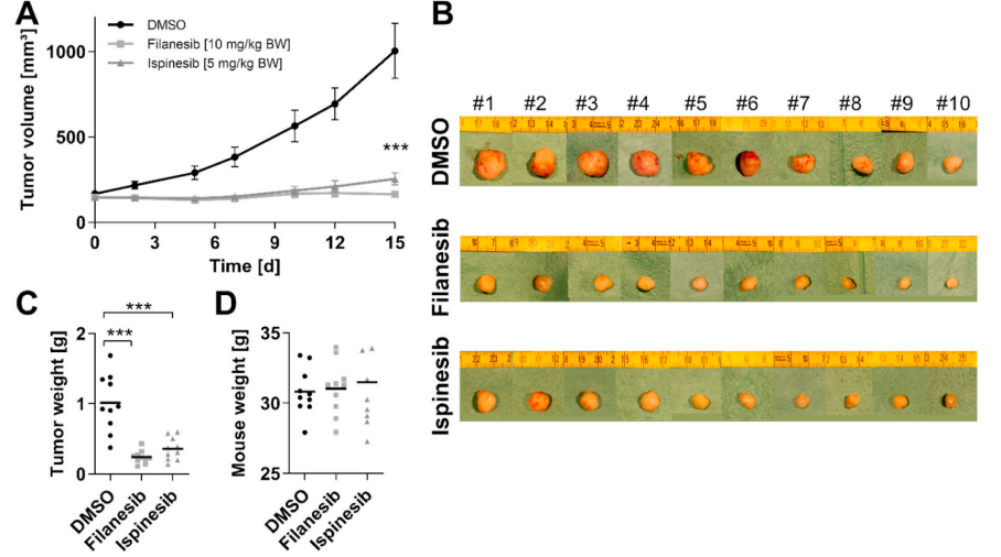
Filanesib (10 mg/kg; i.p.; once every three days on five occasions) inhibited NCH93 tumor growth in NCH93 tumor-bearing mice.
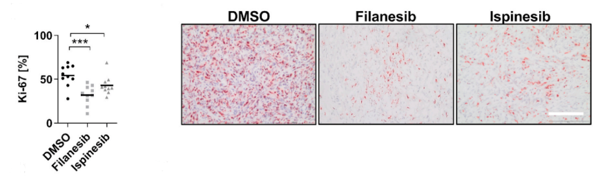
Filanesib TFA purchased from MedChemExpress. Usage Cited in: Cancer Lett. 2021 May 28:506:1-10. [Abstract]
Filanesib (10 mg/kg; i.p.; once every three days on five occasions) showed a significant reduction of Ki-67-positive cells in tumor sections.
-
NPJ Precis Oncol
Functional screening identifies kinesin spindle protein inhibitor filanesib as a potential treatment option for hepatoblastoma. [Abstract]2025 Apr 25;9(1):122. PMID: 40281281 -
Gene
2025 Jul 5:955:149458. PMID: 40187619 -
-
-
Methods Mol Biol
2018:1711:351-398. PMID: 29344898


Purity & Documentation
References
[2]. BZ Carter, et al. Inhibition of KSP by ARRY-520 Induces Cell Cycle Block and Cell Death via the Mitochondrial Pathway in AML Cells. [Content Brief]
[3]. Ki Hyung Kim, et al. KSP inhibitor ARRY-520 as a substitute for Paclitaxel in Type I ovarian cancer cells. J Transl Med. 2009; 7: 63. [Content Brief]

Calculators
Concentration (start) × Volume (start) = Concentration (final) × Volume (final)


Filanesib TFA Related Classifications
HY-15187B Related Classifications
 Powered by Bioz