



ER-Tracker Green (solution)

Based on 1 Customer Validation
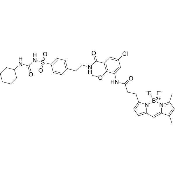
ER-Tracker dye is a derivative of BODIPY series dyes coupled with Glibenclamide (HY-15206) , highly selective binding to the endoplasmic reticulum, non-toxic to cells at low concentrations, this type of dye is an environmentally sensitive probe, and formaldehyde treatment can still retain part of the fluorescence, with high fluorescence life, good extinction coefficient and other characteristics. Glibenclamide is an atp-dependent K+ channel blocker (Kir6, KATP) and CFTR Cl-channel blocker that binds in the endoplasmic reticulum. ER-Tracker is not suitable for staining cells after fixation.
Solvent and concentration: DMSO: 1 mM
Para uso exclusivo en investigación. No vendemos a pacientes.
- No. CAS: 730931-46-1
- Fòrmula: C37H42BClF2N6O6S
- Peso molecular:783.10
-
Almacenamiento:
Please store the product under the recommended conditions in the Certificate of Analysis.


To place orders, for customer services and technical support, please contact: MedChemExpress USA
Tel: 609-228-6898 E-mail: [email protected] [email protected]


-
 Actividad biológica
-
Chemical Information
- Pureza y Documentación
- Referencias
-
 Help & FAQs


-
HY-13753
StreptozotocinDiabetes Modeling Agent -

HY-15763
ErastinFerroptosis Inducer -
HY-135056
MitoTracker Green FMMitochondrial Dye -
HY-D1300
LysoTracker RedCell Death Indicator -

HY-DY1040
LysoTracker Red (solution)Cell Death Indicator -
HY-D1297
ER-Tracker GreenEndoplasmic Reticulum Fluorescent Dye -

HY-DY1032
MitoTracker Green FM (solution)Mitochondrial Dye -
HY-D1116
MitoMark Red IFluorescent Dye


Actividad biológica
Descripciòn
Solvent and concentration: DMSO: 1 mM
In Vitro
Guide (The following is our recommended protocol. This protocol is only a guide and should be modified according to your specific needs).
1. Preparation of ER-Tracker working solution
1.1 Preparation of the stock solution
Stock solution.
It is recommended to store the stock solution at -20°C or -80°C away from light and avoid repetitive freeze-thaw cycles.
1.2 Preparation of ER-Tracker working solution
Dilute the stock solution in serum-free cell culture medium or PBS. The corresponding stock solution can be diluted according to the actual situation. Note that if the solvent is DMSO, the cytotoxicity of DMSO must be considered, and a solvent control should be prepared; if the solvent is pure water, the working solution needs to be filtered and sterilized before adding cells.
Note: Please adjust the concentration of ER-Tracker working solution according to the actual situation.
2. Cell staining
2.1 Suspension cells (6-well plate)
2.1.1 Centrifuge at 1000 g at 4°C for 3-5 minutes and then discard the supernatant. Wash twice with PBS, 5 minutes each time.The cell density is 1×10⁶/mL.
2.1.2 Add 1 mL of working solution, and then incubate at room temperature for 5-30 minutes.
2.1.3 Centrifuge at 400 g at 4°C for 3-4 minutes and then discard the supernatant.
2.1.4 Wash twice with PBS, 5 minutes each time.
2.1.5 Resuspend cells with serum-free cell culture medium or PBS. Observation by fluorescence microscopy or flow cytometry.
2.2 Adherent cells
2.2.1 Culture adherent cells on sterile coverslips.
2.2.2 Remove the coverslip from the medium and aspirate excess medium.
2.2.3 Add 100 μL of working solution, gently shake it to completely cover the cells,and then incubate at room temperature for 5-30 minutes.
2.2.4 Wash twice with medium, 5 minutes each time. Observation by fluorescence microscopy or flow cytometry.
Note: If detection by flow cytometry, cells need to be resuspended before staining.
MedChemExpress (MCE) has not independently confirmed the accuracy of these methods. They are for reference only. .


Chemical Information
-
No. CAS 730931-46-1
-
Appearance Liquid
-
Peso molecular 783.10
-
Fòrmula C37H42BClF2N6O6S
-
Color Light yellow to yellow
-
SMILES
O=C(NS(=O)(C1=CC=C(CCNC(C2=C(OC)C(NC(CCC3=CC=C4C=C5C(C)=CC(C)=[N]5[B+3]([F-])([N-]43)[F-])=O)=CC(Cl)=C2)=O)C=C1)=O)NC6CCCCC6
-
Envío
Room temperature in continental US; may vary elsewhere.
-
Almacenamiento
Please store the product under the recommended conditions in the Certificate of Analysis.


Pureza y Documentación
-
Ficha de datos (277 KB)
-
SDS (251 KB)
- English - EN (251 KB)
- Français - FR (251 KB)
- Deutsch - DE (251 KB)
- Norwegian - NO (251 KB)
- Español - ES (251 KB)
- Swedish - SV (251 KB)
- Italian - IT (251 KB)
- Korean - KR (251 KB)
- Portuguese - PT (251 KB)
-
Instrucciones de manejo (2659 KB)
Referencias
[2]. J Jacob Strouse, et al. Fluorescent substrates for flow cytometric evaluation of efflux inhibition in ABCB1, ABCC1, and ABCG2 transporters. Anal Biochem. 2013 Jun 1;437(1):77-87. [Content Brief]
[3]. Tanuja T Merianda, et al. A functional equivalent of endoplasmic reticulum and Golgi in axons for secretion of locally synthesized proteins. Mol Cell Neurosci. 2009 Feb;40(2):128-42. [Content Brief]

Calculators
Concentration (start) × Volume (start) = Concentration (final) × Volume (final)


ER-Tracker Green (solution) Related Classifications
HY-DY1043 Related Classifications
 Powered by Bioz