



LMD-009
Based on 2 publication(s) in Google Scholar
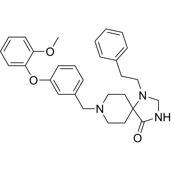
LMD-009 is a selective CCR8 nonpeptide agonist. LMD-009 mediates chemotaxis, inositol phosphate accumulation, and calcium release in high potencies with EC50s from 11 to 87 nM.
Para uso exclusivo en investigación. No vendemos a pacientes.
- Pureza : 99.35%
- No. CAS: 950195-51-4
- Fòrmula: C29H33N3O3
- Peso molecular:471.59
-
Almacenamiento:Powder -20°C, 3 years , 4°C, 2 years ; In solvent -80°C, 2 years , -20°C, 1 year

To place orders, for customer services and technical support, please contact: MedChemExpress USA
Tel: 609-228-6898 E-mail: [email protected] [email protected]


-
 Actividad biológica
-
Chemical Information
-
Solvente y solubilidad
- Protocolo
- Pureza y Documentación
- Referencias
-
 Help & FAQs


-
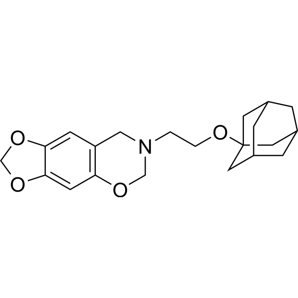
HY-122219
R243CCR8 Antagonist -
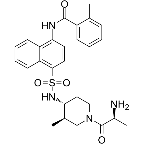
HY-124416
ML604086CCR8 Inhibitor -
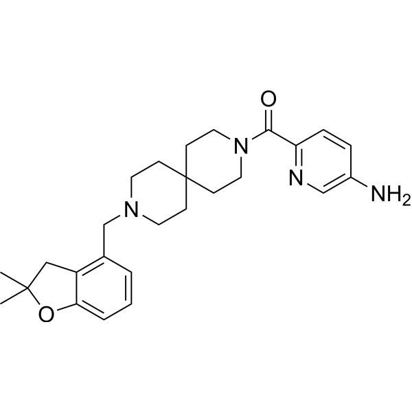
HY-119217
AZ084CCR8 Allosteric Antagonist -
HY-101038A
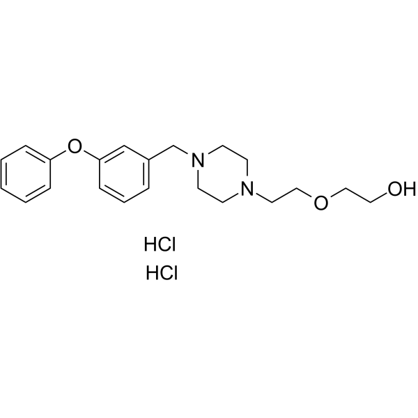
ZK756326 dihydrochlorideCCR8 Agonist -
HY-100183
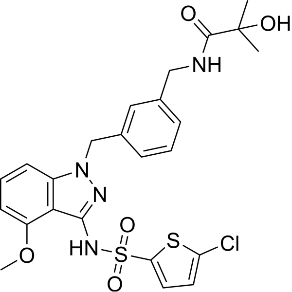
GSK2239633ACCR4 Antagonist -
HY-13004
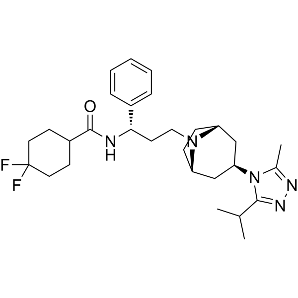
MaravirocCCR5 Antagonist -
HY-12080A
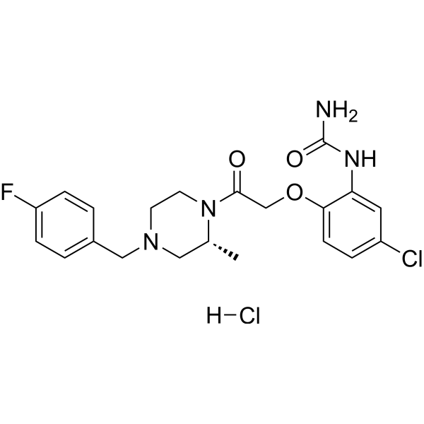
BX471 hydrochlorideCCR Antagonist -
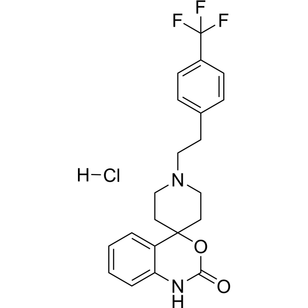
HY-18611
Abaucin hydrochlorideCCR2antagonist -
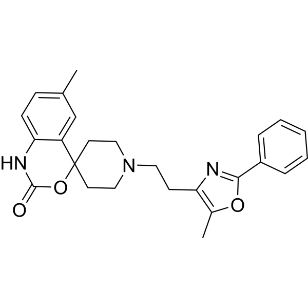
HY-15418
RS 504393CCR2 Antagonist -
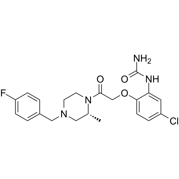
HY-12080
BX471CCR1 Antagonist -
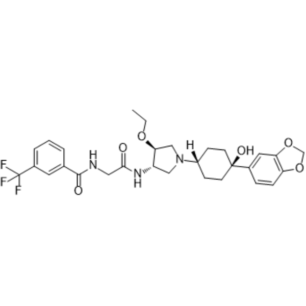
HY-50674
INCB3344CCR2 Antagonis -
HY-101264
CCR2 antagonist 3CCR2 Antagonist
Publications Citing Use of MedChemExpress (MCE) LMD-009
More

Actividad biológica
Descripciòn
IC50 & Target
[1]|
CCR8 11-87 nM (EC50) |
Cellular Effect
|
Cell Line

|
Type | Value | Description | References |
|---|---|---|---|---|
| COS-7 | EC50 |
11 nM
Compound: 71; LMD-009
|
Antiproliferative activity against African green monkey COS7 cells assessed as inhibition of cell growth
Antiproliferative activity against African green monkey COS7 cells assessed as inhibition of cell growth
|
[PMID: 28165741] |
In Vitro
LMD-009 (0-20 nM; 90 min) stimulates inositol phosphate accumulation in COS-7 cells expressing the human CCR8 receptor[1].? LMD-009 (0-100 nM; 1 h) mediates calcium release in Chinese hamster ovary cells[1].? LMD-009 (0.1 nM-100 μM; 40 min) induces L1.2 cells migration[1].? LMD-009 (0-10 μM; 90 min) exhibits different molecular interaction with CCR8 mutations in COS-7 cells[1].? LMD-009 (0-10 nM; 90 min) exhibits no antagonist activity to other human chemokine receptors[1].
MedChemExpress (MCE) has not independently confirmed the accuracy of these methods. They are for reference only. Further protocols information, click here.
-
Cell Line:COS-7 cells
-
Concentration:0-20 nM
-
Incubation Time:90 min
-
Result:Stimulated inositol phosphate accumulation in COS-7 cells expressing the human CCR8 receptor with an EC50 value of 11 nM. Inhibited none of the receptors of other human chemokine receptors.
-
Cell Line:Chinese hamster ovary cells
-
Concentration:0-100 nM
-
Incubation Time:1 hour
-
Result:Regulated calcium release in Chinese hamster ovary cells with an EC50 value 87 nM.
-
Cell Line:Lymphocyte L1.2 cells.
-
Concentration:0.1 nM- 100 μM
-
Incubation Time:40 min
-
Result:Exhibited an Ki value of 66 nM with L1.2 cells and specifically bound 125I-CCL1.


Chemical Information
-
No. CAS 950195-51-4
-
Appearance Solid
-
Peso molecular 471.59
-
Fòrmula C29H33N3O3
-
Color White to off-white
-
SMILES
O=C1NCN(CCC2=CC=CC=C2)C13CCN(CC4=CC=CC(OC5=CC=CC=C5OC)=C4)CC3
-
Envío
Room temperature in continental US; may vary elsewhere.
-
Almacenamiento
Powder -20°C 3 years 4°C 2 years In solvent -80°C 2 years -20°C 1 year


Publications (2)
-
Journal Impact Factor
-
Most Recent
-
Immunity
Spatiotemporal dynamics of CXCL10 encode contextual immune information revealed by the genetically encoded fluorescent sensor. [Abstract]2025 Sep 9;58(9):2320-2335.e9. PMID: 40818452 -


Solvente y solubilidad
In Vitro:
DMSO : 250 mg/mL (530.12 mM; Need ultrasonic; Hygroscopic DMSO has a significant impact on the solubility of product, please use newly opened DMSO)
Please refer to the solubility information to select the appropriate solvent. Once prepared, please aliquot and store the solution to prevent product inactivation from repeated freeze-thaw cycles.
Storage method and period of stock solution: -80°C, 2 years; -20°C, 1 year. When stored at -80°C, please use it within 2 years. When stored at -20°C, please use it within 1 year.
Please refer to the solubility information to select the appropriate solvent. Once prepared, please aliquot and store the solution to prevent product inactivation from repeated freeze-thaw cycles.
Storage method and period of stock solution: -80°C, 2 years; -20°C, 1 year. When stored at -80°C, please use it within 2 years. When stored at -20°C, please use it within 1 year.
Concentration (start) × Volume (start) = Concentration (final) × Volume (final)
In Vivo:
Select the appropriate dissolution method based on your experimental animal and administration route.
- For the following dissolution methods, please ensure to first prepare a clear stock solution using an In Vitro approach and then sequentially add co-solvents:
- To ensure reliable experimental results, the clarified stock solution can be appropriately stored based on storage conditions. As for the working solution for In Vivo experiments, it is recommended to prepare freshly and use it on the same day.
- The percentages shown for the solvents indicate their volumetric ratio in the final prepared solution. If precipitation or phase separation occurs during preparation, heat and/or sonication can be used to aid dissolution.
Add each solvent one by one: 10% DMSO 40% PEG300 5% Tween-80 45% Saline
Solubility: ≥ 2.08 mg/mL (4.41 mM); Clear solution
This protocol yields a clear solution of ≥ 2.08 mg/mL (saturation unknown).
Taking 1 mL working solution as an example, add 100 μL DMSO stock solution (20.8 mg/mL) to 400 μL PEG300, and mix evenly; then add 50 μL Tween-80 and mix evenly; then add 450 μL Saline to adjust the volume to 1 mL.
Preparation of Saline: Dissolve 0.9 g sodium chloride in ddH₂O and dilute to 100 mL to obtain a clear Saline solution.
Add each solvent one by one: 10% DMSO 90% (20% SBE-β-CD in Saline)
Solubility: ≥ 2.08 mg/mL (4.41 mM); Clear solution
This protocol yields a clear solution of ≥ 2.08 mg/mL (saturation unknown).
Taking 1 mL working solution as an example, add 100 μL DMSO stock solution (20.8 mg/mL) to 900 μL 20% SBE-β-CD in Saline, and mix evenly.
Preparation of 20% SBE-β-CD in Saline (4°C, storage for one week): 2 g SBE-β-CD powder is dissolved in 10 mL Saline, completely dissolve until clear.
In Vivo Dissolution Calculator
Please enter the basic information of animal experiments:
-
-
-
-
Recommended: Prepare an additional quantity of animals to account for potential losses during experiments.
Please enter your animal formula composition:
-
%DMSO +
Recommended: Keep the proportion of DMSO in working solution below 2% if your animal is weak.
-
%+
-
+%Tween-80 + +
-
%Saline +
The co-solvents required include: DMSO, . All of co-solvents are available by MedChemExpress (MCE). , Tween 80. All of co-solvents are available by MedChemExpress (MCE).
Working solution concentration: 0.22 mg/mL
Method for preparing stock solution: mg drug dissolved in μL DMSO. Stock solution concentration: mg/mL.
1. Take μL DMSO stock solution;
2. Add μL .
μL , mix evenly;
3. Then add μL Tween 80, mix evenly;
4. Then add μL
Please ensure that the stock solution in the first step is dissolved to a clear state, and add co-solvents in sequence. You can use ultrasonic heating (ultrasonic cleaner, recommended frequency 20-40 kHz), vortexing, etc. to assist dissolution.

Protocolo
-
Cell migration
Cell migration is a method that plays an important role in wound healing, cell differentiation, embryonic development, etc.
-
Chemotaxis Gradient Chamber Assay 1
The chemotaxis gradient chamber assay is based on the principle of observing directional cell migration in response to a stable, linear or quasi-linear concentration gradient of a chemoattractant formed between two fluid reservoirs separated by a narrow observation chamber. Cells placed within the chamber respond to the gradient by polarized movement toward higher chemoattractant concentrations, allowing quantification of chemotactic behavior in real time under microscopy. The classic Zigmond chamber design enables simultaneous visualization of gradient formation and individual cell trajectories, making it suitable for studying leukocyte chemotaxis and other motile cell types in vitro.
-
Under-Agarose Cell Migration Assay
The under-agarose cell migration assay is a classical in vitro chemotaxis method designed to evaluate directed cell movement through a semi-solid agarose matrix toward soluble chemoattractant gradients, enabling visualization and quantification of leukocyte or motile cell migration in a confined 2D-like environment. In this system, cells and chemoattractants are placed in separate wells cut into an agarose gel, allowing diffusion-driven gradient formation that guides directional migration, which is typically assessed by measuring migration distance, cell morphology changes, and accumulation toward the chemoattractant source. This assay has been widely used to study neutrophil and leukocyte chemotaxis as a simple alternative to filter-based migration systems and allows direct microscopic observation of migrating cells under near-physiological confinement conditions.
-
Transwell/Boyden Chamber Migration Assay
The Transwell/Boyden chamber migration assay measures cell movement through a porous membrane separating an upper and lower chamber, usually after a chemoattractant gradient is established by placing cells in the upper chamber and chemoattractant-containing medium in the lower chamber. The readout is generated by quantifying cells that traverse the membrane and appear on the lower membrane surface or in the lower chamber, depending on whether the cell type is adherent or non-adherent. This assay reflects chemotactic or haptotactic migration rather than matrix invasion unless an extracellular-matrix barrier is added to the membrane.
-
Calcium Spark Assay
Calcium sparks are localized, transient increases in intracellular calcium concentration ([Ca2+]i) that occur in cardiac myocytes and represent elementary events underlying excitation-contraction coupling. These events are generated by the coordinated opening of clusters of ryanodine receptors (RyRs) on the sarcoplasmic reticulum membrane, leading to a brief release of Ca2+ into the cytosol. The detection and analysis of calcium sparks provide insights into the mechanisms of calcium handling and signaling in cardiac cells. Imaging techniques using fluorescent calcium indicators such as Fluo-3 are employed to visualize these subcellular calcium transients with high spatial and temporal resolution. The protocol is based on established methodologies described in primary literature for both experimental measurement and automated analysis of calcium sparks.
-
Chemotaxis Gradient Chamber Assay 2
Chemotaxis gradient chamber assays measure directional cell migration in response to a soluble chemical gradient by imaging cells as they move across a defined observation region; the readout is generated from time-lapse cell trajectories, displacement toward the gradient, forward migration index, trajectory plots, rose/polar plots, and statistical tests of non-random directionality. The Dunn chamber is a direct-viewing glass chamber in which cells migrate across a bridge between control and chemoattractant wells, allowing observation of cells in a linear concentration gradient; related direct-viewing formats include the Insall chamber, which supports defined unidirectional gradients and high numerical-aperture microscopy, and the μ-Slide Chemotaxis chamber, which supports long-term live-cell imaging and gradient characterization with fluorescent dye.


Pureza y Documentación
-
Ficha de datos (279 KB)
-
SDS (251 KB)
- English - EN (251 KB)
- Français - FR (251 KB)
- Deutsch - DE (251 KB)
- Norwegian - NO (251 KB)
- Español - ES (251 KB)
- Swedish - SV (251 KB)
- Italian - IT (251 KB)
- Korean - KR (251 KB)
- Portuguese - PT (251 KB)
-
Instrucciones de manejo (2659 KB)
Referencias


Complete Stock Solution Preparation Table
Please refer to the solubility information to select the appropriate solvent. Once prepared, please aliquot and store the solution to prevent product inactivation from repeated freeze-thaw cycles.
Storage method and period of stock solution: -80°C, 2 years; -20°C, 1 year. When stored at -80°C, please use it within 2 years. When stored at -20°C, please use it within 1 year.
| Optional Solvent | Concentration Solvent Mass | 1 mg | 5 mg | 10 mg | 25 mg |
|---|---|---|---|---|---|
| DMSO | 1 mM | 2.1205 mL | 10.6024 mL | 21.2049 mL | 53.0122 mL |
| 5 mM | 0.4241 mL | 2.1205 mL | 4.2410 mL | 10.6024 mL | |
| 10 mM | 0.2120 mL | 1.0602 mL | 2.1205 mL | 5.3012 mL | |
| 15 mM | 0.1414 mL | 0.7068 mL | 1.4137 mL | 3.5341 mL | |
| 20 mM | 0.1060 mL | 0.5301 mL | 1.0602 mL | 2.6506 mL | |
| 25 mM | 0.0848 mL | 0.4241 mL | 0.8482 mL | 2.1205 mL | |
| 30 mM | 0.0707 mL | 0.3534 mL | 0.7068 mL | 1.7671 mL | |
| 40 mM | 0.0530 mL | 0.2651 mL | 0.5301 mL | 1.3253 mL | |
| 50 mM | 0.0424 mL | 0.2120 mL | 0.4241 mL | 1.0602 mL | |
| 60 mM | 0.0353 mL | 0.1767 mL | 0.3534 mL | 0.8835 mL | |
| 80 mM | 0.0265 mL | 0.1325 mL | 0.2651 mL | 0.6627 mL | |
| 100 mM | 0.0212 mL | 0.1060 mL | 0.2120 mL | 0.5301 mL |


LMD-009 Related Classifications
HY-121885 Related Classifications
 Powered by Bioz