



Glucokinase activator 3
Based on 1 publication(s) in Google Scholar
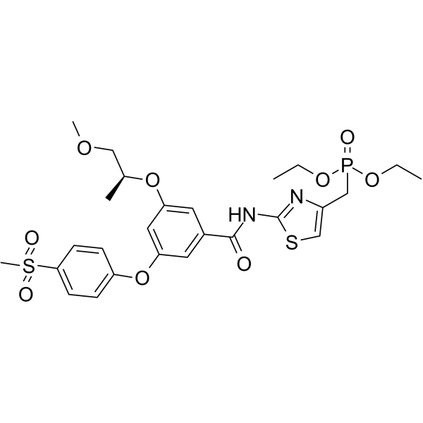
Glucokinase activator 3 is an orally active Glucokinase (GK) activator with an AC50 of 38 nM. Glucokinase activator 3 inhibits hERG channel and sodium channel in patch clamp assays. Glucokinase activator 3 exhibits high efficacy in reducing blood glucose in diet-induced obese (DIO) mice. Glucokinase activator 3 has the potential for the research of type 2 diabetes.
商品は「研究用試薬」です。人や動物の医療用・臨床診断用・食品用の製品ではありません。
研究用途以外に使用した場合、当社は一切の責任を負いかねます。
- 純度 : 98.36%
- CAS 番号: 1001417-92-0
- 分子式: C26H33N2O9PS2
- 分子量:612.65
-
保管条件:Powder -20°C, 3 years , 4°C, 2 years ; In solvent -80°C, 6 months , -20°C, 1 month

To place orders, for customer services and technical support, please contact: MedChemExpress USA
Tel: 609-228-6898 E-mail: [email protected] [email protected]


-
 生物活性
生物活性
-
化学情報
-
溶剤 & 溶解度
- プロトコル
- 純度とドキュメンテーション
- 参考文献
-
 Help & FAQs


-
HY-10595
RO-28-1675Glucokinase Activator -
HY-100221
AM-2394Glucokinase Activator -
HY-15671
GKA50Glucokinase Activator -
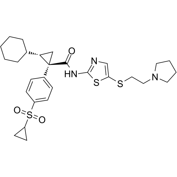
HY-13529
GlobalagliatinGlucokinase Activator -
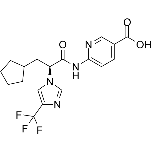
HY-100181
PF-04991532Glucokinase Activator -
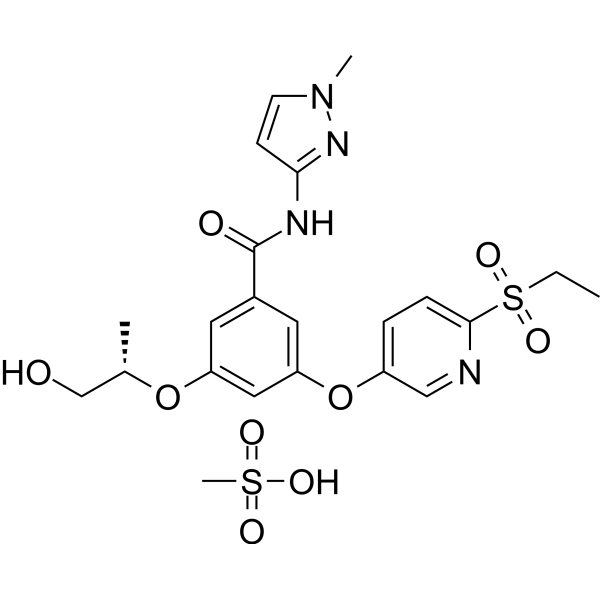
HY-19843
MK-0941Glucokinase Activator -
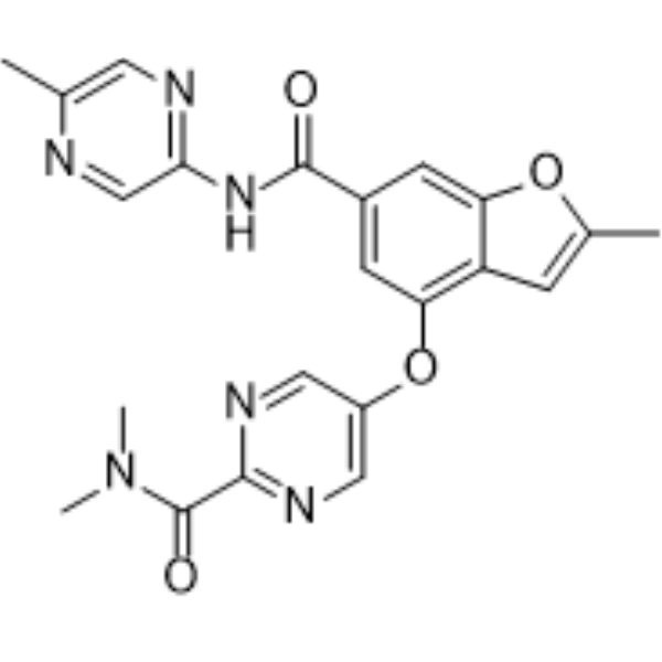
HY-108328
NerigliatinGlucokinase Activator -
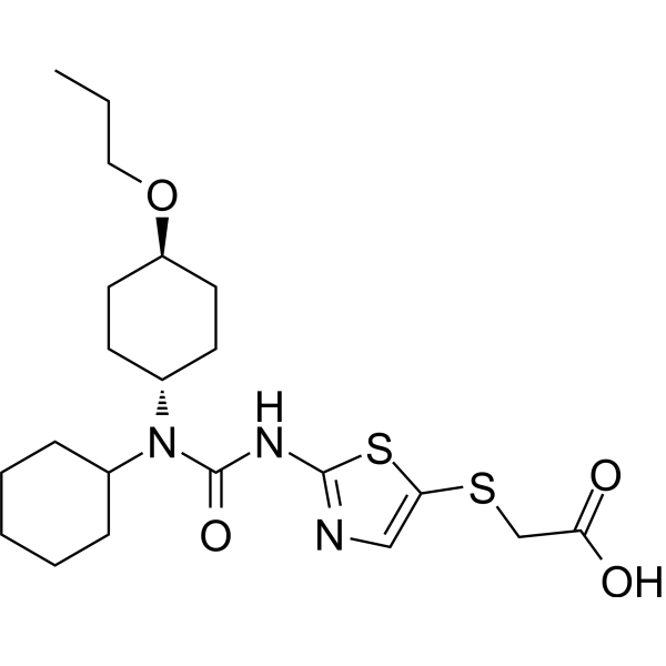
HY-147254
CadisegliatinGK Agent -
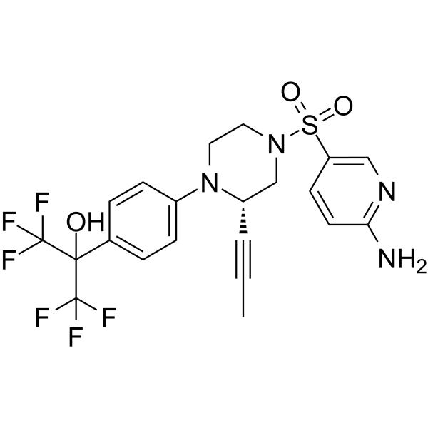
HY-12411
AMG-3969GK-GKRP Disruptor -
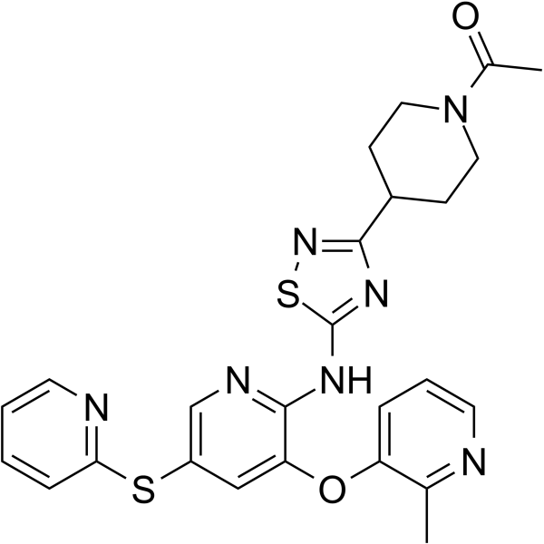
HY-133127
AR453588 -
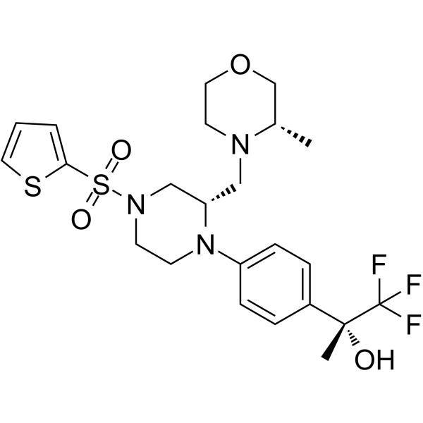
HY-12614
AMG-1694GK-GKRP Disruptor -
HY-117721
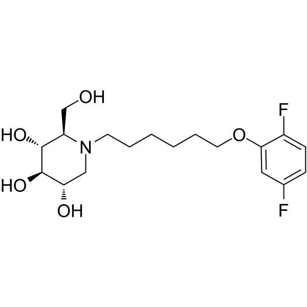
IHVR-11029ER α-glucosidases Inhibitor
MedChemExpress(MCE)の使用を引用している文献 Glucokinase activator 3
More

生物活性
製品説明
IC50 & Target
AC50: 38 nM (GK)[1]
体外実験
Glucokinase activator 3 (Compound 11) (10 μM) inhibits hERG channel by 60% and sodium channel by 40% in patch clamp assays[1].
MedChemExpress (MCE) has not independently confirmed the accuracy of these methods. They are for reference only.
体内実験
MedChemExpress (MCE) has not independently confirmed the accuracy of these methods. They are for reference only.
-
Animal Model:DIO mice were established by maintaining normal C57BL/6J mice on a high-fat diet (HFD) for several weeks, which rendered the mice glucose-intolerant, hyperinsulinemic, and insulin-resistant but not diabetic[1]
-
Dosage:1.83 mg/kg (3 μmol/kg)
-
Administration:p.o. for a single dose
-
Result:Showed highly efficacious in lowering glucose in an oral glucose tolerance test (OGTT).


化学情報
-
CAS 番号 1001417-92-0
-
性状 Solid
-
分子量 612.65
-
分子式 C26H33N2O9PS2
-
Color White to off-white
-
SMILES
O=C(NC1=NC(CP(OCC)(OCC)=O)=CS1)C2=CC(OC3=CC=C(S(=O)(C)=O)C=C3)=CC(O[C@@H](C)COC)=C2
-
輸送条件
Room temperature in continental US; may vary elsewhere.
-
保管条件
Powder -20°C 3 years 4°C 2 years In solvent -80°C 6 months -20°C 1 month


Publications (1)
-
Journal Impact Factor
-
Most Recent


溶剤 & 溶解度
体外:
DMSO : 100 mg/mL (163.23 mM; Need ultrasonic; Hygroscopic DMSO has a significant impact on the solubility of product, please use newly opened DMSO)
Please refer to the solubility information to select the appropriate solvent. Once prepared, please aliquot and store the solution to prevent product inactivation from repeated freeze-thaw cycles.
Storage method and period of stock solution: -80°C, 6 months; -20°C, 1 month. When stored at -80°C, please use it within 6 months. When stored at -20°C, please use it within 1 month.
Please refer to the solubility information to select the appropriate solvent. Once prepared, please aliquot and store the solution to prevent product inactivation from repeated freeze-thaw cycles.
Storage method and period of stock solution: -80°C, 6 months; -20°C, 1 month. When stored at -80°C, please use it within 6 months. When stored at -20°C, please use it within 1 month.
濃度 (開始) × 体積 (開始) = 濃度 (終了) × 体積 (終了)


純度とドキュメンテーション
-
データシート (274 KB)
-
SDS (252 KB)
- English - EN (252 KB)
- Français - FR (252 KB)
- Deutsch - DE (252 KB)
- Norwegian - NO (252 KB)
- Español - ES (252 KB)
- Swedish - SV (252 KB)
- Italian - IT (252 KB)
- Korean - KR (252 KB)
- Portuguese - PT (252 KB)
-
取扱説明書 (2659 KB)
参考文献


Complete Stock Solution Preparation Table
Please refer to the solubility information to select the appropriate solvent. Once prepared, please aliquot and store the solution to prevent product inactivation from repeated freeze-thaw cycles.
Storage method and period of stock solution: -80°C, 6 months; -20°C, 1 month. When stored at -80°C, please use it within 6 months. When stored at -20°C, please use it within 1 month.
| Optional Solvent | Concentration Solvent Mass | 1 mg | 5 mg | 10 mg | 25 mg |
|---|---|---|---|---|---|
| DMSO | 1 mM | 1.6323 mL | 8.1613 mL | 16.3225 mL | 40.8063 mL |
| 5 mM | 0.3265 mL | 1.6323 mL | 3.2645 mL | 8.1613 mL | |
| 10 mM | 0.1632 mL | 0.8161 mL | 1.6323 mL | 4.0806 mL | |
| 15 mM | 0.1088 mL | 0.5441 mL | 1.0882 mL | 2.7204 mL | |
| 20 mM | 0.0816 mL | 0.4081 mL | 0.8161 mL | 2.0403 mL | |
| 25 mM | 0.0653 mL | 0.3265 mL | 0.6529 mL | 1.6323 mL | |
| 30 mM | 0.0544 mL | 0.2720 mL | 0.5441 mL | 1.3602 mL | |
| 40 mM | 0.0408 mL | 0.2040 mL | 0.4081 mL | 1.0202 mL | |
| 50 mM | 0.0326 mL | 0.1632 mL | 0.3265 mL | 0.8161 mL | |
| 60 mM | 0.0272 mL | 0.1360 mL | 0.2720 mL | 0.6801 mL | |
| 80 mM | 0.0204 mL | 0.1020 mL | 0.2040 mL | 0.5101 mL | |
| 100 mM | 0.0163 mL | 0.0816 mL | 0.1632 mL | 0.4081 mL |


Glucokinase activator 3 関連分類
HY-144287 関連分類
 Powered by Bioz