



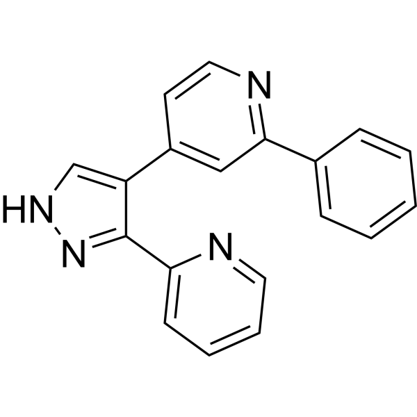
GW-6604

Based on 1 Customer Validation
GW-6604 is an ALK5 inhibitor with an IC50 value of 140 nM for inhibiting its autophosphorylation, and can be used in the study of liver fibrosis.
For research use only. We do not sell to patients.
- Purity : 98.0%
- CAS No.: 452342-37-9
- Formula: C19H14N4
- Molecular Weight:298.34
-
Storage:
4°C, protect from light
* In solvent : -80°C, 6 months; -20°C, 1 month (protect from light)

To place orders, for customer services and technical support, please contact: MedChemExpress USA
Tel: 609-228-6898 E-mail: [email protected] [email protected]


-
 Biological Activity
-
Chemical Information
-
Solvent & Solubility
- Protocols
- Purity & Documentation
- References
-
 Help & FAQs


-
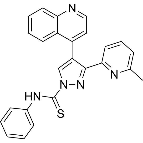
HY-10432
A 83-01ALK4/5/7 Inhibitor -
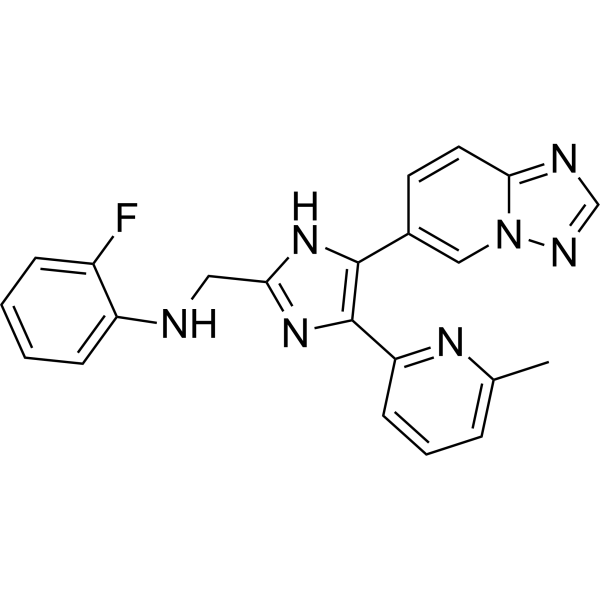
HY-19928
VactosertibALK5 Inhibitor -
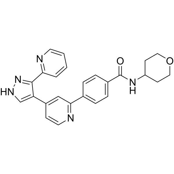
HY-10326
GW788388ALK5 Inhibitor -
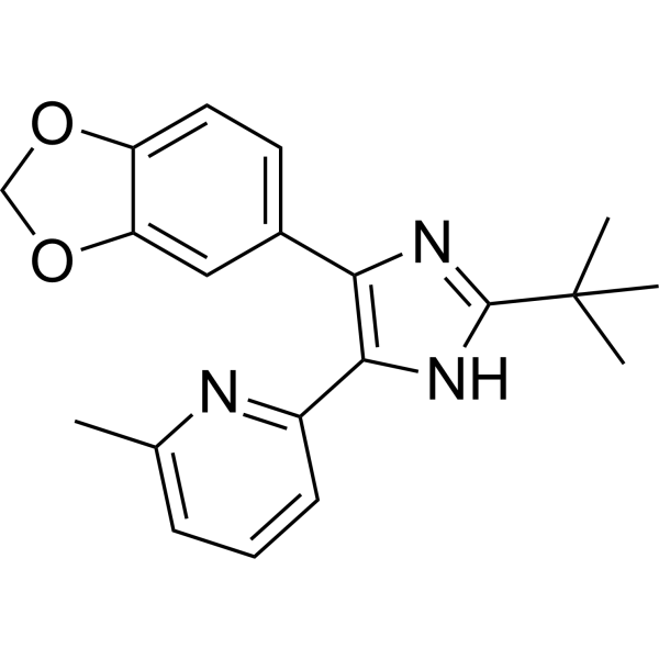
HY-13521
SB-505124ALK4/5/7 Inhibitor -
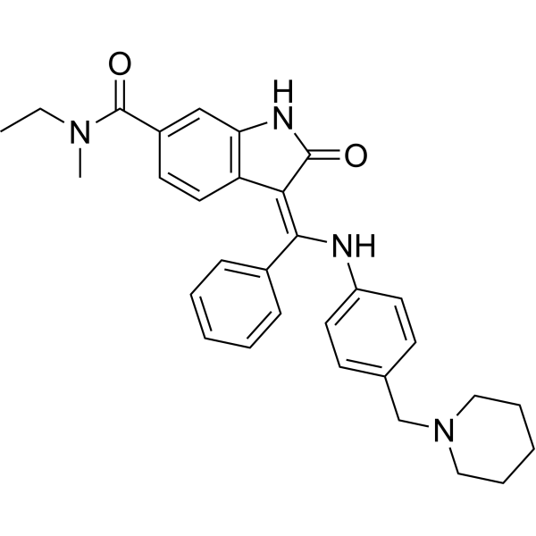
HY-13783
BIBF0775TGF-β Receptor Inhibitor -

HY-10431
SB-431542TGF-β Receptor Kinase Inhibitor -
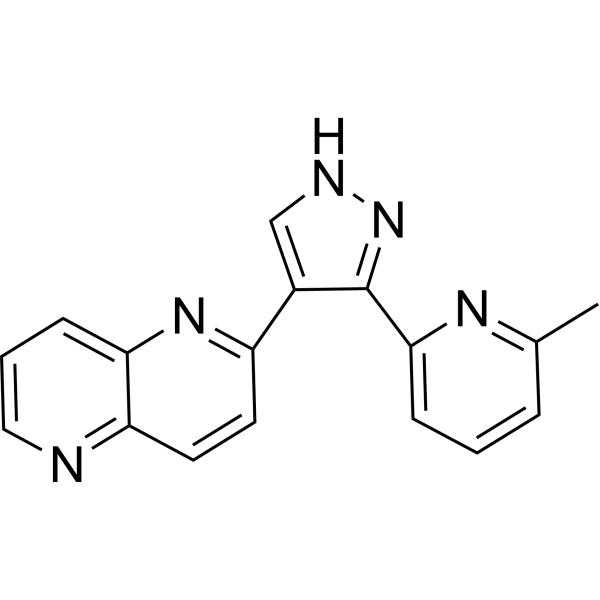
HY-13012
RepSoxTGFβR-1/ALK5 Inhibitor -
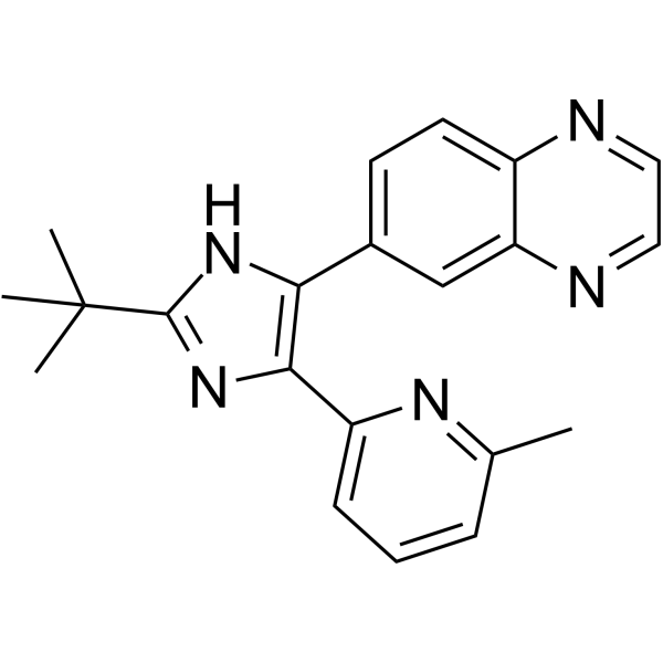
HY-12043
SB 525334ALK5 Inhibitor -
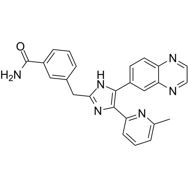
HY-18758
IN-1130ALK5 Inhibitor -
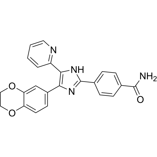
HY-10324
D4476CK1 Inhibitor -
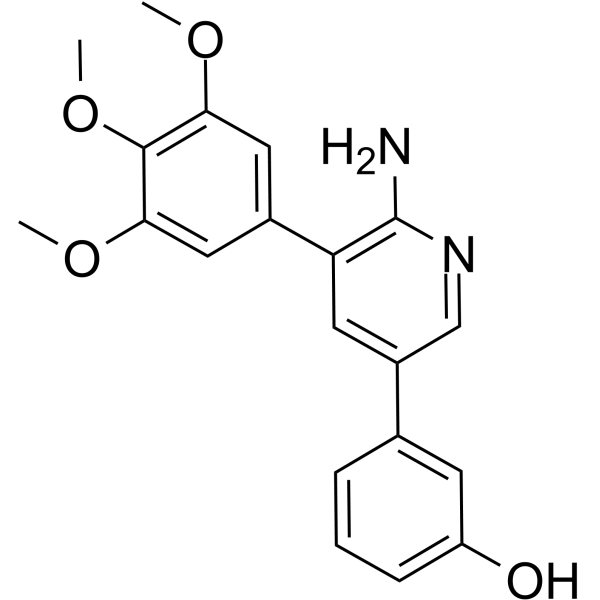
HY-12278
K02288BMPI Inhibitor -
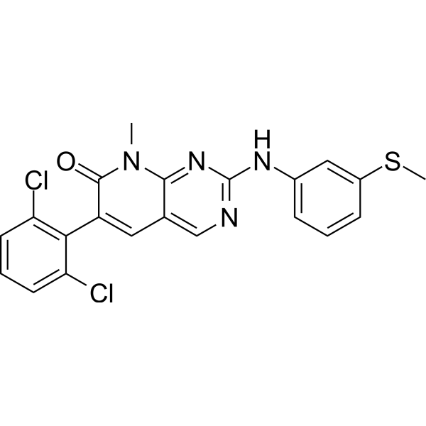
HY-10395
PD173955Src Inhibitor


Biological Activity
Description
Cellular Effect
|
Cell Line

|
Type | Value | Description | References |
|---|---|---|---|---|
| HepG2 | IC50 |
0.519 μM
Compound: 1a
|
Inhibition of TGF beta-induced transcription of firefly luciferase reporter gene in HepG2 cells
Inhibition of TGF beta-induced transcription of firefly luciferase reporter gene in HepG2 cells
|
[PMID: 16570917] |
In Vitro
GW-6604 (0-10 μM, ) inhibits TGF-β-induced PAI-1 transcription and secretion in HepG2 cells with an IC50 value of 500 nM, effectively blocking the Smad-dependent response[1].
MedChemExpress (MCE) has not independently confirmed the accuracy of these methods. They are for reference only. Further protocols information, click here.
In Vivo
MedChemExpress (MCE) has not independently confirmed the accuracy of these methods. They are for reference only.


Chemical Information
-
CAS No. 452342-37-9
-
Appearance Solid
-
Molecular Weight 298.34
-
Formula C19H14N4
-
Color Off-white to light yellow
-
SMILES
C1(C2=NNC=C2C3=CC(C4=CC=CC=C4)=NC=C3)=CC=CC=N1
-
Shipping
Room temperature in continental US; may vary elsewhere.
-
Storage
4°C, protect from light
* In solvent : -80°C, 6 months; -20°C, 1 month (protect from light)


Solvent & Solubility
In Vitro:
DMSO : 100 mg/mL (335.19 mM; Need ultrasonic; Hygroscopic DMSO has a significant impact on the solubility of product, please use newly opened DMSO)
Please refer to the solubility information to select the appropriate solvent. Once prepared, please aliquot and store the solution to prevent product inactivation from repeated freeze-thaw cycles.
Storage method and period of stock solution: -80°C, 6 months; -20°C, 1 month (protect from light). When stored at -80°C, please use it within 6 months. When stored at -20°C, please use it within 1 month.
Please refer to the solubility information to select the appropriate solvent. Once prepared, please aliquot and store the solution to prevent product inactivation from repeated freeze-thaw cycles.
Storage method and period of stock solution: -80°C, 6 months; -20°C, 1 month (protect from light). When stored at -80°C, please use it within 6 months. When stored at -20°C, please use it within 1 month.
Concentration (start) × Volume (start) = Concentration (final) × Volume (final)

Protocols
-
Fibrosis/Collagen Morphometry
Fibrosis and collagen morphometry is based on the quantitative visualization of fibrillar collagen deposition in tissue sections using histochemical stains such as Sirius Red (Picrosirius Red) or Masson's trichrome, followed by image-based or polarization-enhanced analysis to estimate collagen proportional area as a surrogate of extracellular matrix accumulation during fibrotic remodeling. Sirius Red combined with polarized light microscopy enhances detection of collagen fibers due to birefringence properties, enabling more specific visualization of collagen type I and III fibrils compared to conventional bright-field histology, while whole-section or region-restricted digital morphometry reduces field-selection bias in fibrosis assessment. Alternative quantitative approaches include second harmonic generation (SHG) and two-photon excited fluorescence microscopy, which enable label-free detection of fibrillar collagen and have been validated against histological staining and biochemica
-
Collagen: Sirius Red Staining
Sirius Red or picrosirius red staining is a histochemical method for visualizing collagen-rich extracellular matrix in tissue sections, and collagen fibers are detected as red-stained structures under bright-field microscopy with enhanced birefringence under polarized light. Picrosirius red is useful for assessing total collagen organization, distribution, and fibrosis burden, but polarized color should not be interpreted as a definitive collagen type I versus type III readout because color is affected by fiber orientation, thickness, and packing.
-
Cell Cytotoxicity Assay
Cytotoxicity assays are usually based on the assessment of cell membrane damage, which can also be indirectly detected by measuring cell viability. Detection methods include MTT assay, CKK-8 assay, LDH assay and ATP assay, etc.
-
Connective Tissue: Masson's Trichrome/Collagen Trichrome Staining
Masson’s Trichrome (collagen/trichrome staining) is a histological technique that differentially stains tissue compartments using sequential acidic dyes to distinguish collagen from muscle and cytoplasmic components based on dye affinity and tissue permeability differences, enabling visualization of fibrosis and connective tissue architecture in histological sections. The classical formulation typically uses Weigert's iron hematoxylin for nuclear staining, Biebrich scarlet-acid fuchsin for cytoplasm and muscle, and aniline blue (or light green variants) for collagen, producing a characteristic blue/green collagen signal contrasted against red cytoplasm and dark nuclei. The staining principle relies on selective displacement of smaller dye molecules by larger anionic dyes in collagen-rich regions under controlled acidified conditions, which enhances collagen-specific dye retention. This property makes the method widely used for fibrosis assessment in organs such as heart, liver, lung, a
-
Liver Histomorphometry
Liver histomorphometry is a quantitative histological approach used to measure structural alterations in hepatic tissue, including parenchymal loss, steatosis, fibrosis, and vascular remodeling, by combining stained tissue section analysis with stereological or computerized image-based measurements. Classical morphometric frameworks quantify volume fractions of liver compartments and fibrotic regions using systematic sampling and image analysis, enabling objective comparison of pathological changes across experimental groups. These approaches are widely applied in liver cirrhosis and fibrosis studies to reduce subjectivity in histological scoring and improve reproducibility of tissue evaluation. Recent methodological advances integrate automated image analysis and radiomics-based extraction of histological features from standard liver stains (e. g. , H&E and fibrotic stains), enabling quantitative correlation between morphometric features and fibrosis stages in non-alcoholic fatty live


Purity & Documentation
-
Data Sheet (277 KB)
-
SDS (393 KB)
- English - EN (393 KB)
- Français - FR (393 KB)
- Deutsch - DE (393 KB)
- Norwegian - NO (393 KB)
- Español - ES (393 KB)
- Swedish - SV (393 KB)
- Italian - IT (393 KB)
- Korean - KR (393 KB)
- Portuguese - PT (393 KB)
-
Handling Instructions (2659 KB)
References


Complete Stock Solution Preparation Table
Please refer to the solubility information to select the appropriate solvent. Once prepared, please aliquot and store the solution to prevent product inactivation from repeated freeze-thaw cycles.
Storage method and period of stock solution: -80°C, 6 months; -20°C, 1 month (protect from light). When stored at -80°C, please use it within 6 months. When stored at -20°C, please use it within 1 month.
| Optional Solvent | Concentration Solvent Mass | 1 mg | 5 mg | 10 mg | 25 mg |
|---|---|---|---|---|---|
| DMSO | 1 mM | 3.3519 mL | 16.7594 mL | 33.5188 mL | 83.7970 mL |
| 5 mM | 0.6704 mL | 3.3519 mL | 6.7038 mL | 16.7594 mL | |
| 10 mM | 0.3352 mL | 1.6759 mL | 3.3519 mL | 8.3797 mL | |
| 15 mM | 0.2235 mL | 1.1173 mL | 2.2346 mL | 5.5865 mL | |
| 20 mM | 0.1676 mL | 0.8380 mL | 1.6759 mL | 4.1899 mL | |
| 25 mM | 0.1341 mL | 0.6704 mL | 1.3408 mL | 3.3519 mL | |
| 30 mM | 0.1117 mL | 0.5586 mL | 1.1173 mL | 2.7932 mL | |
| 40 mM | 0.0838 mL | 0.4190 mL | 0.8380 mL | 2.0949 mL | |
| 50 mM | 0.0670 mL | 0.3352 mL | 0.6704 mL | 1.6759 mL | |
| 60 mM | 0.0559 mL | 0.2793 mL | 0.5586 mL | 1.3966 mL | |
| 80 mM | 0.0419 mL | 0.2095 mL | 0.4190 mL | 1.0475 mL | |
| 100 mM | 0.0335 mL | 0.1676 mL | 0.3352 mL | 0.8380 mL |


GW-6604 Related Classifications
HY-10327 Related Classifications
 Powered by Bioz