



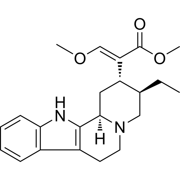
Hirsutine
Based on 1 publication(s) in Google Scholar
Hirsutine, an indole alkaloid of Uncaria rhynchophylla, exhibits anti-cancer activity. Hirsutine induces apoptosis and is a potent Dengue virus inhibitor exhibiting low cytotoxicity.
For research use only. We do not sell to patients.
- Purity : 99.76%
- CAS No.: 7729-23-9
- Formula: C22H28N2O3
- Molecular Weight:368.47
-
Storage:Powder -20°C, 3 years , 4°C, 2 years ; In solvent -80°C, 6 months , -20°C, 1 month

To place orders, for customer services and technical support, please contact: MedChemExpress USA
Tel: 609-228-6898 E-mail: [email protected] [email protected]


-
 Biological Activity
-
Chemical Information
-
Solvent & Solubility
- Protocols
- Purity & Documentation
- References
-
 Help & FAQs


-
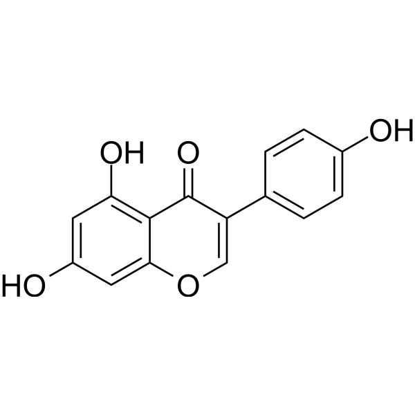
HY-14596
GenisteinAutophagy Inducer -

HY-10182
LaduviglusibGSK-3 Inhibitor -
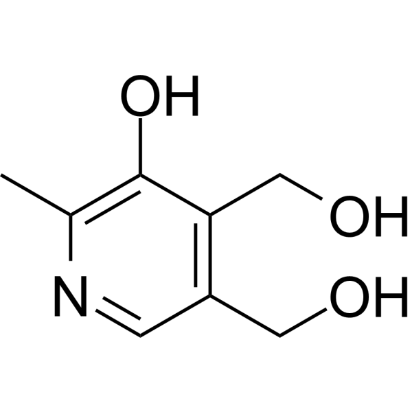
HY-B1328
PyridoxinePyridine Derivative -
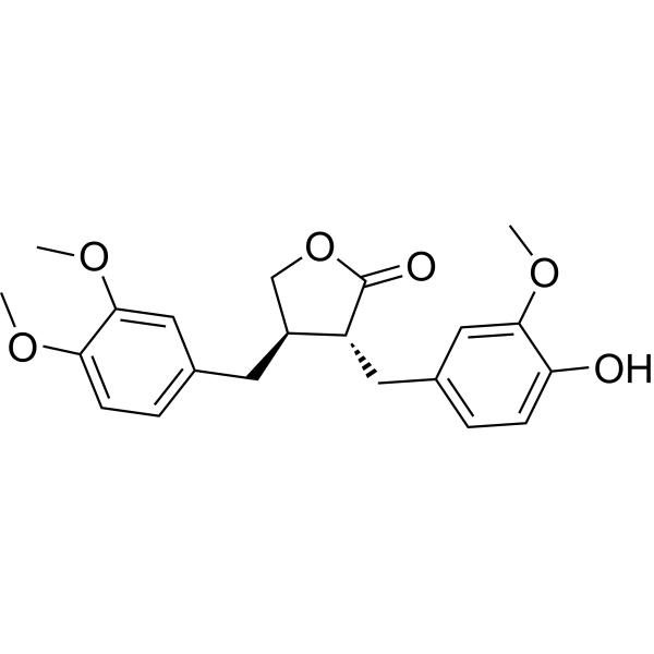
HY-N0035
ArctigeninAntitumor Agent -
HY-15306
EltrombopagTPO Receptor Agonist -
HY-114245
Se-MethylselenocysteineChemopreventive Agent -
HY-10230
MidostaurinMulti-targeted Protein Kinase Inhibitor -

HY-114164
Thrombin (MW 37kDa)Procoagulant/Anticoagulant -
HY-D0803
ThymoquinoneNatural Product -
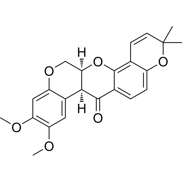
HY-13425
DeguelinChemopreventive Agent -
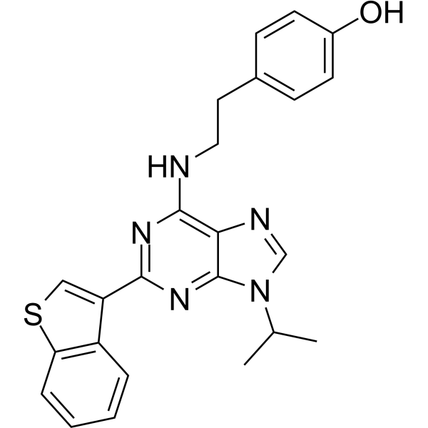
HY-15001
Stemregenin 1AhR Antagonist -
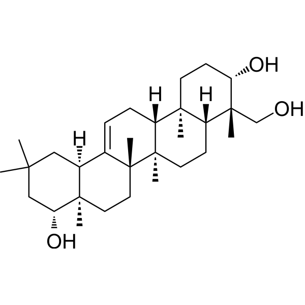
HY-N6074
Soyasapogenol BAutophagy Inducer
Publications Citing Use of MedChemExpress (MCE) Hirsutine
More Customer Validation & Images

-
Histological Imaging/Staining

-
WB

-
RT-PCR

-
IHC

-
IF


Biological Activity
Description
Cellular Effect
|
Cell Line

|
Type | Value | Description | References |
|---|---|---|---|---|
| MDCK | CC50 |
23.5 μg/mL
Compound: Hirsutine 1
|
The compound was evaluated for cytotoxicity by MTT assay in MDCK cells
The compound was evaluated for cytotoxicity by MTT assay in MDCK cells
|
10.1016/S0960-894X(97)10154-8 |
| MDCK | EC50 |
>23.5 μg/mL
Compound: Hirsutine 1
|
The compound was evaluated for inhibitory effect on the replication of influenza A subtype H1N1 Bangkok/10/83 strain in MDCK cells
The compound was evaluated for inhibitory effect on the replication of influenza A subtype H1N1 Bangkok/10/83 strain in MDCK cells
|
10.1016/S0960-894X(97)10154-8 |
| MDCK | EC50 |
>23.5 μg/mL
Compound: Hirsutine 1
|
The compound was evaluated for inhibitory effect on the replication of influenza A subtype H1N1 PR8/34 strain in MDCK cells
The compound was evaluated for inhibitory effect on the replication of influenza A subtype H1N1 PR8/34 strain in MDCK cells
|
10.1016/S0960-894X(97)10154-8 |
| MDCK | EC50 |
>23.5 μg/mL
Compound: Hirsutine 1
|
The compound was evaluated for inhibitory effect on the replication of influenza A subtype H1N1 yamagata/120/86 strain in MDCK cells
The compound was evaluated for inhibitory effect on the replication of influenza A subtype H1N1 yamagata/120/86 strain in MDCK cells
|
10.1016/S0960-894X(97)10154-8 |
| MDCK | EC50 |
>23.5 μg/mL
Compound: Hirsutine 1
|
The compound was evaluated for inhibitory effect on the replication of influenza A subtype H2N2 Adachi/2/57 in MDCK cells
The compound was evaluated for inhibitory effect on the replication of influenza A subtype H2N2 Adachi/2/57 in MDCK cells
|
10.1016/S0960-894X(97)10154-8 |
| MDCK | EC50 |
>23.5 μg/mL
Compound: Hirsutine 1
|
The compound was evaluated for inhibitory effect on the replication of influenza A subtype H2N2 murakami/4/64 strain in MDCK cells
The compound was evaluated for inhibitory effect on the replication of influenza A subtype H2N2 murakami/4/64 strain in MDCK cells
|
10.1016/S0960-894X(97)10154-8 |
| MDCK | EC50 |
>23.5 μg/mL
Compound: Hirsutine 1
|
The compound was evaluated for inhibitory effect on the replication of influenza B norway/1/80 strain in MDCK cells
The compound was evaluated for inhibitory effect on the replication of influenza B norway/1/80 strain in MDCK cells
|
10.1016/S0960-894X(97)10154-8 |
| MDCK | EC50 |
>23.5 μg/mL
Compound: Hirsutine 1
|
The compound was evaluated for inhibitory effect on the replication of influenza B Singapore/222/79 strain in MDCK cells
The compound was evaluated for inhibitory effect on the replication of influenza B Singapore/222/79 strain in MDCK cells
|
10.1016/S0960-894X(97)10154-8 |
| MDCK | EC50 |
0.4 μg/mL
Compound: Hirsutine 1
|
The compound was evaluated for inhibitory effect on the replication of influenza A subtype H3N2 ishikawa/7/82 in MDCK cells
The compound was evaluated for inhibitory effect on the replication of influenza A subtype H3N2 ishikawa/7/82 in MDCK cells
|
10.1016/S0960-894X(97)10154-8 |
| MDCK | EC50 |
0.57 μg/mL
Compound: Hirsutine 1
|
The compound was evaluated for inhibitory effect on the replication of influenza A subtype H3N2 Philippine/2/82 in MDCK cells
The compound was evaluated for inhibitory effect on the replication of influenza A subtype H3N2 Philippine/2/82 in MDCK cells
|
10.1016/S0960-894X(97)10154-8 |
In Vitro
Hirsutine remarkably reduces the viability of MCF-7 and MDA-MB-231 cells in a time- and dose-dependent manner with IC50 values of 447.79 and 179.06 μM, respectively. In the MDA-MB-231 cells, Hirsutine induces apoptosis and depolarization of MMP, releases Cyt C from mitochondria, and activates caspase 9 and caspase 3[2].
MedChemExpress (MCE) has not independently confirmed the accuracy of these methods. They are for reference only. Further protocols information, click here.
In Vivo
MedChemExpress (MCE) has not independently confirmed the accuracy of these methods. They are for reference only.


Chemical Information
-
CAS No. 7729-23-9
-
Appearance Solid
-
Molecular Weight 368.47
-
Formula C22H28N2O3
-
Color Off-white to light yellow
-
SMILES
O=C(OC)/C([C@H]([C@H](CN1CC2)CC)C[C@]1([H])C3=C2C(C=CC=C4)=C4N3)=C/OC
-
Structure Classification
-
Initial Source
-
Shipping
Room temperature in continental US; may vary elsewhere.
-
Storage
Powder -20°C 3 years 4°C 2 years In solvent -80°C 6 months -20°C 1 month


Publications (1)
-
Journal Impact Factor
-
Most Recent
-
J Pharmacol Sci
Hirsutine attenuated oxidative stress and autophagy in diabetic kidney disease through Keap1/Nrf2 pathway. [Abstract]2025 Jun;158(2):143-153. PMID: 40288825
Hirsutine purchased from MedChemExpress. Usage Cited in: J Pharmacol Sci. 2025 Jun;158(2):143-153. [Abstract]
Rat kidney sections were stained with Masson’s trichrome treated with Hirsutine (10, 20 mg/kg, i.g.).
Hirsutine purchased from MedChemExpress. Usage Cited in: J Pharmacol Sci. 2025 Jun;158(2):143-153. [Abstract]
Western blot analysis used to evaluate changes of Vimentin, Collagen-IV and α-SMA treated with Hirsutine (10, 20 mg/kg, i.g.).
Hirsutine purchased from MedChemExpress. Usage Cited in: J Pharmacol Sci. 2025 Jun;158(2):143-153. [Abstract]
The mRNA level of Vimentin, Collagen-IV and α-SMA was measured using RT-qPCR treated with Hirsutine (10, 20 mg/kg, i.g.).
Hirsutine purchased from MedChemExpress. Usage Cited in: J Pharmacol Sci. 2025 Jun;158(2):143-153. [Abstract]
The expression of α-SMA was detected using immunohistochemistry treated with Hirsutine (10, 20 mg/kg, i.g.).
Hirsutine purchased from MedChemExpress. Usage Cited in: J Pharmacol Sci. 2025 Jun;158(2):143-153. [Abstract]
Representative images of ROS changes in rat kidneys visualized by DHE treated with Hirsutine (10, 20 mg/kg, i.g.).

Hirsutine purchased from MedChemExpress. Usage Cited in: J Pharmacol Sci. 2025 Jun;158(2):143-153. [Abstract]
The cell viability was determined using CCK8 assay treated with Hirsutine (0.1, 0.5, 1, 10, 20, 40 μM).


Solvent & Solubility
In Vitro:
DMSO : 100 mg/mL (271.39 mM; Need ultrasonic; Hygroscopic DMSO has a significant impact on the solubility of product, please use newly opened DMSO)
Please refer to the solubility information to select the appropriate solvent. Once prepared, please aliquot and store the solution to prevent product inactivation from repeated freeze-thaw cycles.
Storage method and period of stock solution: -80°C, 6 months; -20°C, 1 month. When stored at -80°C, please use it within 6 months. When stored at -20°C, please use it within 1 month.
Please refer to the solubility information to select the appropriate solvent. Once prepared, please aliquot and store the solution to prevent product inactivation from repeated freeze-thaw cycles.
Storage method and period of stock solution: -80°C, 6 months; -20°C, 1 month. When stored at -80°C, please use it within 6 months. When stored at -20°C, please use it within 1 month.
Concentration (start) × Volume (start) = Concentration (final) × Volume (final)

Protocols
-
Apoptosis
Apoptosis, also called programmed cell death, is generally characterized by distinct morphological characteristics.
-
TUNEL staining for apoptotic DNA fragmentation
TUNEL staining detects DNA strand breaks by using terminal deoxynucleotidyl transferase to add labeled nucleotides to exposed 3′-OH DNA termini, generating either microscopic staining in fixed cells or tissue sections, or fluorescence/cytometric signal in cell suspensions. TUNEL positivity reflects DNA fragmentation but should not be interpreted alone as definitive apoptosis, because TUNEL can also label necrotic, autolytic, mechanically damaged, or DNA-repair-associated DNA breaks.
-
Cell Cytotoxicity Assay
Cytotoxicity assays are usually based on the assessment of cell membrane damage, which can also be indirectly detected by measuring cell viability. Detection methods include MTT assay, CKK-8 assay, LDH assay and ATP assay, etc.
-
Mammalian live/dead viability and cytotoxicity staining
Live/dead viability and cytotoxicity staining assays are based on the simultaneous detection of intracellular esterase activity in metabolically active (viable) cells and membrane integrity loss in non-viable cells. In commonly used dual-staining approaches, membrane-permeant fluorogenic substrates are converted by intracellular esterases into fluorescent products in live cells, while impermeant DNA-binding dyes selectively enter cells with compromised plasma membranes and label nucleic acids in dead or dying cells, enabling discrimination between viable and non-viable populations by fluorescence microscopy or flow cytometry.
-
Annexin V plus membrane-impermeant dye apoptosis staining
Annexin V-based apoptosis assays rely on the detection of phosphatidylserine (PS) externalization from the inner leaflet of the plasma membrane to the outer leaflet, an early biochemical hallmark of apoptosis. Fluorescently labeled Annexin V binds PS in a calcium-dependent manner, enabling identification of early apoptotic cells by flow cytometry or fluorescence microscopy. When combined with a membrane-impermeant DNA-binding dye (e. g. , propidium iodide), this approach allows discrimination between viable (Annexin V−/dye−), early apoptotic (Annexin V+/dye−), and late apoptotic or necrotic (Annexin V+/dye+) cell populations by assessing membrane integrity and PS exposure.
-
Apoptosis Solutions
Apoptosis is a regulated, generally non-lytic cell-death pathway that removes unwanted, damaged, infected, or abnormal cells through coordinated morphological changes, caspase activation, DNA fragmentation, and membrane remodeling. The intrinsic apoptosis pathway is controlled mainly by mitochondrial outer membrane permeabilization, BCL-2 family proteins, cytochrome c release, apoptosome formation, caspase-9 activation, and downstream executioner caspase-3/7 activation. The extrinsic apoptosis pathway is initiated by death receptors such as Fas, TNFR, and TRAIL receptors, which recruit adaptor proteins and activate caspase-8 before engaging executioner caspases or mitochondrial amplification through BID cleavage. Apoptosis is linked to many phenotypes, including cancer cell killing, tissue homeostasis, immune regulation, neurodegeneration, infection response, and treatment-induced cytotoxicity; unresolved questions include how apoptosis interacts with necroptosis, pyroptosis, ferroptos
-
Cell Viability Determination by MTT Colorimetric Assay
The following protocol uses the MTT colorimetric assay as a classic literature-established method for assessing cell viability/metabolic activity in cultured mammalian cells. MTT[3-(4,5-dimethylthiazol-2-yl)-2,5-diphenyltetrazolium bromide] is reduced by metabolically active cells to a colored formazan product; the amount of formazan is quantified spectrophotometrically and provides an indirect measure of metabolically active viable cells. Importantly, MTT reduction reflects cellular oxidoreductase/metabolic activity rather than an absolute direct count of living cells, so changes in cellular metabolism can alter the signal independently of cell number.


Purity & Documentation
-
Data Sheet (277 KB)
-
SDS (254 KB)
- English - EN (254 KB)
- Français - FR (254 KB)
- Deutsch - DE (254 KB)
- Norwegian - NO (254 KB)
- Español - ES (254 KB)
- Swedish - SV (254 KB)
- Italian - IT (254 KB)
- Korean - KR (254 KB)
- Portuguese - PT (254 KB)
-
Handling Instructions (2659 KB)
References
[1]. Hishiki T, et al. Hirsutine, an Indole Alkaloid of Uncaria rhynchophylla, Inhibits Late Step in Dengue Virus Lifecycle. Front Microbiol. 2017 Aug 30;8:1674. [Content Brief]
[2]. Huang QW, et al. [Hirsutine induces apoptosis of human breast cancer MDA-MB-231 cells through mitochondrial pathway]. Sheng Li Xue Bao. 2018 Feb 25;70(1):40-46. [Content Brief]
[3]. Zhang R, et al. Hirsutine induces mPTP-dependent apoptosis through ROCK1/PTEN/PI3K/GSK3β pathway in human lung cancer cells. Cell Death Dis. 2018 May 22;9(6):598. [Content Brief]


Complete Stock Solution Preparation Table
Please refer to the solubility information to select the appropriate solvent. Once prepared, please aliquot and store the solution to prevent product inactivation from repeated freeze-thaw cycles.
Storage method and period of stock solution: -80°C, 6 months; -20°C, 1 month. When stored at -80°C, please use it within 6 months. When stored at -20°C, please use it within 1 month.
| Optional Solvent | Concentration Solvent Mass | 1 mg | 5 mg | 10 mg | 25 mg |
|---|---|---|---|---|---|
| DMSO | 1 mM | 2.7139 mL | 13.5696 mL | 27.1393 mL | 67.8481 mL |
| 5 mM | 0.5428 mL | 2.7139 mL | 5.4279 mL | 13.5696 mL | |
| 10 mM | 0.2714 mL | 1.3570 mL | 2.7139 mL | 6.7848 mL | |
| 15 mM | 0.1809 mL | 0.9046 mL | 1.8093 mL | 4.5232 mL | |
| 20 mM | 0.1357 mL | 0.6785 mL | 1.3570 mL | 3.3924 mL | |
| 25 mM | 0.1086 mL | 0.5428 mL | 1.0856 mL | 2.7139 mL | |
| 30 mM | 0.0905 mL | 0.4523 mL | 0.9046 mL | 2.2616 mL | |
| 40 mM | 0.0678 mL | 0.3392 mL | 0.6785 mL | 1.6962 mL | |
| 50 mM | 0.0543 mL | 0.2714 mL | 0.5428 mL | 1.3570 mL | |
| 60 mM | 0.0452 mL | 0.2262 mL | 0.4523 mL | 1.1308 mL | |
| 80 mM | 0.0339 mL | 0.1696 mL | 0.3392 mL | 0.8481 mL | |
| 100 mM | 0.0271 mL | 0.1357 mL | 0.2714 mL | 0.6785 mL |


Hirsutine Related Classifications
HY-N2193 Related Classifications
 Powered by Bioz