



Negletein

Based on 1 Customer Validation
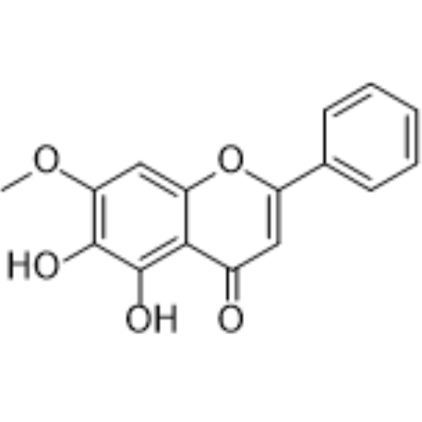
Negletein (5,6-Dihydroxy-7-methoxyflavone) is a flavone found in Scutellaria. Negletein shows anti-inflammatory activity via inhibiting TNF-α and IL-1β with IC50 values of 16.4 and 6.4 μM, respectively. Negletein can activate Nrf2 and inhibit ROS production. Negletein can enhance the neuroprotective effect of nerve growth factor. Negletein can inhibit amyloid beta-peptide release and accumulation. Negletein can inhibit pathogens biofilms formation. Negletein can be used for the researches of cancer, infection, inflammation and neurological disease, such as colon cancer and Alzheimer's disease.
For research use only. We do not sell to patients.
- Purity : 99.55%
- CAS No.: 29550-13-8
- Formula: C16H12O5
- Molecular Weight:284.26
-
Storage:
4°C, sealed storage, away from moisture and light
* In solvent : -80°C, 6 months; -20°C, 1 month (sealed storage, away from moisture and light)

To place orders, for customer services and technical support, please contact: MedChemExpress USA
Tel: 609-228-6898 E-mail: [email protected] [email protected]


-
 Biological Activity
-
Chemical Information
-
Solvent & Solubility
- Protocols
- Purity & Documentation
- References
-
 Help & FAQs


-
HY-14589
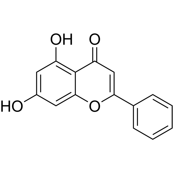
ChrysinEstrogen Receptor/ERR Inhibitor -
HY-N0400
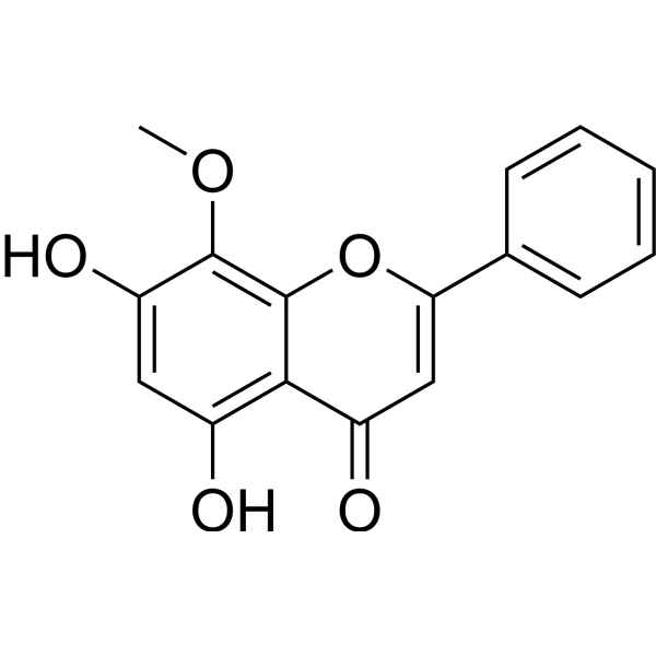
WogoninCDK Inhibitor -
HY-A0003
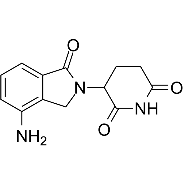
LenalidomideImmunomodulator -

HY-A0003R
Lenalidomide (Standard)Reference Standard -
HY-107390A
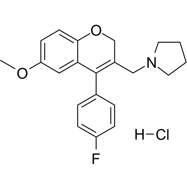
AX-024 hydrochlorideTCR-Nck Interaction Inhibitor -
HY-N4315
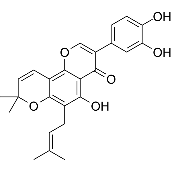
PomiferinHDAC/mTOR Inhibitor -
HY-N0419
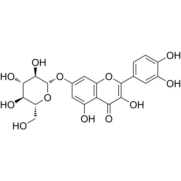
Quercimeritrinα-glucosidase Inhibitor -
HY-14658
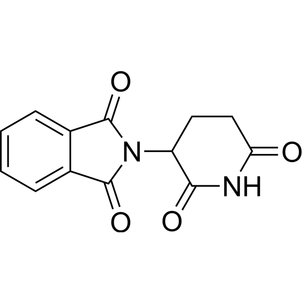
ThalidomideCereblon Inhibitor -
HY-N0455
L-ArginineeNOS Substrate -

HY-14658R
Thalidomide (Standard)Cereblon Inhibitor -

HY-N0455R
L-Arginine (Standard)eNOS Substrate -
HY-15509A
Semapimod tetrahydrochlorideProinflammatory Cytokine Inhibitor


Biological Activity
Description
IC50 & Target
[1]|
IL-1β 6.4 nM (IC50) |
TNF-α 16.4 μM (IC50) |
Cellular Effect
|
Cell Line

|
Type | Value | Description | References |
|---|---|---|---|---|
| DLD-1 | IC50 |
30.93 μM
Compound: 5a
|
Antiproliferative activity against human DLD1 cells after 48 hrs by MTT assay
Antiproliferative activity against human DLD1 cells after 48 hrs by MTT assay
|
10.1039/C5MD00163C |
| H9 | IC50 |
11.5 μg/mL
Compound: 2
|
Concentration that inhibits uninfected H9 cell growth by 50%.
Concentration that inhibits uninfected H9 cell growth by 50%.
|
[PMID: 12729671] |
| HepG2 | IC50 |
>40 μM
Compound: 5a
|
Antiproliferative activity against human HepG2 cells after 48 hrs by MTT assay
Antiproliferative activity against human HepG2 cells after 48 hrs by MTT assay
|
10.1039/C5MD00163C |
| HT-29 | IC50 |
>40 μM
Compound: 5a
|
Antiproliferative activity against human HT-29 cells after 48 hrs by MTT assay
Antiproliferative activity against human HT-29 cells after 48 hrs by MTT assay
|
10.1039/C5MD00163C |
| J774.A1 | IC50 |
0.22 μM
Compound: 47
|
Anti-inflammatory activity in mouse J774.A1 macrophage cells assessed as reduction in LPS-induced TNF-alpha production
Anti-inflammatory activity in mouse J774.A1 macrophage cells assessed as reduction in LPS-induced TNF-alpha production
|
[PMID: 37683361] |
| J774.A1 | IC50 |
6.4 μM
Compound: 47
|
Antiinflammatory activity in LPS-induced mouse J774.A1 macrophage cells assessed as reduction in LPS-induced IL-1 beta level
Antiinflammatory activity in LPS-induced mouse J774.A1 macrophage cells assessed as reduction in LPS-induced IL-1 beta level
|
[PMID: 37683361] |
| SW480 | IC50 |
29.41 μM
Compound: 5a
|
Antiproliferative activity against human SW480 cells after 48 hrs by MTT assay
Antiproliferative activity against human SW480 cells after 48 hrs by MTT assay
|
10.1039/C5MD00163C |
In Vitro
Negletein (10 nM-10 mM) does not induce neurite outgrowth in PC12 cells[1].
Negletein (10 μM, 3 days) trigger PC12 cells to differentiate and extend neuritis companied with nerve growth factor (5 ng/mL)[1].
Negletein (10 μM) increases p-ERK, p-Akt, p-CREB, and GAP-43 levels companied with nerve growth factor (5 ng/mL) in PC12 cells[1].
Negletein (10 nM-10 μM, 24-72 h) increases survival rate (cell viability) of PC12 cells cultured in serum-free medium[1].
Negletein (20 μM) inhibits APP expression, β-Secretase activity and Aβ42 release in iron-induced SHSY5Y cells[3].
Negletein (5-20 μM) inhibits hydroxyl radical formation in iron-induced SHSY5Y cells[3].
Negletein (20 μM, 24 h) prevents oligomerization of amyloid Aβ42 in iron-induced SHSY5Y cells[3].
Negletein (5-50 μM, 4 h) increases Nrf2 expression in HCT116 cells[4].
Negletein (0-30 μM, 4 h) reduces ROS production in HCT116 cells[4].
Negletein shows MIC of 32 μg/mL against S. aureus and Bacillus sp.[5].
MedChemExpress (MCE) has not independently confirmed the accuracy of these methods. They are for reference only. Further protocols information, click here.


Chemical Information
-
CAS No. 29550-13-8
-
Appearance Solid
-
Molecular Weight 284.26
-
Formula C16H12O5
-
Color Light yellow to green yellow
-
SMILES
O=C1C=C(C2=CC=CC=C2)OC3=CC(OC)=C(O)C(O)=C13
-
Synonyms
5,6-Dihydroxy-7-methoxyflavone
-
Structure Classification
-
Initial Source
-
Shipping
Room temperature in continental US; may vary elsewhere.
-
Storage
4°C, sealed storage, away from moisture and light
* In solvent : -80°C, 6 months; -20°C, 1 month (sealed storage, away from moisture and light)


Solvent & Solubility
In Vitro:
DMSO : 100 mg/mL (351.79 mM; Need ultrasonic; Hygroscopic DMSO has a significant impact on the solubility of product, please use newly opened DMSO)
Please refer to the solubility information to select the appropriate solvent. Once prepared, please aliquot and store the solution to prevent product inactivation from repeated freeze-thaw cycles.
Storage method and period of stock solution: -80°C, 6 months; -20°C, 1 month (sealed storage, away from moisture and light). When stored at -80°C, please use it within 6 months. When stored at -20°C, please use it within 1 month.
Please refer to the solubility information to select the appropriate solvent. Once prepared, please aliquot and store the solution to prevent product inactivation from repeated freeze-thaw cycles.
Storage method and period of stock solution: -80°C, 6 months; -20°C, 1 month (sealed storage, away from moisture and light). When stored at -80°C, please use it within 6 months. When stored at -20°C, please use it within 1 month.
Concentration (start) × Volume (start) = Concentration (final) × Volume (final)
In Vivo:
Select the appropriate dissolution method based on your experimental animal and administration route.
- For the following dissolution methods, please ensure to first prepare a clear stock solution using an In Vitro approach and then sequentially add co-solvents:
- To ensure reliable experimental results, the clarified stock solution can be appropriately stored based on storage conditions. As for the working solution for In Vivo experiments, it is recommended to prepare freshly and use it on the same day.
- The percentages shown for the solvents indicate their volumetric ratio in the final prepared solution. If precipitation or phase separation occurs during preparation, heat and/or sonication can be used to aid dissolution.
In Vivo Dissolution Calculator
Please enter the basic information of animal experiments:
-
-
-
-
Recommended: Prepare an additional quantity of animals to account for potential losses during experiments.
Please enter your animal formula composition:
-
%DMSO +
Recommended: Keep the proportion of DMSO in working solution below 2% if your animal is weak.
-
%+
-
+%Tween-80 + +
-
%Saline +
The co-solvents required include: DMSO, . All of co-solvents are available by MedChemExpress (MCE). , Tween 80. All of co-solvents are available by MedChemExpress (MCE).
Working solution concentration: 0.22 mg/mL
Method for preparing stock solution: mg drug dissolved in μL DMSO. Stock solution concentration: mg/mL. * In solvent : -80°C, 6 months; -20°C, 1 month (sealed storage, away from moisture and light)
1. Take μL DMSO stock solution;
2. Add μL .
μL , mix evenly;
3. Then add μL Tween 80, mix evenly;
4. Then add μL
Please ensure that the stock solution in the first step is dissolved to a clear state, and add co-solvents in sequence. You can use ultrasonic heating (ultrasonic cleaner, recommended frequency 20-40 kHz), vortexing, etc. to assist dissolution.

Protocols
-
Research Protocol for Infectious Diseases
Infectious-disease experiments test how pathogens interact with host barriers, innate immune receptors, inflammatory signaling, pathogen replication, and tissue injury; pattern-recognition receptors such as TLRs, RIG-I-like receptors, NOD-like receptors, and inflammasomes detect microbial molecules and activate NF-κB, interferon, and cytokine responses. The central hypothesis is that infection severity reflects the balance between pathogen burden and host response: protective inflammation restricts pathogen growth, whereas excessive or mislocalized inflammation contributes to tissue damage and disease phenotype. Unresolved questions include which host pathways are protective versus pathogenic, why some infection models fail to translate to human disease, and which combined readouts best predict clinically relevant infection outcomes.
-
Cell Cytotoxicity Assay
Cytotoxicity assays are usually based on the assessment of cell membrane damage, which can also be indirectly detected by measuring cell viability. Detection methods include MTT assay, CKK-8 assay, LDH assay and ATP assay, etc.
-
Cotton Pellet Granuloma
Cotton pellet granuloma is a classical in vivo chronic inflammation model used to evaluate the anti-inflammatory potential of test substances by measuring their ability to inhibit granuloma tissue formation around an implanted foreign body (cotton pellet) in rodents. The method is based on the biological response to a sterile implanted material, which induces proliferative phase inflammation characterized by fibroblast proliferation and collagen-rich granuloma formation, and the final readout reflects the extent of chronic inflammatory tissue growth surrounding the pellet. In multiple preclinical pharmacological evaluations, inhibition of cotton pellet-induced granuloma formation has been used as an indicator of anti-inflammatory activity in both synthetic and natural product screening contexts.
-
ROS/oxidative-stress fluorescent staining
ROS/oxidative-stress fluorescent staining uses cell-permeant fluorogenic probes that become fluorescent after oxidation inside cells or tissues; commonly used examples include DCFH-DA/DCFDA for broad cellular oxidant detection, DHE for superoxide-related signal detection, MitoSOX for mitochondrial superoxide-related signal detection, and CellROX probes for oxidative-stress-associated fluorescence readouts. The assay detects probe oxidation rather than a single ROS species unless the probe and analysis method have been chemically validated for that species. DCFH-DA enters cells, is deacetylated by intracellular esterases to DCFH, and produces fluorescent DCF after oxidation, so the readout is used as an operational measure of total cellular oxidative stress rather than a species-specific ROS measurement. DHE and MitoSOX can report superoxide-related oxidation, but red fluorescence alone can include non-specific ethidium-like oxidation products; HPLC or optimized spectral approaches are
-
Carrageenan-Induced Paw Edema
Carrageenan-induced paw edema is an acute inflammation model in which intraplantar injection of carrageenan induces localized inflammatory swelling characterized by vascular permeability, leukocyte infiltration, and production of inflammatory mediators such as prostaglandins and cytokines, making it widely used to evaluate anti-inflammatory agents in vivo. The resulting paw volume or thickness increase is quantified over time as a direct readout of inflammatory intensity and drug efficacy, typically reflecting cyclooxygenase-mediated prostaglandin-driven edema formation and immune cell recruitment in peripheral tissue[20].
-
Research Protocol for Inflammation-related Diseases
The NLRP3 inflammasome is a cytosolic innate immune signaling platform that integrates priming signals and danger-signal activation to promote caspase-1 activation, maturation of IL-1β and IL-18, and gasdermin D-mediated pyroptotic cell death. The core experimental logic is to determine whether inflammatory disease phenotypes are driven by increased NLRP3 expression, ASC-containing inflammasome assembly, caspase-1 cleavage, GSDMD cleavage, and extracellular release of IL-1β/IL-18 rather than by nonspecific cell injury alone. The pathway is strongly linked to inflammation-related disease phenotypes because monosodium urate crystals activate NALP3/NLRP3 inflammasome signaling in gout-like crystal inflammation, cholesterol crystals activate NLRP3 inflammasomes in atherogenesis models, and DSS-induced intestinal inflammation has been reported to involve NLRP3 inflammasome activity. However, experimental colitis studies also show context-dependent protective effects of NLRP3 inflammasome co
-
Research Protocol for Neurological Diseases
PINK1/Parkin-mediated mitophagy pathway is a mitochondrial quality-control signaling axis in which mitochondrial depolarization stabilizes PINK1 on damaged mitochondria, activates Parkin recruitment and E3 ubiquitin ligase activity, promotes ubiquitination of outer mitochondrial membrane proteins, recruits selective autophagy adaptors, and drives lysosomal degradation of damaged mitochondria. In neurological disease research, this pathway is experimentally important because neurons, especially dopaminergic neurons, are highly dependent on mitochondrial integrity, and defective mitochondrial turnover can lead to mitochondrial dysfunction, oxidative stress, impaired neuronal survival, α-synuclein accumulation, and neuroinflammatory damage-associated signals. The genetic disease link is strongest in Parkinson’s disease because mutations in PRKN/parkin cause autosomal recessive juvenile parkinsonism, mutations in PINK1 cause hereditary early-onset Parkinson’s disease, and Drosophila studie
-
Amyloid: Congo Red Amyloid Staining
Congo red amyloid staining is a histochemical method used to detect extracellular amyloid deposits in tissue sections based on the affinity of Congo red dye for β-pleated sheet-rich protein aggregates. When bound to amyloid, Congo red produces characteristic apple-green birefringence under polarized light microscopy, which is widely regarded as a diagnostic feature of amyloid deposition in histopathology. The diagnostic principle relies on the combination of dye binding (congophilia) and optical anisotropy under polarized illumination, which distinguishes amyloid from most non-amyloid eosinophilic extracellular deposits in routine histological evaluation. Amyloid identification by Congo red staining remains a cornerstone in diagnostic pathology despite the availability of adjunct methods such as immunohistochemistry and mass spectrometry, particularly because of its ability to localize deposits directly within tissue architecture. The specificity of Congo red-positive deposits is incre
-
Pyroptosis Solutions
Pyroptosis is a lytic inflammatory cell-death pathway executed by gasdermin pores, most classically through inflammasome-mediated activation of caspase-1, cleavage of gasdermin D, membrane pore formation, LDH release, and secretion of IL-1β and IL-18. The canonical pathway is commonly modeled by priming cells with an inflammatory signal such as LPS to induce pro-IL-1β and inflammasome components, followed by an activation signal such as ATP or nigericin to activate NLRP3, ASC speck formation, caspase-1 cleavage, GSDMD cleavage, cytokine release, and pyroptotic membrane rupture. The non-canonical pathway is triggered when cytosolic LPS activates mouse caspase-11 or human caspase-4/5, leading to GSDMD cleavage and pyroptosis, and this can secondarily activate NLRP3-dependent IL-1β release. Pyroptosis is linked to inflammatory injury, infection, cancer, liver disease, ocular disease, placental inflammation, and other disease phenotypes, but unresolved questions include which gasdermin fam
-
Alzheimer’s Disease Modeling
Alzheimer’s Disease (AD) is a neurodegenerative disorder characterized by a progressive decline in cognitive functions and loss of specific types of neurons and synapses. Alzheimer's symptoms can be simulated in mice by injecting drugs (such as Aβ) or genetically modified.


Purity & Documentation
-
Data Sheet (277 KB)
-
SDS (393 KB)
- English - EN (393 KB)
- Français - FR (393 KB)
- Deutsch - DE (393 KB)
- Norwegian - NO (393 KB)
- Español - ES (393 KB)
- Swedish - SV (393 KB)
- Italian - IT (393 KB)
- Korean - KR (393 KB)
- Portuguese - PT (393 KB)
-
Handling Instructions (2659 KB)
References
[1]. Phan CW, et al. Negletein as a neuroprotectant enhances the action of nerve growth factor and induces neurite outgrowth in PC12 cells. Biofactors. 2016 Nov 12;42(6):591-599. [Content Brief]
[2]. Singh B, et al. Anti-inflammatory and immunomodulatory flavones from Actinocarya tibetica Benth. Nat Prod Res. 2013;27(23):2227-30. [Content Brief]
[3]. Banerjee P, et al. Multiple mechanisms of iron-induced amyloid beta-peptide accumulation in SHSY5Y cells: protective action of negletein. Neuromolecular Med. 2014 Dec;16(4):787-98. [Content Brief]
[4]. Havermann S, et al. Modulation of the Nrf2 signalling pathway in Hct116 colon carcinoma cells by baicalein and its methylated derivative negletein. Pharm Biol. 2016 Sep;54(9):1491-502. [Content Brief]
[5]. Rajendran N, et al. Antimicrobial flavonoids isolated from Indian medicinal plant Scutellaria oblonga inhibit biofilms formed by common food pathogens. Nat Prod Res. 2016 Sep;30(17):2002-6. [Content Brief]


Complete Stock Solution Preparation Table
Please refer to the solubility information to select the appropriate solvent. Once prepared, please aliquot and store the solution to prevent product inactivation from repeated freeze-thaw cycles.
Storage method and period of stock solution: -80°C, 6 months; -20°C, 1 month (sealed storage, away from moisture and light). When stored at -80°C, please use it within 6 months. When stored at -20°C, please use it within 1 month.
| Optional Solvent | Concentration Solvent Mass | 1 mg | 5 mg | 10 mg | 25 mg |
|---|---|---|---|---|---|
| DMSO | 1 mM | 3.5179 mL | 17.5895 mL | 35.1791 mL | 87.9477 mL |
| 5 mM | 0.7036 mL | 3.5179 mL | 7.0358 mL | 17.5895 mL | |
| 10 mM | 0.3518 mL | 1.7590 mL | 3.5179 mL | 8.7948 mL | |
| 15 mM | 0.2345 mL | 1.1726 mL | 2.3453 mL | 5.8632 mL | |
| 20 mM | 0.1759 mL | 0.8795 mL | 1.7590 mL | 4.3974 mL | |
| 25 mM | 0.1407 mL | 0.7036 mL | 1.4072 mL | 3.5179 mL | |
| 30 mM | 0.1173 mL | 0.5863 mL | 1.1726 mL | 2.9316 mL | |
| 40 mM | 0.0879 mL | 0.4397 mL | 0.8795 mL | 2.1987 mL | |
| 50 mM | 0.0704 mL | 0.3518 mL | 0.7036 mL | 1.7590 mL | |
| 60 mM | 0.0586 mL | 0.2932 mL | 0.5863 mL | 1.4658 mL | |
| 80 mM | 0.0440 mL | 0.2199 mL | 0.4397 mL | 1.0993 mL | |
| 100 mM | 0.0352 mL | 0.1759 mL | 0.3518 mL | 0.8795 mL |


Negletein Related Classifications
HY-N4285 Related Classifications
 Powered by Bioz