



Isopsoralenoside

Based on 1 Customer Validation
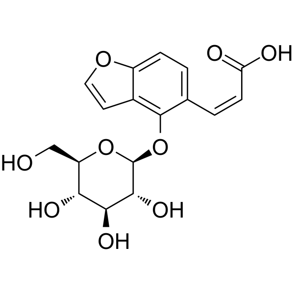
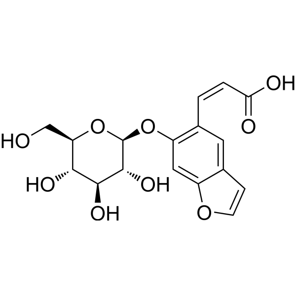
Isopsoralenoside is a benzofuran glycoside and coumarin-derived compound. Isopsoralenoside exists in the dried mature fruits of Psoralea corylifolia L., and acts as a metabolite precursor, toxicant and inducer, with estrogen-like, osteoblast proliferation-promoting, anti-tumor and antibacterial activities. Isopsoralenoside can be deglycosylated by intestinal flora to form isopsoralen. Isopsoralenoside induces oocyte death in zebrafish, activates oxidative stress and cell death-related pathways in zebrafish follicles, and is the main effector substance responsible for the reproductive toxicity induced by Fructus Psoraleae. Isopsoralenoside can be used in studies related to reproductive toxicity (ovarian dysfunction, follicular atresia, impaired fertility).
For research use only. We do not sell to patients.
- Purity : 98.57%
- CAS No.: 905954-18-9
- Formula: C17H18O9
- Molecular Weight:366.32
-
Storage:
-20°C, sealed storage, away from moisture and light
* In solvent : -80°C, 6 months; -20°C, 1 month (sealed storage, away from moisture and light)

To place orders, for customer services and technical support, please contact: MedChemExpress USA
Tel: 609-228-6898 E-mail: [email protected] [email protected]


-
 Biological Activity
-
Chemical Information
-
Solvent & Solubility
- Protocols
- Purity & Documentation
- References
-
 Help & FAQs




Biological Activity
Description
In Vitro
Isopsoralenoside (IPO) (0.572 mg/mL; 0-20 h) is completely metabolized to isopsoralen by rat intestinal microflora within 4 h via de-glucosylation[1].
Isopsoralenoside (0.202 mg/mL; 0-12 h) is not significantly affected by gastric acid, pepsin, and pancreatin in its metabolism to isopsoralen[1].
Isopsoralenoside (24 h) induces dose-dependent cytotoxicity in isolated zebrafish oocytes with an LC50 of 6.52 μM, and its measured ovarian accumulation concentration in zebrafish exceeds its BMDL threshold, identifying it as a key reproductive toxicant[3].
Isopsoralenoside (0.21 μM) induces widespread transcriptional perturbations in cultured zebrafish follicles, activating pathways linked to reproductive toxicity, and contributes 0.961 normalized weight to Fructus Psoraleae-induced reproductive toxicity[3].
MedChemExpress (MCE) has not independently confirmed the accuracy of these methods. They are for reference only. Further protocols information, click here.
Parmacokinetics
In Vivo
Isopsoralenoside (60.05 mg/kg; p.o.) undergoes in vivo biotransformation to isopsoralen within the gastrointestinal tract of healthy rats[1].
Isopsoralenoside (9 g/kg; p.o.; single dose) exhibits high plasma absorption and moderate bone tissue distribution, with rapid clearance from both plasma and bone tissue[2].
Isopsoralenoside (0.21 µM; waterborne; daily; 21 days) is a dominant contributor to Fructus Psoraleae-induced reproductive toxicity in zebrafish, with a normalized toxicity contribution weight of 0.961, and has a predicted maximum permissible 21-day daily oral intake of 0.021 µg/kg for adult females[3].
MedChemExpress (MCE) has not independently confirmed the accuracy of these methods. They are for reference only.
-
Animal Model:Sprague-Dawley (male, 200-220 g)[1]
-
Dosage:39.62 mg/kg (as part of PCE); 60.05 mg/kg (as part of PBGF)
-
Administration:p.o.
-
Result:Reached a maximum plasma concentration (Cₘₐₓ) of 2.58 μg/mL at 2.58 h (Tₘₐₓ), with an elimination half-life (t1/2) of 5.64 h, area under the plasma concentration-time curve from 0 to last measurable time (AUC0-ₜ) of 16.82 μg h/mL, and AUC from 0 to infinity (AUC0-∞) of 17.21 μg h/mL after oral administration of PCE.
Reached a Cₘₐₓ of 1.22 μg/mL at 3.00 h (Tₘₐₓ), with a t1/2 of 9.31 h, AUC0-ₜ of 10.57 μg h/mL, and AUC0-∞ of 11.54 μg h/mL after oral administration of PBGF.
Was detected in the contents and homogenized tissue of the stomach, small intestine, cecum, and colon at 1, 3, and 9 h after oral PBGF administration.
Was completely metabolized to isopsoralen within 4 h in anaerobic rat fecal suspension incubation.
-
Animal Model:Sprague-Dawley (male, 200-220 g)[1]
-
Dosage:60.05 mg/kg (as part of PBGF)
-
Administration:p.o.
-
Result:Was detected in the contents and homogenized tissue of the stomach, small intestine, cecum, and colon at 1, 3, and 9 h post-administration.
Isopsoralen was concurrently detected in these samples, confirming in vivo biotransformation.
-
Animal Model:Sprague-Dawley (SD) (female, 8 weeks old, 200 g)[2]
-
Dosage:9 g/kg (equivalent crude drug dose of Psoraleae Fructus extract containing isopsoralenoside)
-
Administration:p.o.; single dose
-
Result:Reached a plasma Cmax of 7486.24 ng/mL, with a fast absorption, fast elimination pharmacokinetic profile.
Reached a bone tissue Cmax of 310.26 ng/g at 1 h post-administration.
Was no longer detectable in bone tissue at 12 h post-administration.
-
Animal Model:wild-type (healthy adult females)[3]
-
Dosage:0.21 µM (ovarian accumulation)
-
Administration:waterborne; daily; 21 days
-
Result:Exceeded its benchmark dose limit (BMDL) of 0.018 µM.
Had a normalized toxicity contribution weight of 0.961 to Fructus Psoraleae-induced reproductive toxicity pathways.
Predicted a maximum permissible 21-day daily oral intake of 0.021 µg/kg for adult females, corresponding to a human ovarian safety threshold of 3.9×10⁻⁵ μM.


Chemical Information
-
CAS No. 905954-18-9
-
Appearance Solid
-
Molecular Weight 366.32
-
Formula C17H18O9
-
Color White to light yellow
-
SMILES
O=C(O)/C=C\C1=CC=C(OC=C2)C2=C1O[C@H]3[C@@H]([C@H]([C@@H]([C@@H](CO)O3)O)O)O
-
Structure Classification
-
Initial Source
-
Shipping
Room temperature in continental US; may vary elsewhere.
-
Storage
-20°C, sealed storage, away from moisture and light
* In solvent : -80°C, 6 months; -20°C, 1 month (sealed storage, away from moisture and light)


Solvent & Solubility
In Vitro:
DMSO : 100 mg/mL (272.99 mM; Need ultrasonic; Hygroscopic DMSO has a significant impact on the solubility of product, please use newly opened DMSO)
Please refer to the solubility information to select the appropriate solvent. Once prepared, please aliquot and store the solution to prevent product inactivation from repeated freeze-thaw cycles.
Storage method and period of stock solution: -80°C, 6 months; -20°C, 1 month (sealed storage, away from moisture and light). When stored at -80°C, please use it within 6 months. When stored at -20°C, please use it within 1 month.
Please refer to the solubility information to select the appropriate solvent. Once prepared, please aliquot and store the solution to prevent product inactivation from repeated freeze-thaw cycles.
Storage method and period of stock solution: -80°C, 6 months; -20°C, 1 month (sealed storage, away from moisture and light). When stored at -80°C, please use it within 6 months. When stored at -20°C, please use it within 1 month.
Concentration (start) × Volume (start) = Concentration (final) × Volume (final)
In Vivo:
Select the appropriate dissolution method based on your experimental animal and administration route.
- For the following dissolution methods, please ensure to first prepare a clear stock solution using an In Vitro approach and then sequentially add co-solvents:
- To ensure reliable experimental results, the clarified stock solution can be appropriately stored based on storage conditions. As for the working solution for In Vivo experiments, it is recommended to prepare freshly and use it on the same day.
- The percentages shown for the solvents indicate their volumetric ratio in the final prepared solution. If precipitation or phase separation occurs during preparation, heat and/or sonication can be used to aid dissolution.
Add each solvent one by one: 10% DMSO 40% PEG300 5% Tween-80 45% Saline
Solubility: ≥ 2.5 mg/mL (6.82 mM); Clear solution
This protocol yields a clear solution of ≥ 2.5 mg/mL (saturation unknown).
Taking 1 mL working solution as an example, add 100 μL DMSO stock solution (25.0 mg/mL) to 400 μL PEG300, and mix evenly; then add 50 μL Tween-80 and mix evenly; then add 450 μL Saline to adjust the volume to 1 mL.
Preparation of Saline: Dissolve 0.9 g sodium chloride in ddH₂O and dilute to 100 mL to obtain a clear Saline solution.
Add each solvent one by one: 10% DMSO 90% (20% SBE-β-CD in Saline)
Solubility: ≥ 2.5 mg/mL (6.82 mM); Clear solution
This protocol yields a clear solution of ≥ 2.5 mg/mL (saturation unknown).
Taking 1 mL working solution as an example, add 100 μL DMSO stock solution (25.0 mg/mL) to 900 μL 20% SBE-β-CD in Saline, and mix evenly.
Preparation of 20% SBE-β-CD in Saline (4°C, storage for one week): 2 g SBE-β-CD powder is dissolved in 10 mL Saline, completely dissolve until clear.
In Vivo Dissolution Calculator
Please enter the basic information of animal experiments:
-
-
-
-
Recommended: Prepare an additional quantity of animals to account for potential losses during experiments.
Please enter your animal formula composition:
-
%DMSO +
Recommended: Keep the proportion of DMSO in working solution below 2% if your animal is weak.
-
%+
-
+%Tween-80 + +
-
%Saline +
The co-solvents required include: DMSO, . All of co-solvents are available by MedChemExpress (MCE). , Tween 80. All of co-solvents are available by MedChemExpress (MCE).
Working solution concentration: 0.22 mg/mL
Method for preparing stock solution: mg drug dissolved in μL DMSO. Stock solution concentration: mg/mL. * In solvent : -80°C, 6 months; -20°C, 1 month (sealed storage, away from moisture and light)
1. Take μL DMSO stock solution;
2. Add μL .
μL , mix evenly;
3. Then add μL Tween 80, mix evenly;
4. Then add μL
Please ensure that the stock solution in the first step is dissolved to a clear state, and add co-solvents in sequence. You can use ultrasonic heating (ultrasonic cleaner, recommended frequency 20-40 kHz), vortexing, etc. to assist dissolution.

Protocols
-
Reproductive and Developmental Toxicity Study
Reproductive and developmental toxicity studies detect adverse effects of prenatal or peri/postnatal exposure on maternal condition, pregnancy maintenance, embryo-fetal survival, fetal growth, structural development, and offspring reproductive or developmental endpoints; classic rat protocols generate readouts by comparing treated groups with vehicle, pair-fed, or untreated controls for implantation, resorption, fetal weight, crown-rump length, external morphology, visceral morphology, skeletal ossification, anogenital distance, nipple/areola retention, and postnatal cohort outcomes.
-
Research Protocol for Infectious Diseases
Infectious-disease experiments test how pathogens interact with host barriers, innate immune receptors, inflammatory signaling, pathogen replication, and tissue injury; pattern-recognition receptors such as TLRs, RIG-I-like receptors, NOD-like receptors, and inflammasomes detect microbial molecules and activate NF-κB, interferon, and cytokine responses. The central hypothesis is that infection severity reflects the balance between pathogen burden and host response: protective inflammation restricts pathogen growth, whereas excessive or mislocalized inflammation contributes to tissue damage and disease phenotype. Unresolved questions include which host pathways are protective versus pathogenic, why some infection models fail to translate to human disease, and which combined readouts best predict clinically relevant infection outcomes.
-
Cell Cytotoxicity Assay
Cytotoxicity assays are usually based on the assessment of cell membrane damage, which can also be indirectly detected by measuring cell viability. Detection methods include MTT assay, CKK-8 assay, LDH assay and ATP assay, etc.
-
Mammalian live/dead viability and cytotoxicity staining
Live/dead viability and cytotoxicity staining assays are based on the simultaneous detection of intracellular esterase activity in metabolically active (viable) cells and membrane integrity loss in non-viable cells. In commonly used dual-staining approaches, membrane-permeant fluorogenic substrates are converted by intracellular esterases into fluorescent products in live cells, while impermeant DNA-binding dyes selectively enter cells with compromised plasma membranes and label nucleic acids in dead or dying cells, enabling discrimination between viable and non-viable populations by fluorescence microscopy or flow cytometry.
-
ROS/oxidative-stress fluorescent staining
ROS/oxidative-stress fluorescent staining uses cell-permeant fluorogenic probes that become fluorescent after oxidation inside cells or tissues; commonly used examples include DCFH-DA/DCFDA for broad cellular oxidant detection, DHE for superoxide-related signal detection, MitoSOX for mitochondrial superoxide-related signal detection, and CellROX probes for oxidative-stress-associated fluorescence readouts. The assay detects probe oxidation rather than a single ROS species unless the probe and analysis method have been chemically validated for that species. DCFH-DA enters cells, is deacetylated by intracellular esterases to DCFH, and produces fluorescent DCF after oxidation, so the readout is used as an operational measure of total cellular oxidative stress rather than a species-specific ROS measurement. DHE and MitoSOX can report superoxide-related oxidation, but red fluorescence alone can include non-specific ethidium-like oxidation products; HPLC or optimized spectral approaches are
-
Research Protocol for Endocrine Diseases
Endocrine diseases often arise from disrupted hormone production, hormone signaling, or target-tissue responsiveness; for diabetes-focused endocrine disease models, insulin signaling regulates glucose uptake, hepatic glucose output, lipid metabolism, and β-cell compensation. Type 2 diabetes develops through interacting defects in insulin resistance, β-cell dysfunction, adipose inflammation, hepatic glucose overproduction, altered incretin signaling, and ectopic lipid metabolism. A major unresolved question is whether endocrine dysfunction is driven primarily by target-tissue insulin resistance, intrinsic β-cell failure, immune/inflammatory stress, or combined multi-organ failure that differs by disease stage.
-
Apoptosis Solutions
Apoptosis is a regulated, generally non-lytic cell-death pathway that removes unwanted, damaged, infected, or abnormal cells through coordinated morphological changes, caspase activation, DNA fragmentation, and membrane remodeling. The intrinsic apoptosis pathway is controlled mainly by mitochondrial outer membrane permeabilization, BCL-2 family proteins, cytochrome c release, apoptosome formation, caspase-9 activation, and downstream executioner caspase-3/7 activation. The extrinsic apoptosis pathway is initiated by death receptors such as Fas, TNFR, and TRAIL receptors, which recruit adaptor proteins and activate caspase-8 before engaging executioner caspases or mitochondrial amplification through BID cleavage. Apoptosis is linked to many phenotypes, including cancer cell killing, tissue homeostasis, immune regulation, neurodegeneration, infection response, and treatment-induced cytotoxicity; unresolved questions include how apoptosis interacts with necroptosis, pyroptosis, ferroptos
-
Cell Viability Determination by MTT Colorimetric Assay
The following protocol uses the MTT colorimetric assay as a classic literature-established method for assessing cell viability/metabolic activity in cultured mammalian cells. MTT[3-(4,5-dimethylthiazol-2-yl)-2,5-diphenyltetrazolium bromide] is reduced by metabolically active cells to a colored formazan product; the amount of formazan is quantified spectrophotometrically and provides an indirect measure of metabolically active viable cells. Importantly, MTT reduction reflects cellular oxidoreductase/metabolic activity rather than an absolute direct count of living cells, so changes in cellular metabolism can alter the signal independently of cell number.


Purity & Documentation
-
Data Sheet (296 KB)
-
SDS (393 KB)
- English - EN (393 KB)
- Français - FR (393 KB)
- Deutsch - DE (393 KB)
- Norwegian - NO (393 KB)
- Español - ES (393 KB)
- Swedish - SV (393 KB)
- Italian - IT (393 KB)
- Korean - KR (393 KB)
- Portuguese - PT (393 KB)
-
Handling Instructions (2659 KB)
References
[1]. Wang YF, et al. A UPLC-MS/MS method for in vivo and in vitro pharmacokinetic studies of psoralenoside, isopsoralenoside, psoralen and isopsoralen from Psoralea corylifolia extract. Journal of ethnopharmacology. 2014;151(1):609-17. [Content Brief]
[2]. Xu Q, et al. Comprehensive studies of plasma pharmacokinetic and bone tissue distribution in rats to elucidate pharmacodynamic material basis of Psoraleae Fructus treating osteoporosis. Journal of ethnopharmacology. 2026 Apr 06;360:121145. [Content Brief]
[3]. Shen X, et al. Reproductive toxicity of Fructus Psoraleae in zebrafish: material basis and implications for clinical safety dosing. Phytomedicine : international journal of phytotherapy and phytopharmacology. 2026 Jun;155:158194. [Content Brief]


Complete Stock Solution Preparation Table
Please refer to the solubility information to select the appropriate solvent. Once prepared, please aliquot and store the solution to prevent product inactivation from repeated freeze-thaw cycles.
Storage method and period of stock solution: -80°C, 6 months; -20°C, 1 month (sealed storage, away from moisture and light). When stored at -80°C, please use it within 6 months. When stored at -20°C, please use it within 1 month.
| Optional Solvent | Concentration Solvent Mass | 1 mg | 5 mg | 10 mg | 25 mg |
|---|---|---|---|---|---|
| DMSO | 1 mM | 2.7299 mL | 13.6493 mL | 27.2985 mL | 68.2463 mL |
| 5 mM | 0.5460 mL | 2.7299 mL | 5.4597 mL | 13.6493 mL | |
| 10 mM | 0.2730 mL | 1.3649 mL | 2.7299 mL | 6.8246 mL | |
| 15 mM | 0.1820 mL | 0.9100 mL | 1.8199 mL | 4.5498 mL | |
| 20 mM | 0.1365 mL | 0.6825 mL | 1.3649 mL | 3.4123 mL | |
| 25 mM | 0.1092 mL | 0.5460 mL | 1.0919 mL | 2.7299 mL | |
| 30 mM | 0.0910 mL | 0.4550 mL | 0.9100 mL | 2.2749 mL | |
| 40 mM | 0.0682 mL | 0.3412 mL | 0.6825 mL | 1.7062 mL | |
| 50 mM | 0.0546 mL | 0.2730 mL | 0.5460 mL | 1.3649 mL | |
| 60 mM | 0.0455 mL | 0.2275 mL | 0.4550 mL | 1.1374 mL | |
| 80 mM | 0.0341 mL | 0.1706 mL | 0.3412 mL | 0.8531 mL | |
| 100 mM | 0.0273 mL | 0.1365 mL | 0.2730 mL | 0.6825 mL |


Isopsoralenoside Related Classifications
HY-N7504 Related Classifications
 Powered by Bioz