



APTSTAT3-9R
Based on 1 publication(s) in Google Scholar
APTSTAT3-9R, a specific STAT3-binding peptide, inhibits STAT3 activation and downstream signaling by specifically blocking STAT3 phosphorylation. APTSTAT3-9R exerts antiproliferative effects and antitumor activity.
For research use only. We do not sell to patients.
- Purity : 98.82%
- Formula: C223H330N80O51
- Molecular Weight:4947.51
-
Storage:
Sealed storage, away from moisture and light, under nitrogen.
Powder -80°C, 2 years , -20°C, 1 year* In solvent : -80°C, 6 months; -20°C, 1 month (sealed storage, away from moisture and light, under nitrogen)

To place orders, for customer services and technical support, please contact: MedChemExpress USA
Tel: 609-228-6898 E-mail: [email protected] [email protected]


-
 Biological Activity
-
Chemical Information
-
Solvent & Solubility
- Protocols
- Purity & Documentation
- References
-
 Help & FAQs


Publications Citing Use of MedChemExpress (MCE) APTSTAT3-9R
More

Biological Activity
Description
IC50 & Target
[1]|
STAT3 |
In Vitro
APTSTAT3-9R (7.5, 15, and 30 μmol/L; 6 hours) significantly reduces STAT3–DNA-binding activity in a dose-dependent manner in human lung carcinoma cells (A549)[1].
APTSTAT3-9R (30 μM; for 2 weeks) suppresses cell viability and proliferation of cancer cells and significantly suppresses colony formation. APTSTAT3-9R has IC50s of 10 to 20 μM in A549, B16F1 and HepG2 cells[1].
APTSTAT3-9R (7.5, 15, and 30 μmol/L; 6 hours) effectively inhibits phosphorylation of STAT3 but does not affect the level of AKT phosphorylation, indicating specificity of the aptide[1].
MedChemExpress (MCE) has not independently confirmed the accuracy of these methods. They are for reference only.
In Vivo
MedChemExpress (MCE) has not independently confirmed the accuracy of these methods. They are for reference only.


Chemical Information
-
Appearance Solid
-
Molecular Weight 4947.51
-
Formula C223H330N80O51
-
Color White to off-white
-
Sequence
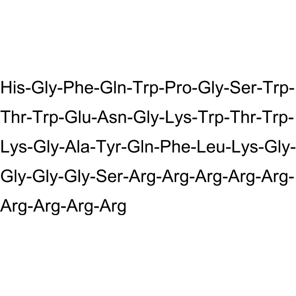
His-Gly-Phe-Gln-Trp-Pro-Gly-Ser-Trp-Thr-Trp-Glu-Asn-Gly-Lys-Trp-Thr-Trp-Lys-Gly-Ala-Tyr-Gln-Phe-Leu-Lys-Gly-Gly-Gly-Gly-Ser-Arg-Arg-Arg-Arg-Arg-Arg-Arg-Arg-Arg
-
Sequence Shortening
HGFQWPGSWTWENGKWTWKGAYQFLKGGGGSRRRRRRRRR
-
Shipping
Room temperature in continental US; may vary elsewhere.
-
Storage
Sealed storage, away from moisture and light, under nitrogen
Powder -80°C 2 years -20°C 1 year * In solvent : -80°C, 6 months; -20°C, 1 month (sealed storage, away from moisture and light, under nitrogen)


Publications (1)
-
Journal Impact Factor
-
Most Recent
-
Arch Oral Biol
Parasympathetic-macrophages-ductal epithelial cells axis promotes female rat submandibular gland regeneration after excretory duct ligation/deligation. [Abstract]2023 Jan:145:105586. PMID: 36410162


Solvent & Solubility
In Vitro:
H2O : ≥ 50 mg/mL (10.11 mM)
* "≥" means soluble, but saturation unknown.
Please refer to the solubility information to select the appropriate solvent. Once prepared, please aliquot and store the solution to prevent product inactivation from repeated freeze-thaw cycles.
Storage method and period of stock solution: -80°C, 6 months; -20°C, 1 month (sealed storage, away from moisture and light, under nitrogen). When stored at -80°C, please use it within 6 months. When stored at -20°C, please use it within 1 month.
* Note: If you choose water as the stock solution, please dilute it to the working solution, then filter and sterilize it with a 0.22 μm filter before use.
Please refer to the solubility information to select the appropriate solvent. Once prepared, please aliquot and store the solution to prevent product inactivation from repeated freeze-thaw cycles.
Storage method and period of stock solution: -80°C, 6 months; -20°C, 1 month (sealed storage, away from moisture and light, under nitrogen). When stored at -80°C, please use it within 6 months. When stored at -20°C, please use it within 1 month.
* Note: If you choose water as the stock solution, please dilute it to the working solution, then filter and sterilize it with a 0.22 μm filter before use.
Concentration (start) × Volume (start) = Concentration (final) × Volume (final)


Purity & Documentation
-
Data Sheet (287 KB)
-
SDS (252 KB)
- English - EN (252 KB)
- Français - FR (252 KB)
- Deutsch - DE (252 KB)
- Norwegian - NO (252 KB)
- Español - ES (252 KB)
- Swedish - SV (252 KB)
- Italian - IT (252 KB)
- Korean - KR (252 KB)
- Portuguese - PT (252 KB)
-
Handling Instructions (2659 KB)
References


Complete Stock Solution Preparation Table
Please refer to the solubility information to select the appropriate solvent. Once prepared, please aliquot and store the solution to prevent product inactivation from repeated freeze-thaw cycles.
Storage method and period of stock solution: -80°C, 6 months; -20°C, 1 month (sealed storage, away from moisture and light, under nitrogen). When stored at -80°C, please use it within 6 months. When stored at -20°C, please use it within 1 month.
| Optional Solvent | Concentration Solvent Mass | 1 mg | 5 mg | 10 mg | 25 mg |
|---|---|---|---|---|---|
| H2O | 1 mM | 0.2021 mL | 1.0106 mL | 2.0212 mL | 5.0530 mL |
| 5 mM | 0.0404 mL | 0.2021 mL | 0.4042 mL | 1.0106 mL | |
| 10 mM | 0.0202 mL | 0.1011 mL | 0.2021 mL | 0.5053 mL |
* Note: If you choose water as the stock solution, please dilute it to the working solution, then filter and sterilize it with a 0.22 μm filter before use.


APTSTAT3-9R Related Classifications
HY-P2282 Related Classifications
 Powered by Bioz