



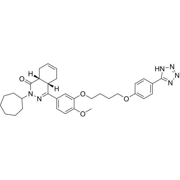
(4aS,8aR)-NPD-001
(4aS,8aR)-NPD-001 is a potent and allosteric inhibitor of DNMT3A. (4aS,8aR)-NPD-001 inhibits DNMT3A activity by disrupting protein-protein interactions. (4aS,8aR)-NPD-001 induces apoptosis of acute myeloid leukemia (AML) cell lines. (4aS,8aR)-NPD-001 induces differentiation of distinct AML cell lines including cells with mutated DNMT3A R882.
For research use only. We do not sell to patients.
- CAS No.: 2366272-43-5
- Formula: C33H40N6O4
- Molecular Weight:584.71
-
Storage:
Please store the product under the recommended conditions in the Certificate of Analysis.

To place orders, for customer services and technical support, please contact: MedChemExpress USA
Tel: 609-228-6898 E-mail: [email protected] [email protected]


-
 Biological Activity
-
Chemical Information
- Protocols
- Purity & Documentation
- References
-
 Help & FAQs


All DNA Methyltransferase Isoforms
More

Biological Activity
Description
IC50 & Target
|
DNMT3A |
Cellular Effect
|
Cell Line

|
Type | Value | Description | References |
|---|---|---|---|---|
| HeLa | IC50 |
>25 μM
Compound: 15; MMV690027
|
Cytotoxicity against human HeLa cells assessed as reduction in cell viability after 48 hrs by resazurin dye based fluorescence assay
Cytotoxicity against human HeLa cells assessed as reduction in cell viability after 48 hrs by resazurin dye based fluorescence assay
|
[PMID: 30647879] |
| Sf21 | IC50 |
0.004 μM
Compound: 3, PPS54019
|
Inhibition of Trypanosoma brucei full length PDEB1 expressed in insect SF21 cells using cAMP as substrate by scintillation proximity assay
Inhibition of Trypanosoma brucei full length PDEB1 expressed in insect SF21 cells using cAMP as substrate by scintillation proximity assay
|
[PMID: 23409953] |
In Vitro
(4aS,8aR)-NPD-001 (compound 2) (60 μM) disrupts DNMT3A-DNMT3L interactions at the DNMT3A tetramer interface, inhibits the stimulation of DNMT3A_WT activity by DNMT3L, but does not inhibit the activation of DNMT3A_WT by H3 peptides[1].
(4aS,8aR)-NPD-001 (0-120 μM, 100 min) inhibits the activation of DNMT3A_R882H by DNMT3L[1].
(4aS,8aR)-NPD-001 (0-30 μM, 72 h) induces apoptosis and and differentiation in AML cell lines in a concentration-dependent manner[1].
(4aS,8aR)-NPD-001 (5 μM, 20 days) leads to a time-dependent decrease of global 5-methylcytosine[1].
MedChemExpress (MCE) has not independently confirmed the accuracy of these methods. They are for reference only.
-
Cell Line:Human AML cell lines
-
Concentration:0, 1, 5, 10, 20, and 30 μM
-
Incubation Time:72 h
-
Result:Showed a dose-response effect with a marked increase in apoptosis in the 7-12 μM range. Led to a concentration-dependent increase in the myeloid differentiation marker, CD11b, in multiple PI-negative AML cell lines: MV411 (biphenotypic B myelomonocytic leukemia), MOLM13 (acute monocytic leukemia), THP-1 (acute monocytic leukemia), OCI-AML3 (DNMT3A R882 mutant, AML), KASUMI (acute myeloblastic leukemia), HL60 (acute promyelocytic leukemia), and K562 (chronic myelogenous leukemia).


Chemical Information
-
CAS No. 2366272-43-5
-
Molecular Weight 584.71
-
Formula C33H40N6O4
-
SMILES
O=C1N(C2CCCCCC2)N=C(C3=CC=C(OC)C(OCCCCOC4=CC=C(C5=NN=NN5)C=C4)=C3)[C@@]6([H])CC=CC[C@@]16[H]
-
Shipping
Room temperature in continental US; may vary elsewhere.
-
Storage
Please store the product under the recommended conditions in the Certificate of Analysis.


Purity & Documentation
References

Calculators
Concentration (start) × Volume (start) = Concentration (final) × Volume (final)


(4aS,8aR)-NPD-001 Related Classifications
HY-150025 Related Classifications
 Powered by Bioz