



Corr4A

Based on 1 Customer Validation
Corr4A is a chemical corrector, which can be used for cystic fibrosis. Corr4A interacts directly with the cystic fibrosis transmembrane conductance regulator (CFTR) or affects indirectly its folding process. Corr4A increases the expression of CFTR ΔF508 on the cell surface, thereby improving its transport to the plasma membrane and increasing the stability of the rescued mutant protein.
商品は「研究用試薬」です。人や動物の医療用・臨床診断用・食品用の製品ではありません。
研究用途以外に使用した場合、当社は一切の責任を負いかねます。
- 純度 : 99.58%
- CAS 番号: 421580-53-2
- 分子式: C21H17ClN4O2S2
- 分子量:456.97
-
保管条件:Powder -20°C, 3 years , 4°C, 2 years ; In solvent -80°C, 6 months , -20°C, 1 month


To place orders, for customer services and technical support, please contact: MedChemExpress USA
Tel: 609-228-6898 E-mail: [email protected] [email protected]


-
 生物活性
生物活性
-
化学情報
- プロトコル
- 純度とドキュメンテーション
- 参考文献
-
 Help & FAQs


-
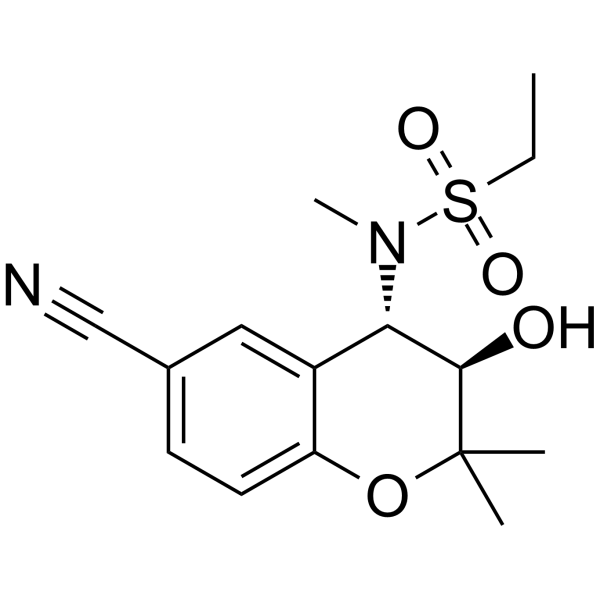
HY-108575
Chromanol 293BIKs Blocker -
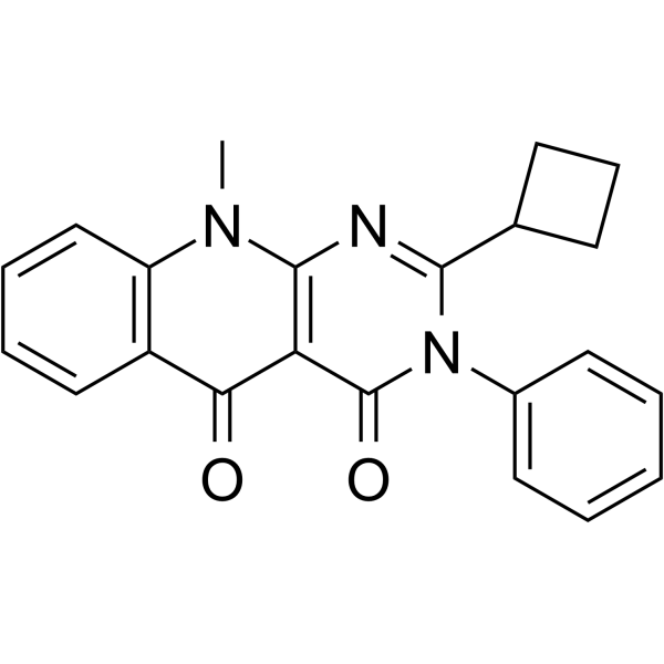
HY-150090
SRI-41315CFTR Activator -
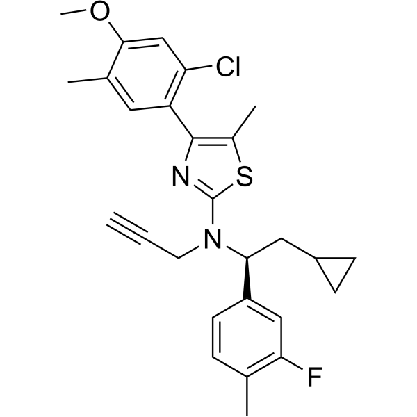
HY-106203
CrinecerfontCRF1 Receptor Antagonist -

HY-119229
VRT-325CFTR Modulator -
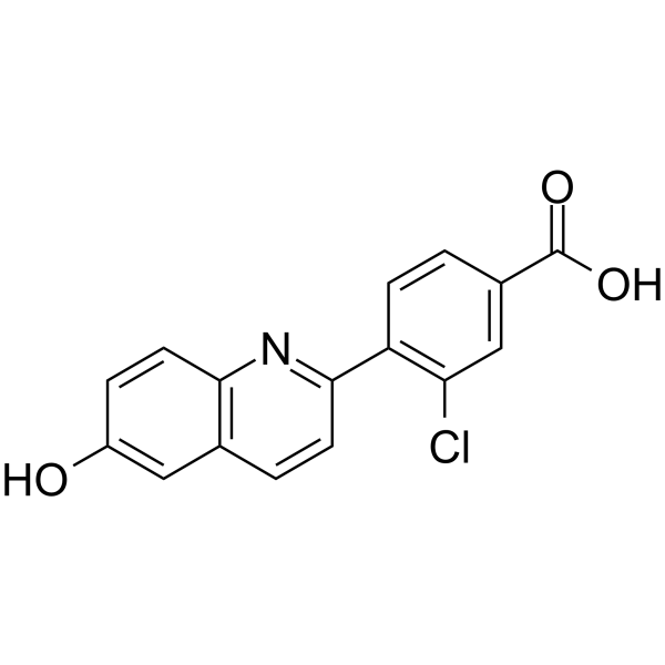
HY-109027
CavosonstatGSNOR Inhibitor -
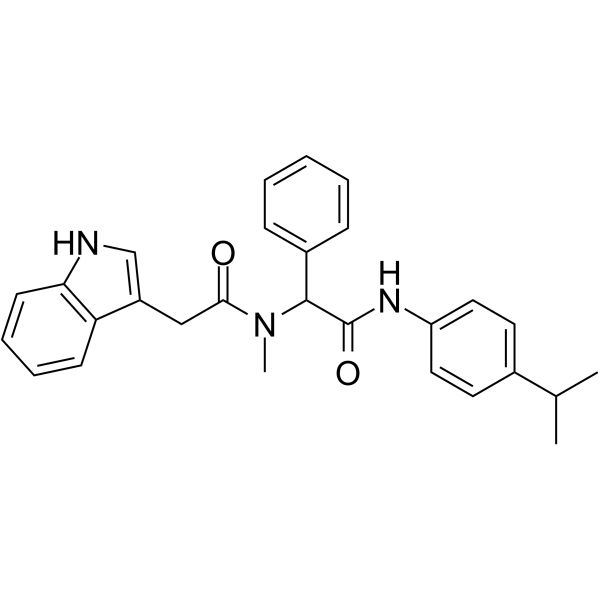
HY-103369
PG01CFTR Potentiator -
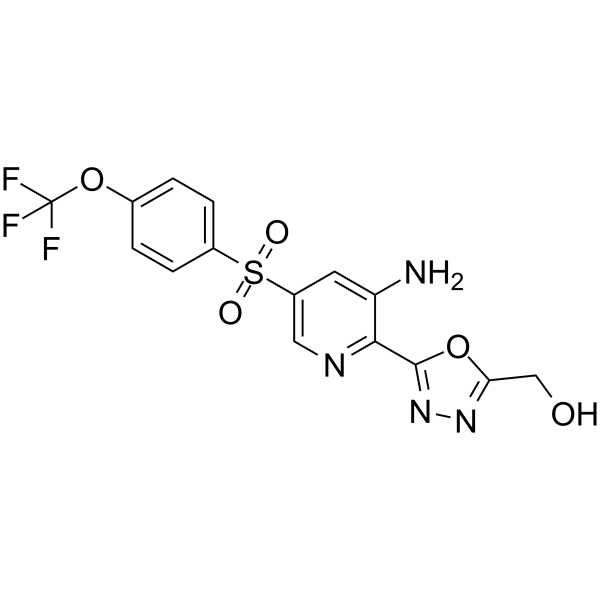
HY-109152
NavocaftorCystic Fibrosis Transmembrane Regulator -
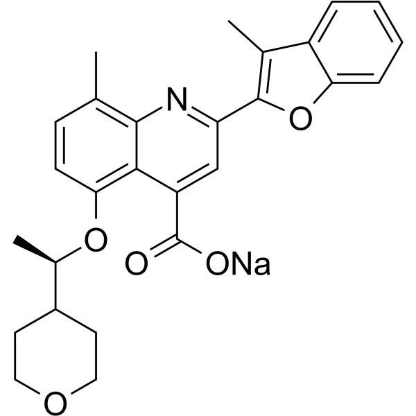
HY-109187A
Posenacaftor sodiumCFTR Modulator -
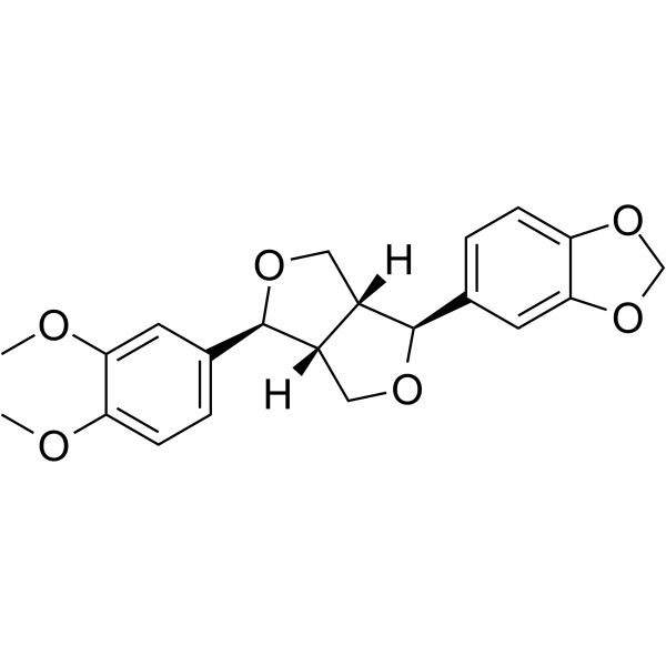
HY-N5101
KobusinCalmodulin inhibitor -
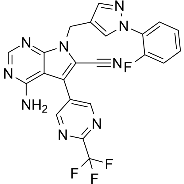
HY-136939
CFTR corrector 6CFTR Activator -
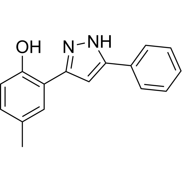
HY-114725
VRT-532CFTR Modulator -
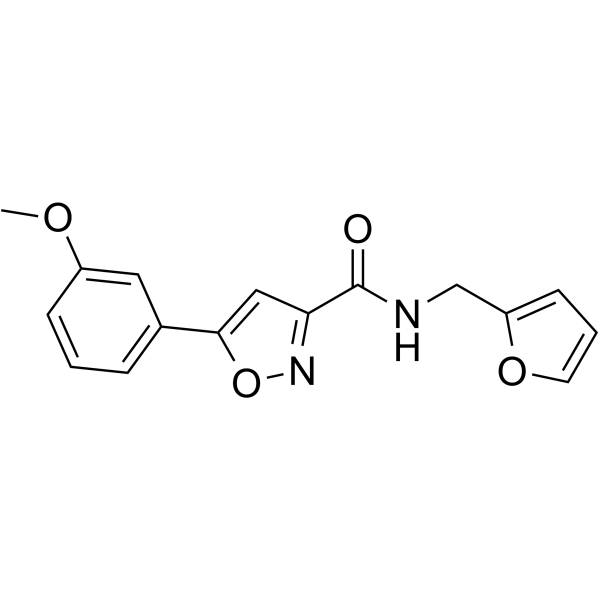
HY-147315
CFTR corrector 9CFTR Modulator


生物活性
製品説明
Cellular Effect
|
Cell Line

|
Type | Value | Description | References |
|---|---|---|---|---|
| Epithelial cell | IC50 |
6 μM
Compound: Corr4a
|
Correction of defective trafficking of human deltaF508 mutant CFTR protein expressed in FRT epithelial cells coexpressing YFP-H148Q/I152L protein assessed as iodide influx for 16 to 20 hrs by fluorescence assay
Correction of defective trafficking of human deltaF508 mutant CFTR protein expressed in FRT epithelial cells coexpressing YFP-H148Q/I152L protein assessed as iodide influx for 16 to 20 hrs by fluorescence assay
|
[PMID: 25061695] |


化学情報
-
CAS 番号 421580-53-2
-
性状 Solid
-
分子量 456.97
-
分子式 C21H17ClN4O2S2
-
Color White to off-white
-
SMILES
O=C(NC1=NC(C)=C(C2=CSC(NC3=CC(Cl)=CC=C3OC)=N2)S1)C4=CC=CC=C4
-
輸送条件
Room temperature in continental US; may vary elsewhere.
-
保管条件
Powder -20°C 3 years 4°C 2 years In solvent -80°C 6 months -20°C 1 month


純度とドキュメンテーション
-
データシート (266 KB)
-
SDS (251 KB)
- English - EN (251 KB)
- Français - FR (251 KB)
- Deutsch - DE (251 KB)
- Norwegian - NO (251 KB)
- Español - ES (251 KB)
- Swedish - SV (251 KB)
- Italian - IT (251 KB)
- Korean - KR (251 KB)
- Portuguese - PT (251 KB)
-
取扱説明書 (2659 KB)
参考文献

Calculators
濃度 (開始) × 体積 (開始) = 濃度 (終了) × 体積 (終了)


Corr4A 関連分類
HY-117187 関連分類
 Powered by Bioz