
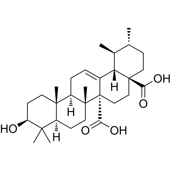
Quinovic acid
Based on 1 Customer Validation
Quinovic acid is triterpene. Quinovic acid can ameliorate the Amyloid-β burden, p53 expression and cholesterol accumulation by deterring the oxidative stress through upregulating the Nrf2/HO-1 pathway. Quinovic acid can induce cancer cells apoptosis by upregulating death receptor 5 (DR5). Quinovic acid can be used for the researches of cancer, inflammation, metabolic and neurological disease, such as lung cancer and Alzheimer’s disease (AD).
商品は「研究用試薬」です。人や動物の医療用・臨床診断用・食品用の製品ではありません。
研究用途以外に使用した場合、当社は一切の責任を負いかねます。
- 純度 : 96.0%
- CAS 番号: 465-74-7
- 分子式: C30H46O5
- 分子量:486.68
-
保管条件:
-20°C, protect from light
* In solvent : -80°C, 6 months; -20°C, 1 month (protect from light)
生物活性
製品説明
体外実験
Quinovic acid (5-115 μM, 24 h) shows low cytotoxicity and alleviates oligomeric Aβ (AβO)-induced cell death in SH-SY5Y cells[1].
Quinovic acid (85 μM, 24 h) downregulates AβO-induced expressions of p53 and 3-hydroxy-3-methylglutaryl-CoA reductase (HMGCR) in SH-SY5Y cells[1].
Quinovic acid (QA) (5-40 μM, 24-72 h) inhibits the proliferation and viability of A549, H1299, MCF-7 and MDA-MB-231 cells[2].
Quinovic acid (QA) (25-35 μM, 24 h) induces apoptosis in MDA-MB-231 and A549 cells[2].
Quinovic acid (25 μM, 4-24 h) upregulates the mRNA and protein levels of death receptor 5 (DR5) in A549, MCF-7, and MDA-MB-231 cells[2].
MedChemExpress (MCE) has not independently confirmed the accuracy of these methods. They are for reference only. Further protocols information, click here.
-
Cell Line:MDA-MB-231 and A549 cells
-
Concentration:25, 30 and 35 μM
-
Incubation Time:24 h
-
Result:Reduced procaspase 3 and 8 levels.
Increased cleaved PARP levels.
体内実験
MedChemExpress (MCE) has not independently confirmed the accuracy of these methods. They are for reference only.
-
Animal Model:C57BL/6J mice treated with Aβ(1-42)[1]
-
Dosage:50 mg/kg
-
Administration:Intraperitoneally injection, every other day for 3 weeks
-
Result:Reduced Aβ oligomers, monomers, and BACE1 expression in cortex/hippocampus and decreased Aβ burden.
Downregulated p53 and HMGCR expressions and reduced total and free cholesterol levels.
Activated the Nrf2/HO-1 pathway, reduced ROS levels and restored GSH/GSSG ratio.
Downregulated GFAP, Iba-1, p-NF-κB, and IL-1β.
Downregulated Bax, cytochrome c, cleaved caspase-3; upregulates Bcl-2 and upregulated PSD-95 and synaptophysin.
Enhanced cognitive ability.
化学情報
-
CAS 番号 465-74-7
-
性状 Solid
-
分子量 486.68
-
分子式 C30H46O5
-
Color White to off-white
-
SMILES
OC([C@@]12C([C@@]3([H])[C@@](CC2)(CC[C@H]([C@@H]3C)C)C(O)=O)=CC[C@@]4([H])[C@]1(CC[C@]5([H])[C@@]4(CC[C@@H](C5(C)C)O)C)C)=O
-
Structure Classification
-
Initial Source
-
輸送条件
Room temperature in continental US; may vary elsewhere.
-
保管条件
-20°C, protect from light
* In solvent : -80°C, 6 months; -20°C, 1 month (protect from light)
プロトコル
-
RNA extraction experimental
By lysing cells, releasing RNA, and removing impurities such as proteins and DNA, high-purity RNA products are finally obtained. The commonly used traditional method is the guanidine isothiocyanate/phenol/chloroform method (Trizol), which is suitable for a variety of animal materials including animal tissues, microorganisms, cultured cells, etc., and most plant materials.
-
Detection of Apoptosis
Apoptosis, also called programmed cell death, is generally characterized by distinct morphological characteristics.
-
TUNEL staining for apoptotic DNA fragmentation
TUNEL staining detects DNA strand breaks by using terminal deoxynucleotidyl transferase to add labeled nucleotides to exposed 3′-OH DNA termini, generating either microscopic staining in fixed cells or tissue sections, or fluorescence/cytometric signal in cell suspensions. TUNEL positivity reflects DNA fragmentation but should not be interpreted alone as definitive apoptosis, because TUNEL can also label necrotic, autolytic, mechanically damaged, or DNA-repair-associated DNA breaks.
-
Annexin V plus membrane-impermeant dye apoptosis staining
Annexin V-based apoptosis assays rely on the detection of phosphatidylserine (PS) externalization from the inner leaflet of the plasma membrane to the outer leaflet, an early biochemical hallmark of apoptosis. Fluorescently labeled Annexin V binds PS in a calcium-dependent manner, enabling identification of early apoptotic cells by flow cytometry or fluorescence microscopy. When combined with a membrane-impermeant DNA-binding dye (e. g. , propidium iodide), this approach allows discrimination between viable (Annexin V−/dye−), early apoptotic (Annexin V+/dye−), and late apoptotic or necrotic (Annexin V+/dye+) cell populations by assessing membrane integrity and PS exposure.
-
ROS/oxidative-stress fluorescent staining
ROS/oxidative-stress fluorescent staining uses cell-permeant fluorogenic probes that become fluorescent after oxidation inside cells or tissues; commonly used examples include DCFH-DA/DCFDA for broad cellular oxidant detection, DHE for superoxide-related signal detection, MitoSOX for mitochondrial superoxide-related signal detection, and CellROX probes for oxidative-stress-associated fluorescence readouts. The assay detects probe oxidation rather than a single ROS species unless the probe and analysis method have been chemically validated for that species. DCFH-DA enters cells, is deacetylated by intracellular esterases to DCFH, and produces fluorescent DCF after oxidation, so the readout is used as an operational measure of total cellular oxidative stress rather than a species-specific ROS measurement. DHE and MitoSOX can report superoxide-related oxidation, but red fluorescence alone can include non-specific ethidium-like oxidation products; HPLC or optimized spectral approaches are
-
Apoptosis Solutions
Apoptosis is a regulated, generally non-lytic cell-death pathway that removes unwanted, damaged, infected, or abnormal cells through coordinated morphological changes, caspase activation, DNA fragmentation, and membrane remodeling. The intrinsic apoptosis pathway is controlled mainly by mitochondrial outer membrane permeabilization, BCL-2 family proteins, cytochrome c release, apoptosome formation, caspase-9 activation, and downstream executioner caspase-3/7 activation. The extrinsic apoptosis pathway is initiated by death receptors such as Fas, TNFR, and TRAIL receptors, which recruit adaptor proteins and activate caspase-8 before engaging executioner caspases or mitochondrial amplification through BID cleavage. Apoptosis is linked to many phenotypes, including cancer cell killing, tissue homeostasis, immune regulation, neurodegeneration, infection response, and treatment-induced cytotoxicity; unresolved questions include how apoptosis interacts with necroptosis, pyroptosis, ferroptos
-
Research Protocol for Inflammation-related Diseases
The NLRP3 inflammasome is a cytosolic innate immune signaling platform that integrates priming signals and danger-signal activation to promote caspase-1 activation, maturation of IL-1β and IL-18, and gasdermin D-mediated pyroptotic cell death. The core experimental logic is to determine whether inflammatory disease phenotypes are driven by increased NLRP3 expression, ASC-containing inflammasome assembly, caspase-1 cleavage, GSDMD cleavage, and extracellular release of IL-1β/IL-18 rather than by nonspecific cell injury alone. The pathway is strongly linked to inflammation-related disease phenotypes because monosodium urate crystals activate NALP3/NLRP3 inflammasome signaling in gout-like crystal inflammation, cholesterol crystals activate NLRP3 inflammasomes in atherogenesis models, and DSS-induced intestinal inflammation has been reported to involve NLRP3 inflammasome activity. However, experimental colitis studies also show context-dependent protective effects of NLRP3 inflammasome co
-
Research Protocol for Neurological Diseases
PINK1/Parkin-mediated mitophagy pathway is a mitochondrial quality-control signaling axis in which mitochondrial depolarization stabilizes PINK1 on damaged mitochondria, activates Parkin recruitment and E3 ubiquitin ligase activity, promotes ubiquitination of outer mitochondrial membrane proteins, recruits selective autophagy adaptors, and drives lysosomal degradation of damaged mitochondria. In neurological disease research, this pathway is experimentally important because neurons, especially dopaminergic neurons, are highly dependent on mitochondrial integrity, and defective mitochondrial turnover can lead to mitochondrial dysfunction, oxidative stress, impaired neuronal survival, α-synuclein accumulation, and neuroinflammatory damage-associated signals. The genetic disease link is strongest in Parkinson’s disease because mutations in PRKN/parkin cause autosomal recessive juvenile parkinsonism, mutations in PINK1 cause hereditary early-onset Parkinson’s disease, and Drosophila studie
-
Amyloid: Congo Red Amyloid Staining
Congo red amyloid staining is a histochemical method used to detect extracellular amyloid deposits in tissue sections based on the affinity of Congo red dye for β-pleated sheet-rich protein aggregates. When bound to amyloid, Congo red produces characteristic apple-green birefringence under polarized light microscopy, which is widely regarded as a diagnostic feature of amyloid deposition in histopathology. The diagnostic principle relies on the combination of dye binding (congophilia) and optical anisotropy under polarized illumination, which distinguishes amyloid from most non-amyloid eosinophilic extracellular deposits in routine histological evaluation. Amyloid identification by Congo red staining remains a cornerstone in diagnostic pathology despite the availability of adjunct methods such as immunohistochemistry and mass spectrometry, particularly because of its ability to localize deposits directly within tissue architecture. The specificity of Congo red-positive deposits is incre
-
Alzheimer’s Disease Modeling
Alzheimer’s Disease (AD) is a neurodegenerative disorder characterized by a progressive decline in cognitive functions and loss of specific types of neurons and synapses. Alzheimer's symptoms can be simulated in mice by injecting drugs (such as Aβ) or genetically modified.
純度とドキュメンテーション
-
データシート (277 KB)
-
SDS (252 KB)
- English - EN (252 KB)
- Français - FR (252 KB)
- Deutsch - DE (252 KB)
- Norwegian - NO (252 KB)
- Español - ES (252 KB)
- Swedish - SV (252 KB)
- Italian - IT (252 KB)
- Korean - KR (252 KB)
- Portuguese - PT (252 KB)
-
取扱説明書 (2659 KB)
参考文献
[1]. Saeed K, et al. Quinovic Acid Impedes Cholesterol Dyshomeostasis, Oxidative Stress, and Neurodegeneration in an Amyloid-β-Induced Mouse Model. Oxid Med Cell Longev. 2020 Nov 20;2020:9523758. [Content Brief]
[2]. Khayam AU, et al. Quinovic acid purified from medicinal plant Fagonia indica mediates anticancer effects via death receptor 5. Mol Cell Biochem. 2020 Nov;474(1-2):159-169. [Content Brief]
Calculators
濃度 (開始) × 体積 (開始) = 濃度 (終了) × 体積 (終了)

