



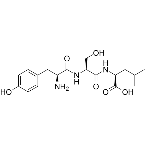
Tyroserleutide
Tyroserleutide (YSL), isolated from the degradation products of porcine spleen, is a small molecular tripeptide which inhibits tumor growth both in vitro and in vivo.
商品は「研究用試薬」です。人や動物の医療用・臨床診断用・食品用の製品ではありません。
研究用途以外に使用した場合、当社は一切の責任を負いかねます。
- CAS 番号: 138168-48-6
- 分子式: C18H27N3O6
- 分子量:381.42
-
保管条件:
Please store the product under the recommended conditions in the Certificate of Analysis.

To place orders, for customer services and technical support, please contact: MedChemExpress USA
Tel: 609-228-6898 E-mail: [email protected] [email protected]


-
 生物活性
生物活性
-
化学情報
-
溶剤 & 溶解度
- プロトコル
- 純度とドキュメンテーション
- 参考文献
-
 Help & FAQs




生物活性
製品説明
IC50 & Target
体外実験
Tyroserleutide (YSL) exhibits immuno-modulating effects, such as enhancing concanavalin (ConA) induced proliferation of mouse spleen lymphocytes, phagocytosis of mouse peritoneal macrophages, and the activity of natural killer (NK) cells[1].
Tyroserleutide (YSL), an immunologically therapeutic tripeptide, can promote hepatocarcinoma cell (H22) apoptosis through downregulating Bcl-2 and cyclin D1 expression[2].
Tyroserleutide is an ideal choice for inducing apoptosis of liver tumor cells[2].
Tyroserleutide inhibits tumor growth and does not cause severe toxicities in the major organs. Tyroserleutide can inhibit tumor cell migration[2].
MedChemExpress (MCE) has not independently confirmed the accuracy of these methods. They are for reference only. Further protocols information, click here.
体内実験
MedChemExpress (MCE) has not independently confirmed the accuracy of these methods. They are for reference only.
-
Animal Model:Female Kun-Ming mice (18-22 g, 6 week old) with H22 tumor model[1]
-
Dosage:10, 20, 40, and 80 μg/kg
-
Administration:Injection (i.p.) one time every day until mice were dead.
-
Result:Survival times are 25.53±14.14, 25.82±14.29, 30.47±17.89, 35.06±20.90 days for 10, 20, 40, and 80 μg/kg, respectively.
臨床実験
| NCT Number | Sponsor | Condition | Start Date |
Phase

|
|---|---|---|---|---|
| NCT01329991 | Plexxikon| | 2011-05 | PHASE1 |


化学情報
-
CAS 番号 138168-48-6
-
分子量 381.42
-
分子式 C18H27N3O6
-
輸送条件
Room temperature in continental US; may vary elsewhere.
-
保管条件
Please store the product under the recommended conditions in the Certificate of Analysis.


溶剤 & 溶解度
体外
H2O
Peptide Solubility and Storage Guidelines:
1. Calculate the length of the peptide.
2. Calculate the overall charge of the entire peptide according to the following table:
| Contents | Assign value | |
|---|---|---|
| Acidic amino acid | Asp (D), Glu (E), and the C-terminal -COOH. | -1 |
| Basic amino acid | Arg (R), Lys (K), His (H), and the N-terminal -NH2 | +1 |
| Neutral amino acid | Gly (G), Ala (A), Leu (L), Ile (I), Val (V), Cys (C), Met (M), Thr (T), Ser (S), Phe (F), Tyr (Y), Trp (W), Pro (P), Asn (N), Gln (Q) | 0 |
3. Recommended solution:
| Overall charge of peptide | Details |
|---|---|
| Negative (<0) |
1. Try to dissolve the peptide in water first. 2. If water fails, add NH4OH (<50 μL). 3. If the peptide still does not dissolve, add DMSO (50-100 μL) to solubilize the peptide. |
| Positive (>0) |
1. Try to dissolve the peptide in water first. 2. If water fails, try dissolving the peptide in a 10%-30% acetic acid solution. 3. If the peptide still does not dissolve, try dissolving the peptide in a small amount of DMSO. |
| Zero (=0) |
1. Try to dissolve the peptide in organic solvent (acetonitrile, methanol, etc.) first. 2. For very hydrophobic peptides, try dissolving the peptide in a small amount of DMSO, and then dilute the solution with water to the desired concentration. |

プロトコル
-
Patient-Derived Xenograft (PDX)
Patient-derived xenograft (PDX) models are generated by engrafting primary human tumor tissue directly into immunodeficient mice, allowing in vivo propagation of patient tumor biology without initial in vitro adaptation. These models are used to preserve key histopathological and molecular characteristics of the original tumor and enable assessment of tumor growth dynamics and therapeutic response in a living organism. The biological readout is tumor engraftment and subsequent growth in the murine host, which reflects the ability of human tumor cells to survive, vascularize, and expand in an immunocompromised microenvironment.
-
Patient-Derived Orthotopic Xenograft (PDOX)
Patient-derived orthotopic xenograft (PDOX) modeling implants fresh patient tumor tissue or patient-derived tumor cells into the anatomically corresponding organ or tissue site of immunodeficient mice, usually by surgical orthotopic implantation, to preserve patient tumor histology, local microenvironmental context, invasion, metastatic behavior, and treatment-response features better than subcutaneous implantation. PDOX readouts include tumor engraftment, orthotopic tumor growth, local invasion, metastasis, recurrence after resection, histologic similarity to the donor tumor, biomarker retention, molecular concordance, survival, and response or resistance to therapy. PDOX models are used for preclinical drug testing and individualized therapy evaluation, but engraftment success varies by tumor type and specimen quality.
-
Subcutaneous Cell-Line-Derived Xenograft
Subcutaneous cell-line-derived xenograft (CDX) models are established by implanting cultured human cancer cell lines into immunodeficient mice, where the injected cells form localized tumors that can be monitored in vivo as a measure of tumorigenic potential, growth kinetics, and treatment response. These models are widely used in oncology research because they allow reproducible tumor formation and enable comparative assessment of tumor growth between different cell lines or genetic manipulations in a controlled in vivo microenvironment. Subcutaneous implantation of cancer cells in immunodeficient mice is a standard approach for evaluating tumor growth behavior and therapeutic response across multiple cancer types, including prostate, esophageal, pancreatic, and colon cancer models.
-
Orthotopic Cell-Line Xenograft
Orthotopic cell-line xenograft models involve implantation of human cancer cell lines into the anatomically corresponding organ of immunodeficient mice to reproduce tumor growth within a native microenvironment, enabling more clinically relevant tumor behavior compared with subcutaneous models. These models are widely used because orthotopic placement better recapitulates tumor progression, including invasion and metastatic spread, which are often underrepresented in heterotopic implantation systems. Compared with conventional xenografts, orthotopic implantation is described as more technically complex but provides improved simulation of tumor-microenvironment interactions and metastatic behavior, making it particularly valuable for translational oncology research. Surgical orthotopic implantation approaches have been emphasized as enabling faithful reproduction of clinical cancer features, including metastasis and disease progression patterns that align with the tumor’s organ of origi


純度とドキュメンテーション
参考文献
[1]. Wang C, et al. Studies on the large scale synthesis and anti-tumor activity of YSL. Prep Biochem Biotechnol. 2003 Aug;33(3):189-95. [Content Brief]
[2]. Liang P, et al. pH-Triggered Conformational Change of Antp-Based Drug Delivery Platform for Tumor Treatment with Combined Photothermal Therapy and Chemotherapy. Adv Healthc Mater. 2019 Aug;8(15):e1900306. [Content Brief]
[3]. Yao Z, Qiu S, Wang L, et al. Tripeptide tyroserleutide enhances the antitumor effects of macrophages and stimulates macrophage secretion of IL-1beta, TNF-alpha, and NO in vitro. Cancer Immunol Immunother. 2006;55(1):56-60. [Content Brief]
[4]. Ma C, Wei T, Hua Y, Wang Z, Zhang L. Effective Antitumor of Orally Intestinal Targeting Penetrating Peptide-Loaded Tyroserleutide/PLGA Nanoparticles in Hepatocellular Carcinoma. Int J Nanomedicine. 2021;16:4495-4513. [Content Brief]
[5]. Che X, Lu R, Fu Z, et al. Therapeutic effects of tyroserleutide on lung metastasis of human hepatocellular carcinoma SK-HEP-1 and its mechanism affecting ICAM-1 and MMP-2 and -9. Drug Des Devel Ther. 2018;12:3357-3368. [Content Brief]

Calculators
濃度 (開始) × 体積 (開始) = 濃度 (終了) × 体積 (終了)


Tyroserleutide 関連分類
HY-106263 関連分類
 Powered by Bioz