



CAY10512

Based on 1 Customer Validation
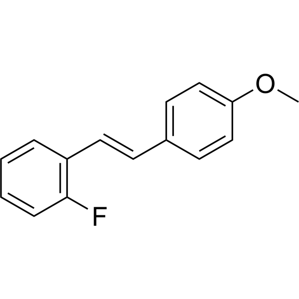
CAY10512 is a NF-κB inhibitor. CAY10512 can suppress the upregulation of NF-κB-sensitive proinflammatory miRNAs (miRNA-9, miRNA-125b, miRNA-146a, miRNA-155) in cerebrospinal fluid and extracellular fluid. CAY10512 significantly reduces the release of pro-inflammatory cytokines (such as TNF-α, MCP-1, IL-8, IL-6). CAY10512 can be used for researchs on neuroinflammation, islet transplantation and microRNA regulation.
For research use only. We do not sell to patients.
- Purity : 97.0%
- CAS No.: 139141-12-1
- Formula: C15H13FO
- Molecular Weight:228.26
-
Storage:Powder -20°C, 3 years ; In solvent -80°C, 6 months , -20°C, 1 month

To place orders, for customer services and technical support, please contact: MedChemExpress USA
Tel: 609-228-6898 E-mail: [email protected] [email protected]


-
 Biological Activity
-
Chemical Information
- Protocols
- Purity & Documentation
- References
-
 Help & FAQs


-
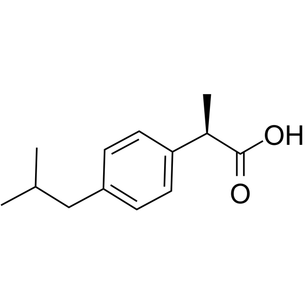
HY-78131B
(R)-(-)-IbuprofenNF-κB Inhibitor -
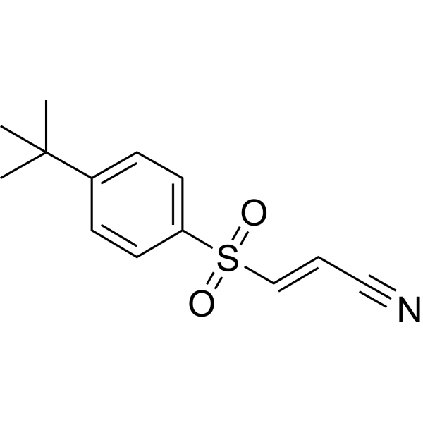
HY-10257
BAY 11-7085NF-κB Activation Inhibitor -
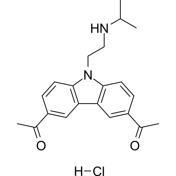
HY-18935A
CBL0137 hydrochlorideNF-κB Inhibitor -
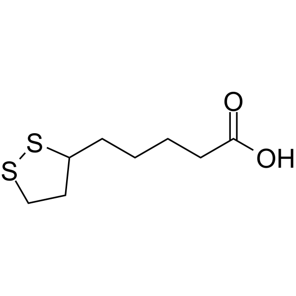
HY-N0492
α-Lipoic AcidAntioxidant -
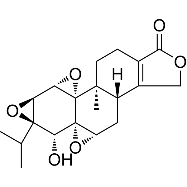
HY-32735
TriptolideImmunosuppressive Agent -
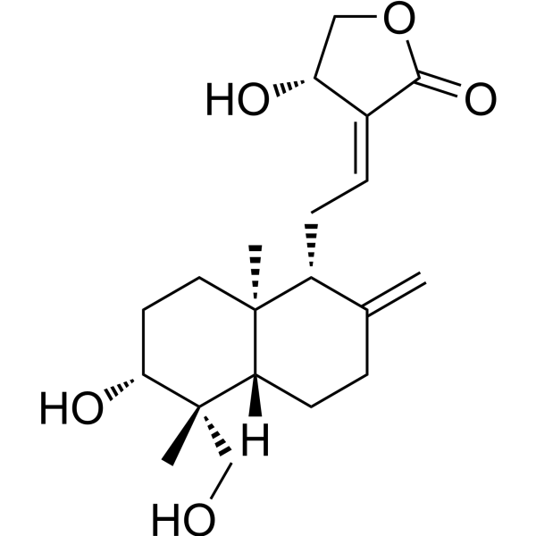
HY-N0191
AndrographolideNF-κB Inhibitor -
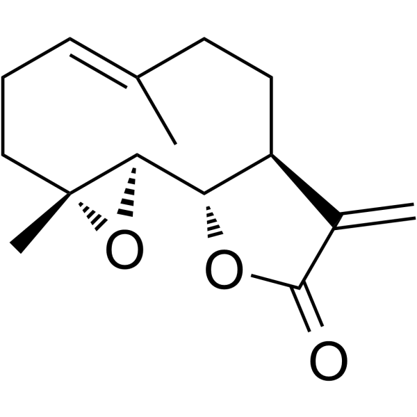
HY-N0141
ParthenolideNF-κB Inhibitor -
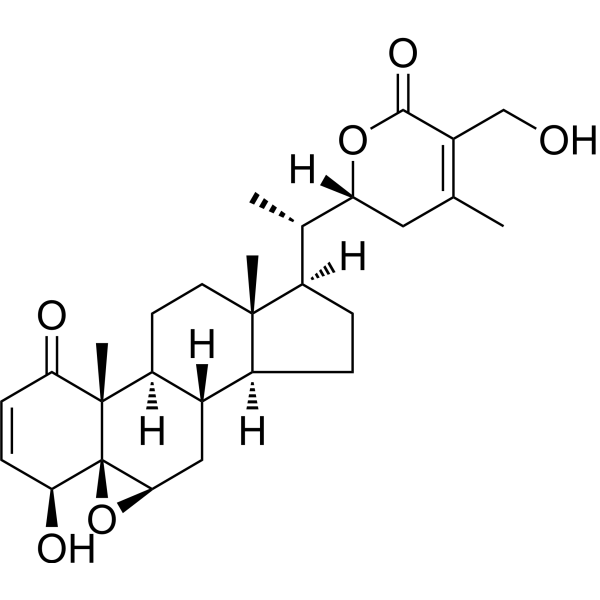
HY-N2065
Withaferin ANF-κB Inhibitor -
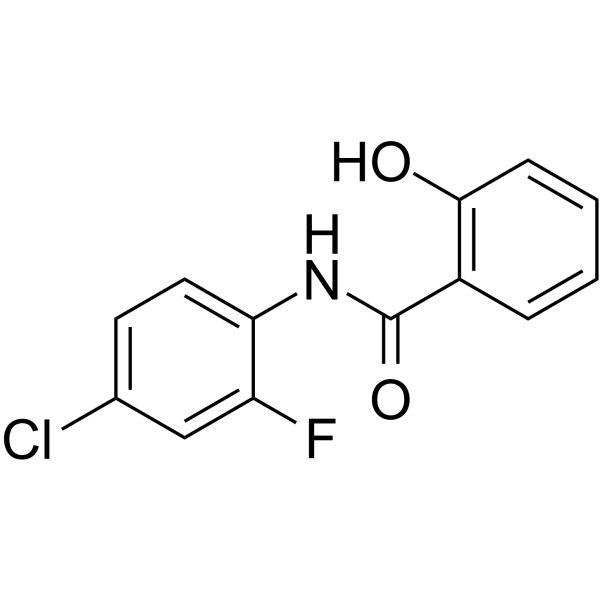
HY-124958
NDMC101Osteoclastogenesis Inhibitor -
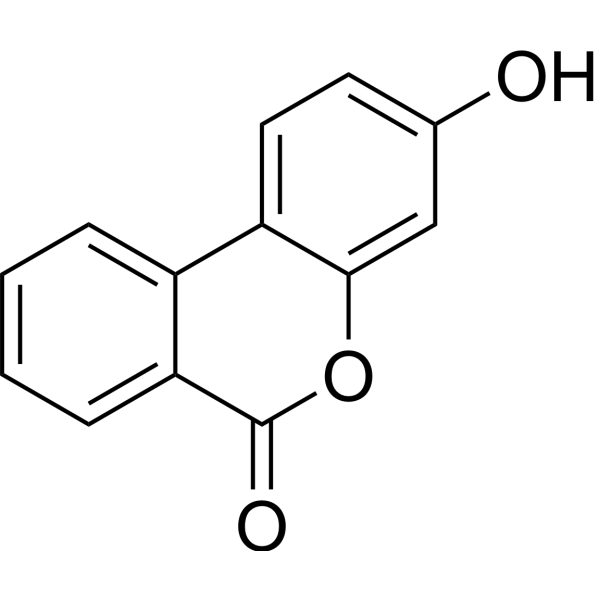
HY-126307
Urolithin BNF-κB Inhibitor -
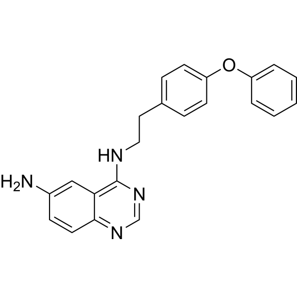
HY-13812
QNZTNF Receptor Inhibitor -

HY-78131BR
(R)-(-)-Ibuprofen (Standard)NF-κB Inhibitor


Biological Activity
Description
|
NF-κB |
IL-6 |
IL-8 |
Cellular Effect
|
Cell Line

|
Type | Value | Description | References |
|---|---|---|---|---|
| HEK-293T | IC50 |
0.15 μM
Compound: 6p
|
Inhibition of TNF-alpha-induced NF-kappaB activation in HEK293T cells
Inhibition of TNF-alpha-induced NF-kappaB activation in HEK293T cells
|
[PMID: 17125270] |
| HepG2 | EC50 |
5.4 μM
Compound: 1; LD55
|
Activation of Nrf2 (unknown origin) expressed in human HepG2 cells after 5 hrs by ARE-driven luciferase reporter gene assay
Activation of Nrf2 (unknown origin) expressed in human HepG2 cells after 5 hrs by ARE-driven luciferase reporter gene assay
|
[PMID: 28126440] |
In Vitro
CAY10512 (5 μM, 0-96 h) effectively inhibits miRNA-9, miRNA-125b, miRNA146a and miRNA-155 upregulation in HNG cells stimulated by AD-ECF[1].
CAY10512 (0.15 μM, 1-7 h) significantly protects pancreatic islet viability and effectively alleviates pancreatic islet cell damage by inhibiting the increase of pro-inflammatory factors and the expression of tissue factor TF in primary human islets[2].
CAY10512 (0.25 μM, 25 h) significantly inhibits the gene and protein expressions of pro-inflammatory factor IL-6 and inflammatory receptor TNFR2 induced by MR agonists (Aldosterone (HY-113313), 11-dehydrocorticosterone (HY-113447)) in BV-2 cells[3].
MedChemExpress (MCE) has not independently confirmed the accuracy of these methods. They are for reference only. Further protocols information, click here.
-
Cell Line:Primary human islets
-
Concentration:0.15 μM
-
Incubation Time:Pre-treat for one hour, then mix with autologous blood and incubate for another 1, 3, 6 h
-
Result:Prevented the increase of TNF-α, MCP-1, IL-8 and IL-6.
-
Cell Line:BV-2 cells
-
Concentration:0.25 μM
-
Incubation Time:Pre-treatment for 1 hour, then add the stimulus and incubate for 24 hours
-
Result:Completely blocked the upregulation of IL-6 mRNA induced by Aldosterone and the upregulation of TNFR2 mRNA induced by 11-dehydrocorticosterone.
-
Cell Line:BV-2 cells
-
Concentration:0.25 μM
-
Incubation Time:Pre-treatment for 1 hour, then add the stimulus and incubate for 24 hours
-
Result:Completely blocked the phosphorylation of ILp65 induced by aldosterone and its translocation to the nucleus, as well as the upregulation of TNFR2 protein levels induced by 11-dehydrocorticosterone.


Chemical Information
-
CAS No. 139141-12-1
-
Appearance Solid
-
Molecular Weight 228.26
-
Formula C15H13FO
-
Color White to off-white
-
SMILES
COC(C=C1)=CC=C1/C=C/C2=C(C=CC=C2)F
-
Shipping
Room temperature in continental US; may vary elsewhere.
-
Storage
Powder -20°C 3 years In solvent -80°C 6 months -20°C 1 month

Protocols
-
LPS-Induced Endotoxemia/Systemic Inflammation
Lipopolysaccharide (LPS)-induced endotoxemia is a widely used in vivo model of acute systemic inflammation in which LPS, a Gram-negative bacterial endotoxin, activates innate immune signaling primarily through TLR4, leading to rapid and transient induction of pro-inflammatory cytokines such as TNF-α, IL-6, and IL-1β in circulation and tissues. This cytokine surge is commonly used as a measurable readout of systemic inflammatory activation and immune dysregulation, and is typically assessed within hours after intraperitoneal LPS administration in mouse models of endotoxemia. The model captures key features of systemic inflammatory response syndrome, including cytokine release, immune cell activation, and downstream tissue responses, and has been used to evaluate anti-inflammatory interventions such as cytokine modulation, lipid mediators, and immune cell-targeting therapies.
-
Research Protocol for Inflammation-related Diseases
The NLRP3 inflammasome is a cytosolic innate immune signaling platform that integrates priming signals and danger-signal activation to promote caspase-1 activation, maturation of IL-1β and IL-18, and gasdermin D-mediated pyroptotic cell death. The core experimental logic is to determine whether inflammatory disease phenotypes are driven by increased NLRP3 expression, ASC-containing inflammasome assembly, caspase-1 cleavage, GSDMD cleavage, and extracellular release of IL-1β/IL-18 rather than by nonspecific cell injury alone. The pathway is strongly linked to inflammation-related disease phenotypes because monosodium urate crystals activate NALP3/NLRP3 inflammasome signaling in gout-like crystal inflammation, cholesterol crystals activate NLRP3 inflammasomes in atherogenesis models, and DSS-induced intestinal inflammation has been reported to involve NLRP3 inflammasome activity. However, experimental colitis studies also show context-dependent protective effects of NLRP3 inflammasome co
-
Research Protocol for Neurological Diseases
PINK1/Parkin-mediated mitophagy pathway is a mitochondrial quality-control signaling axis in which mitochondrial depolarization stabilizes PINK1 on damaged mitochondria, activates Parkin recruitment and E3 ubiquitin ligase activity, promotes ubiquitination of outer mitochondrial membrane proteins, recruits selective autophagy adaptors, and drives lysosomal degradation of damaged mitochondria. In neurological disease research, this pathway is experimentally important because neurons, especially dopaminergic neurons, are highly dependent on mitochondrial integrity, and defective mitochondrial turnover can lead to mitochondrial dysfunction, oxidative stress, impaired neuronal survival, α-synuclein accumulation, and neuroinflammatory damage-associated signals. The genetic disease link is strongest in Parkinson’s disease because mutations in PRKN/parkin cause autosomal recessive juvenile parkinsonism, mutations in PINK1 cause hereditary early-onset Parkinson’s disease, and Drosophila studie


Purity & Documentation
-
Data Sheet (273 KB)
-
SDS (393 KB)
- English - EN (393 KB)
- Français - FR (393 KB)
- Deutsch - DE (393 KB)
- Norwegian - NO (393 KB)
- Español - ES (393 KB)
- Swedish - SV (393 KB)
- Italian - IT (393 KB)
- Korean - KR (393 KB)
- Portuguese - PT (393 KB)
-
Handling Instructions (2659 KB)
References
[1]. Alexandrov P N, et al. microRNA (miRNA) speciation in Alzheimer’s disease (AD) cerebrospinal fluid (CSF) and extracellular fluid (ECF)[J]. International journal of biochemistry and molecular biology, 2012, 3(4): 365. [Content Brief]
[2]. Kanak MA, et al. Alleviation of instant blood-mediated inflammatory reaction in autologous conditions through treatment of human islets with NF-κB inhibitors. Transplantation. 2014 Sep 15;98(5):578-84. [Content Brief]
[3]. Chantong B, et al. Mineralocorticoid and glucocorticoid receptors differentially regulate NF-kappaB activity and pro-inflammatory cytokine production in murine BV-2 microglial cells. J Neuroinflammation. 2012 Nov 28;9:260. [Content Brief]

Calculators
Concentration (start) × Volume (start) = Concentration (final) × Volume (final)


CAY10512 Related Classifications
HY-120269 Related Classifications
 Powered by Bioz