



Hm1a
Hm1a is a venom peptide and a selective hNaV1.1 activator with an EC50 of 7.5 nM. Hm1a enhances hNaV1.1 and hNaV1.3 channel currents via delayed inactivation. Hm1a restores action potential firing in Dravet syndrome GABAergic inhibitory interneurons, reduces interictal epileptiform discharges and whole-brain hyperexcitability, lowers seizure frequency, and rescues premature death in Dravet syndrome mice. Hm1a can be used for the research of neurological disease, such as Dravet syndrome.
연구목적의 판매만을 진행합니다. 환자를 대상으로 한 판매는 하지 않습니다.
- 화학식: C170H239N47O54S6
- 분자량:3997.39
-
보관:
Please store the product under the recommended conditions in the Certificate of Analysis.

To place orders, for customer services and technical support, please contact: MedChemExpress USA
Tel: 609-228-6898 E-mail: [email protected] [email protected]


-
 Biological Activity
-
Chemical Information
- Protocol
- 순도&문서
- References
-
 Help & FAQs




Biological Activity
제품 설명
IC50 & Target
[1]|
hNav1.1 7.5 nM (EC50) |
hNav1.3 39.5 nM (EC50) |
In Vitro
Hm1a potently and selectively modulates hNaV1.1 and hNaV1.3 channels with efficacy EC50 values of 7.5 and 39.5 nM in HEK293T cells[1].
Hm1a (50 nM; 2 min) has no functional effect on hNaV1.2, hNaV1.4, hNaV1.5, hNaV1.6, hNaV1.7, or hNaV1.8 channels in HEK293T cells[1].
Hm1a (300 nM) has no functional effect on Kv1.7, Kv10.1, Kv11.1, KCa1.1, KCa2.2, or KCa3.1 channels[1].
MedChemExpress (MCE) has not independently confirmed the accuracy of these methods. They are for reference only. Further protocols information, click here.
In Vivo
Hm1a (0.5 μM; continuous ICV infusion; 0.2 μL/min; up to 5 days) reduces seizure frequency to near zero over 4 days and maintains 90% survival at day 3 in Dravet syndrome mice[1].
MedChemExpress (MCE) has not independently confirmed the accuracy of these methods. They are for reference only.
-
Animal Model:Scn1a (R1407X) Dravet syndrome mice (N2 backcross generation on C57BL/6J background; aged postnatal day 18-26)[1]
-
Dosage:0.5 μM
-
Administration:ICV infusion; 0.1-0.2 μL/min; 1 h
-
Result:Reduced mean interictal spike frequency from 27.6 spikes/h to 11.7 spikes/h.
Achieved a significant reduction in peak 0.5-2 Hz activity via power spectrum analysis.
Induced a spike-free period of ~1 hour following infusion cessation.
-
Animal Model:Scn1a (R1407X) Dravet syndrome mice (N2 backcross generation on C57BL/6J background; aged postnatal day 18-26)[1]
-
Dosage:0.5 μM
-
Administration:continuous ICV infusion; 0.2 μL/min; up to 5 days
-
Result:Reduced mean seizure count from 3.50 seizures/day at day 0 to near 0 seizures/day by day 4.
Led 67% of treated mice to have significantly reduced or completely abolished seizures after 3 days.
Maintained 90% survival of treated mice to day 3, compared to 0% survival of vehicle-treated controls.


Chemical Information
-
분자량 3997.39
-
화학식 C170H239N47O54S6
-
Sequence
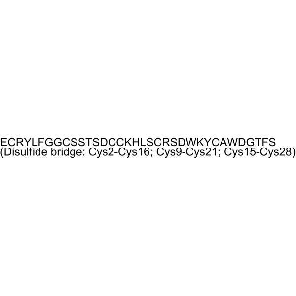
Glu-Cys-Arg-Tyr-Leu-Phe-Gly-Gly-Cys-Ser-Ser-Thr-Ser-Asp-Cys-Cys-Lys-His-Leu-Ser-Cys-Arg-Ser-Asp-Trp-Lys-Tyr-Cys-Ala-Trp-Asp-Gly-Thr-Phe-Ser (Disulfide bridge: Cys2-Cys16; Cys9-Cys21; Cys15-Cys28)
-
Sequence Shortening
ECRYLFGGCSSTSDCCKHLSCRSDWKYCAWDGTFS (Disulfide bridge: Cys2-Cys16; Cys9-Cys21; Cys15-Cys28)
-
선적
Room temperature in continental US; may vary elsewhere.
-
보관
Please store the product under the recommended conditions in the Certificate of Analysis.

Protocol
-
Cardiac voltage-sensitive optical mapping
Cardiac voltage-sensitive optical mapping records changes in transmembrane potential from cardiac tissue by staining the preparation with a voltage-sensitive dye and imaging fluorescence changes during electrical activation; the resulting optical action potentials can be used to map activation time, action potential duration, conduction velocity, wavefront propagation, and arrhythmia dynamics. The optical signal represents a relative fluorescence change from a tissue volume rather than a single-cell intracellular recording, so spatial resolution, sampling rate, voltage resolution, optical magnification, light penetration, and motion control must be considered together when interpreting optical action potentials.
-
Neuronal voltage-sensitive dye imaging
Neuronal voltage-sensitive dye imaging detects membrane-potential-dependent optical changes from dyes associated with neuronal membranes, enabling optical recording of electrical activity from single neurons, dendrites, axons, spines, or neuronal populations in brain slices and cultured neurons. VSD signals are typically reported as fractional fluorescence or absorbance changes over baseline, such as ΔF/F or ΔI/I, and published protocols use high-speed cameras or photodiode arrays because neuronal voltage signals occur on millisecond time scales. Fast VSD imaging can be applied at two common scales: bulk staining of brain slices to measure circuit-level spatiotemporal activity, and single-cell loading or biolistic delivery to record membrane-potential transients from individual neuronal compartments. Optical signals should be interpreted as membrane-potential-related readouts, and validation by simultaneous electrophysiology or pharmacological controls is recommended when the experimen
-
Acute brain-slice whole-cell patch-clamp recording
Acute brain-slice whole-cell patch-clamp recording measures membrane voltage or ionic current from visually targeted cells in living brain slices; after giga-seal formation, the membrane under the pipette is ruptured to provide low-resistance electrical access to the cell interior, enabling current-clamp analysis of excitability and voltage-clamp analysis of synaptic or membrane currents. Acute slices preserve local tissue architecture better than dissociated preparations and allow visually guided recording from defined brain regions or fluorescently labeled cells; however, whole-cell access also permits exchange between pipette solution and cytoplasm, so intracellular dialysis must be considered when interpreting signaling-dependent phenomena.
-
Cell-attached patch-clamp recording
Cell-attached patch-clamp recording measures ionic current through one or more ion channels in a small membrane patch that remains attached to an intact cell; the readout is a time-resolved current trace generated when channels in the sealed patch open and close under controlled pipette voltage or stimulus conditions. Classic applications include single acetylcholine receptor currents in frog skeletal muscle, single sodium-channel currents in cultured rat muscle cells, one-channel NMDA receptor recordings, and mechanically activated PIEZO-channel recordings. The method depends on forming a high-resistance pipette-membrane seal, commonly described as a gigaohm seal, which reduces leak and noise sufficiently to resolve picoampere-scale single-channel currents. In the cell-attached configuration, the patch membrane is not ruptured, so cytosolic composition is not directly dialyzed by the pipette solution.
-
Research Protocol for Neurological Diseases
PINK1/Parkin-mediated mitophagy pathway is a mitochondrial quality-control signaling axis in which mitochondrial depolarization stabilizes PINK1 on damaged mitochondria, activates Parkin recruitment and E3 ubiquitin ligase activity, promotes ubiquitination of outer mitochondrial membrane proteins, recruits selective autophagy adaptors, and drives lysosomal degradation of damaged mitochondria. In neurological disease research, this pathway is experimentally important because neurons, especially dopaminergic neurons, are highly dependent on mitochondrial integrity, and defective mitochondrial turnover can lead to mitochondrial dysfunction, oxidative stress, impaired neuronal survival, α-synuclein accumulation, and neuroinflammatory damage-associated signals. The genetic disease link is strongest in Parkinson’s disease because mutations in PRKN/parkin cause autosomal recessive juvenile parkinsonism, mutations in PINK1 cause hereditary early-onset Parkinson’s disease, and Drosophila studie


순도&문서
References

Calculators
Concentration (start) × Volume (start) = Concentration (final) × Volume (final)


Hm1a Related Classifications
HY-P5183 Related Classifications
 Powered by Bioz