



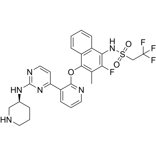
PAIR2

Based on 1 Customer Validation
PAIR2 is a highly selective inhibitor targeting the kinase domain of human IRE1α, with a Ki value of 8.8 nM against human IRE1α. PAIR2 fully occupies the ATP-binding site of the IRE1α kinase domain, partially antagonizes the ribonuclease activity of IRE1α, specifically inhibits regulated IRE1α-dependent decay (RIDD) and its mediated substrate cleavage, while preserving the splicing function of Xbp1 mRNA. PAIR2 also promotes the differentiation of B cells into plasma cells, blocks IRE1α-induced cell apoptosis, and restores the expression of Fgfr2 mRNA in AT2 cells. PAIR2 effectively reaches a steady-state concentration in the lung tissues of Mus musculus, and serves as an important tool for investigating the function of the IRE1α signaling pathway in diseases such as pulmonary fibrosis.
연구목적의 판매만을 진행합니다. 환자를 대상으로 한 판매는 하지 않습니다.
- Purity : 99.56%
- CAS No.: 2771006-54-1
- 화학식: C27H26F4N6O3S
- 분자량:590.59
-
보관:Powder -20°C, 3 years , 4°C, 2 years ; In solvent -80°C, 6 months , -20°C, 1 month

To place orders, for customer services and technical support, please contact: MedChemExpress USA
Tel: 609-228-6898 E-mail: [email protected] [email protected]


-
 Biological Activity
-
Chemical Information
-
용액&용해도
- Protocol
- 순도&문서
- References
-
 Help & FAQs




Biological Activity
제품 설명
IC50 & Target
|
IRE1α 8.8 nM (Ki) |
FGFR2 |
In Vitro
PAIR2 (1-10 μM; 1 h + 6 h doxycycline (HY-N0565)) dose-dependently blocks doxycycline-induced autophosphorylation of IRE1α in INS-1::PCMV/2xTetO cells[1].
PAIR2 (0.5-10 μM; 1 h+2 h DTT (HY-15917)) maintains XBP1 mRNA splicing in parental INS-1 cells subjected to DTT stress[1].
PAIR2 (2 μM; 24 h Thapsigargin (HY-13433)) rescues Fgfr2 mRNA expression in serum-free, feeder-free primary mouse AT2 organoids under Thapsigargin stress, while preserving IRE1α-mediated Xbp1 splicing[2].
PAIR2 (2 μM; 3-d) fails to prevent the conversion of primary serum-free, feeder-free mouse AT2 cells to DATCs when pemigatinib directly blocks the Fgfr signaling pathway[2].
MedChemExpress (MCE) has not independently confirmed the accuracy of these methods. They are for reference only.
-
Cell Line:INS-1::PCMV/2xTetO rat insulinoma cells (doxycycline-inducible IRE1α overexpression)
-
Concentration:1, 3, 10 μM
-
Incubation Time:1 h (PAIR2 pre-incubation); 6 h (doxycycline treatment)
-
Result:Dose-dependently blocked doxycycline-induced IRE1α autophosphorylation, reducing phosphorylated IRE1α levels to near baseline at concentrations ≥3 μM, with equivalent potency to KIRA8.
-
Cell Line:parent INS-1 rat insulinoma cells
-
Concentration:0.5, 1, 3, 10 μM
-
Incubation Time:1 h (PAIR2 pre-incubation); 2 h (DTT treatment)
-
Result:Preserved DTT-induced XBP1 mRNA splicing at all tested concentrations, with no significant reduction in splicing percentage relative to DTT-only treated cells (all P values > 0.05, non-significant).
In Vivo
MedChemExpress (MCE) has not independently confirmed the accuracy of these methods. They are for reference only.
-
Animal Model:C57BL/6 (10-20 week-old, both male and female, bleomycin-induced pulmonary fibrosis)[2]
-
Dosage:30 mg/kg
-
Administration:i.p.; twice daily; starting 1 day before bleomycin exposure and continuing through study end
-
Result:Reduced fraction of trace+Krt8+ / trace+ cells compared to vehicle control at day 10 post-bleomycin exposure.
Significantly lowered lung hydroxyproline content and reduced fibrillar collagen deposition via picrosirius red staining at day 14 post-bleomycin exposure.
Derepressed expression of canonical RIDD target Bloc1s1 in lung tissue.
Did not significantly affect Xbp1 splicing.
- Fibrosis/Collagen Morphometry
- RNA extraction experimental
- Collagen: Sirius Red Staining
- Detection of Apoptosis
- TUNEL staining for apoptotic DNA fragmentation
- Annexin V plus membrane-impermeant dye apoptosis staining
- Connective Tissue: Masson's Trichrome/Collagen Trichrome Staining
- Apoptosis Solutions


Chemical Information
-
CAS No. 2771006-54-1
-
Appearance Solid
-
분자량 590.59
-
화학식 C27H26F4N6O3S
-
Color White to off-white
-
SMILES
CC(C(F)=C(NS(CC(F)(F)F)(=O)=O)C1=C2C=CC=C1)=C2OC3=NC=CC=C3C4=NC(N[C@H]5CCCNC5)=NC=C4
-
선적
Room temperature in continental US; may vary elsewhere.
-
보관
Powder -20°C 3 years 4°C 2 years In solvent -80°C 6 months -20°C 1 month


용액&용해도
In Vitro:
DMSO : 100 mg/mL (169.32 mM; Need ultrasonic; Hygroscopic DMSO has a significant impact on the solubility of product, please use newly opened DMSO)
Please refer to the solubility information to select the appropriate solvent. Once prepared, please aliquot and store the solution to prevent product inactivation from repeated freeze-thaw cycles.
Storage method and period of stock solution: -80°C, 6 months; -20°C, 1 month. When stored at -80°C, please use it within 6 months. When stored at -20°C, please use it within 1 month.
Please refer to the solubility information to select the appropriate solvent. Once prepared, please aliquot and store the solution to prevent product inactivation from repeated freeze-thaw cycles.
Storage method and period of stock solution: -80°C, 6 months; -20°C, 1 month. When stored at -80°C, please use it within 6 months. When stored at -20°C, please use it within 1 month.
Concentration (start) × Volume (start) = Concentration (final) × Volume (final)
In Vivo:
Select the appropriate dissolution method based on your experimental animal and administration route.
- For the following dissolution methods, please ensure to first prepare a clear stock solution using an In Vitro approach and then sequentially add co-solvents:
- To ensure reliable experimental results, the clarified stock solution can be appropriately stored based on storage conditions. As for the working solution for In Vivo experiments, it is recommended to prepare freshly and use it on the same day.
- The percentages shown for the solvents indicate their volumetric ratio in the final prepared solution. If precipitation or phase separation occurs during preparation, heat and/or sonication can be used to aid dissolution.
Add each solvent one by one: 10% DMSO 40% PEG300 5% Tween-80 45% Saline
Solubility: ≥ 2.5 mg/mL (4.23 mM); Clear solution
This protocol yields a clear solution of ≥ 2.5 mg/mL (saturation unknown).
Taking 1 mL working solution as an example, add 100 μL DMSO stock solution (25.0 mg/mL) to 400 μL PEG300, and mix evenly; then add 50 μL Tween-80 and mix evenly; then add 450 μL Saline to adjust the volume to 1 mL.
Preparation of Saline: Dissolve 0.9 g sodium chloride in ddH₂O and dilute to 100 mL to obtain a clear Saline solution.
In Vivo Dissolution Calculator
Please enter the basic information of animal experiments:
-
-
-
-
Recommended: Prepare an additional quantity of animals to account for potential losses during experiments.
Please enter your animal formula composition:
-
%DMSO +
Recommended: Keep the proportion of DMSO in working solution below 2% if your animal is weak.
-
%+
-
+%Tween-80 + +
-
%Saline +
The co-solvents required include: DMSO, . All of co-solvents are available by MedChemExpress (MCE). , Tween 80. All of co-solvents are available by MedChemExpress (MCE).
Working solution concentration: 0.22 mg/mL
Method for preparing stock solution: mg drug dissolved in μL DMSO. Stock solution concentration: mg/mL.
1. Take μL DMSO stock solution;
2. Add μL .
μL , mix evenly;
3. Then add μL Tween 80, mix evenly;
4. Then add μL
Please ensure that the stock solution in the first step is dissolved to a clear state, and add co-solvents in sequence. You can use ultrasonic heating (ultrasonic cleaner, recommended frequency 20-40 kHz), vortexing, etc. to assist dissolution.


순도&문서
-
Data Sheet (279 KB)
-
SDS (252 KB)
- English - EN (252 KB)
- Français - FR (252 KB)
- Deutsch - DE (252 KB)
- Norwegian - NO (252 KB)
- Español - ES (252 KB)
- Swedish - SV (252 KB)
- Italian - IT (252 KB)
- Korean - KR (252 KB)
- Portuguese - PT (252 KB)
-
Handling Instructions (2659 KB)
References
[1]. Feldman HC, et al. ATP-competitive partial antagonists of the IRE1α RNase segregate outputs of the UPR. Nat Chem Biol. 2021;17(11):1148-1156. [Content Brief]
[2]. Auyeung VC, et al. Pharmacologic inhibition of IRE1α-dependent decay protects alveolar epithelial identity and prevents pulmonary fibrosis in mice. J Clin Invest. 2025;135(20):e184522. Published 2025 Oct 15. [Content Brief]


Complete Stock Solution Preparation Table
Please refer to the solubility information to select the appropriate solvent. Once prepared, please aliquot and store the solution to prevent product inactivation from repeated freeze-thaw cycles.
Storage method and period of stock solution: -80°C, 6 months; -20°C, 1 month. When stored at -80°C, please use it within 6 months. When stored at -20°C, please use it within 1 month.
| Optional Solvent | Concentration Solvent Mass | 1 mg | 5 mg | 10 mg | 25 mg |
|---|---|---|---|---|---|
| DMSO | 1 mM | 1.6932 mL | 8.4661 mL | 16.9322 mL | 42.3305 mL |
| 5 mM | 0.3386 mL | 1.6932 mL | 3.3864 mL | 8.4661 mL | |
| 10 mM | 0.1693 mL | 0.8466 mL | 1.6932 mL | 4.2331 mL | |
| 15 mM | 0.1129 mL | 0.5644 mL | 1.1288 mL | 2.8220 mL | |
| 20 mM | 0.0847 mL | 0.4233 mL | 0.8466 mL | 2.1165 mL | |
| 25 mM | 0.0677 mL | 0.3386 mL | 0.6773 mL | 1.6932 mL | |
| 30 mM | 0.0564 mL | 0.2822 mL | 0.5644 mL | 1.4110 mL | |
| 40 mM | 0.0423 mL | 0.2117 mL | 0.4233 mL | 1.0583 mL | |
| 50 mM | 0.0339 mL | 0.1693 mL | 0.3386 mL | 0.8466 mL | |
| 60 mM | 0.0282 mL | 0.1411 mL | 0.2822 mL | 0.7055 mL | |
| 80 mM | 0.0212 mL | 0.1058 mL | 0.2117 mL | 0.5291 mL | |
| 100 mM | 0.0169 mL | 0.0847 mL | 0.1693 mL | 0.4233 mL |


PAIR2 Related Classifications
HY-145425 Related Classifications
 Powered by Bioz