



Leukotriene B5
Leukotriene B5 (LTB5) is a leukotriene with diverse biological activities. It is a metabolite of eicosapentaenoic acid formed through the 5-lipoxygenase (5-LO) pathway. LTB5 increases contraction of bullfrog lung strips ex vivo in a concentration-dependent manner. In vivo, LTB5 (100 nM) reduces tumor volume in mice injected with Tm1 murine melanoma cells. LTB5 also elicits chemokinesis and lysosomal enzyme release from polymorphonuclear leukocytes (PMNLs) 20- to 30-fold less, and induces platelet aggregation 8-fold less, potently than LTB4.
For research use only. We do not sell to patients.
- CAS No.: 80445-66-5
- Formula: C20H30O4
- Molecular Weight:334.45
-
Storage:
Please store the product under the recommended conditions in the Certificate of Analysis.


To place orders, for customer services and technical support, please contact: MedChemExpress USA
Tel: 609-228-6898 E-mail: [email protected] [email protected]


-
 Biological Activity
-
Chemical Information
- Protocols
- Purity & Documentation
- References
-
 Help & FAQs


-
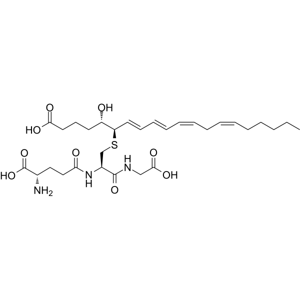
HY-113446
Leukotriene C4Endogenous Metabolite -
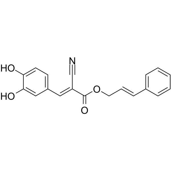
HY-138688
Cinnamyl-3,4-dihydroxy-α-cyanocinnamate12/15-Lipoxygenases Inhibitor -
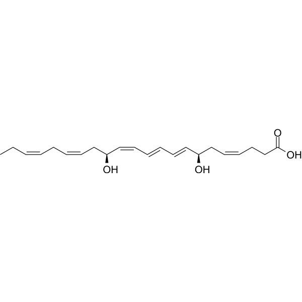
HY-116429
Maresin 1Ca2+ Stimulator -
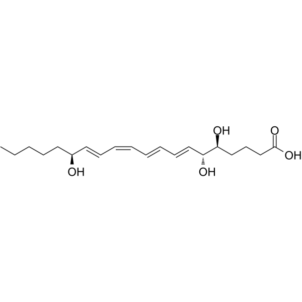
HY-113509
Lipoxin A4Anti-inflammatory Agent -
HY-126716
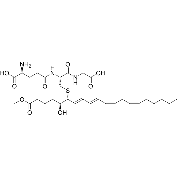
Leukotriene C4 methyl esterLeukotriene -
HY-130413
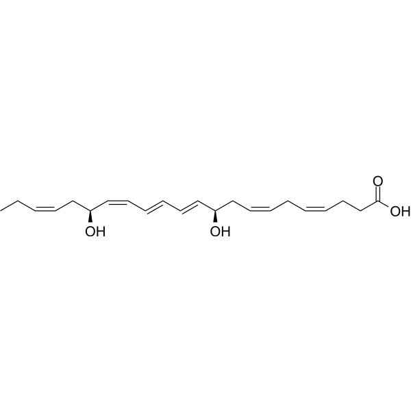
Protectin D1Neuroprotectin D1 -
HY-128171
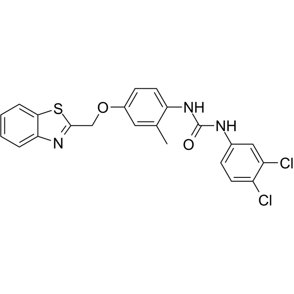
DiflapolinFLAP/sEH Inhibitor -
HY-N11630
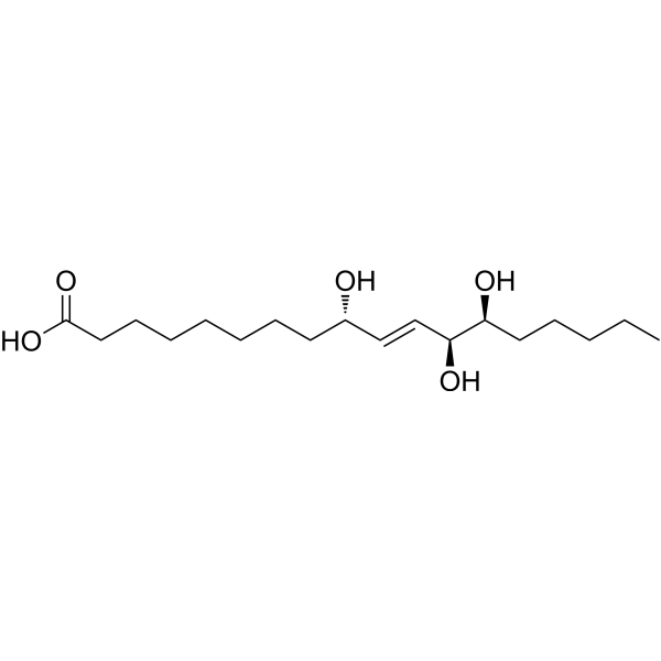
Pinellic acidAdjuvant for Nasal Influenza Vaccine -
HY-113456
Leukotriene D4Bronchoconstrictor -
HY-119684
Maresin 2Anti-inflammatory And Pro-resolving Mediator -
HY-116124
17(S)-HpDHAHydrolyzate -

HY-116763
COX-2/5-LOX-IN-4COX-2/LOX Inhibitor
All Leukotriene Receptor Isoforms
More

Biological Activity
Description


Chemical Information
-
CAS No. 80445-66-5
-
Molecular Weight 334.45
-
Formula C20H30O4
-
SMILES
CC/C=C\C/C=C\C[C@@H](O)/C=C/C=C/C=C\[C@@H](O)CCCC(O)=O
-
Synonyms
LTB5
-
Shipping
Room temperature in continental US; may vary elsewhere.
-
Storage
Please store the product under the recommended conditions in the Certificate of Analysis.

Protocols
-
Lysosome and acidic-vesicle live-cell staining
Lysosome and acidic-vesicle live-cell staining detects acidic intracellular compartments by using membrane-permeant acidotropic probes that accumulate in low-pH vesicles, including lysosomes, late endosomes, autolysosomes, and acidic phagosomes. LysoTracker staining is commonly used as an intensity-based readout of acidic lysosomal compartment abundance or enlargement, while acridine orange produces green fluorescence in less concentrated compartments and red fluorescence after concentration-dependent accumulation in acidic vesicular organelles. Loss or reduction of acridine-orange red signal can be used as a readout of lysosomal membrane permeabilization or reduced acidic-vesicle integrity. This protocol is designed for live cultured cells and can be adapted for fluorescence microscopy, high-content imaging, plate-reader readout, or flow cytometry when the selected literature supports the readout. Because these dyes report acidotropic accumulation rather than lysosome identity alone,


Purity & Documentation
References

Calculators
Concentration (start) × Volume (start) = Concentration (final) × Volume (final)


Leukotriene B5 Related Classifications
HY-113484 Related Classifications
 Powered by Bioz