



Ligelizumab
Based on 1 publication(s) in Google Scholar
Ligelizumab (QGE 031) is a humanized high-affinity anti-immunoglobulin IgE monoclonal antibody. Ligelizumab selectively inhibits the binding of IgE to the high-affinity receptor FcεRI, while the inhibitory effect on the low-affinity receptor CD23 is weak. Ligelizumab can inhibit the activation of effector cells such as mast cells and Basophil, while reducing the production of IgE by B cells, and restoring the IFN-α production and regulatory T cell (Treg) induction function of plasmacytoid dendritic cells (pDC). Ligelizumab can be used in the study of allergic diseases (such as chronic spontaneous urticaria, allergic asthma).
For research use only. We do not sell to patients.
- Purity : 99.85%
- CAS No.: 1322627-61-1
- Molecular Weight:146.32 kDa
-
Storage:
Please store the product under the recommended conditions in the Certificate of Analysis.




To place orders, for customer services and technical support, please contact: MedChemExpress USA
Tel: 609-228-6898 E-mail: [email protected] [email protected]


-
 Biological Activity
-
Chemical Information
- Protocols
- Purity & Documentation
- References
-
 Help & FAQs




Publications Citing Use of MedChemExpress (MCE) Ligelizumab
More

Biological Activity
Description
Isotype
Human IgG1 kappa
Recommend Isotype Controls
Species Reactivity
Human
IC50 & Target
IGHE
In Vitro
Ligelizumab is more effective than Omalizumab (HY-P9950) in blocking the binding of free IgE to FcεRI on plasmacytoid dendritic cells (pDCs)[2].
In vitro, Ligelizumab can restore the ability of pDCs to produce IFN-α stimulated by TLR9-L, and the effect is better than Omalizumab[2].
In in vitro co-culture experiments, Ligelizumab can restore the ability of pDCs to induce the generation of regulatory T cells (Tregs) like Omalizumab[2].
MedChemExpress (MCE) has not independently confirmed the accuracy of these methods. They are for reference only. Further protocols information, click here.
In Vivo
MedChemExpress (MCE) has not independently confirmed the accuracy of these methods. They are for reference only.
-
Animal Model:Human FcεRIα transgenic (huFcεRIα tg) mice on a mixed C57BL/6J-C57BL/6N background[1]
-
Dosage:10 μg of Ligelizumab
-
Administration:Intraperitoneal injection once, administered on day 0 before sensitization with IgE, and antigen challenge on day 1
-
Result:Completely protected the mice from antigen-induced systemic anaphylaxis, as evidenced by no significant decrease in core body temperature after challenge.
Significantly lowered the levels of IgE on peritoneal mast cells compared to omalizumab-treated mice, which only showed partial protection.
Clinical Trial
| NCT Number | Sponsor | Condition | Start Date |
Phase

|
|---|---|---|---|---|
| NCT01329991 | Plexxikon| | 2011-05 | PHASE1 |
Accession
Conjugated
Unconjugated
Reconsititution
The product can be reconstituted/diluted with sterile PBS or saline.
Format
-
Human IgG1 kappa
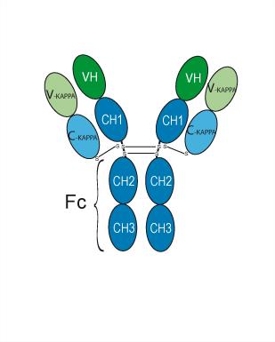
Application
ELISA, FACS, Functional assay
Verified Bioactivity


Chemical Information
-
CAS No. 1322627-61-1
-
Appearance Liquid
-
Molecular Weight 146.32 kDa
-
Color Colorless to light yellow
-
SMILES
[Ligelizumab]
-
Synonyms
QGE 031; Anti-IGHE Recombinant Antibody
-
Shipping
Shipping with dry ice.
-
Formulation
Please refer to the lot-specific COA for specific buffer information.
-
Storage
Please store the product under the recommended conditions in the Certificate of Analysis.


Publications (1)
-
Journal Impact Factor
-
Most Recent
-
Arch Dermatol Res
From wheal to wellness: efficacy and safety of ligelizumab in chronic spontaneous urticaria: a systematic review and meta-analysis. [Abstract]2025 Feb 27;317(1):503. PMID: 40014098

Protocols
-
Naïve CD4+ T-cell subset differentiation/polarization
Naïve CD4+ T-cell subset differentiation/polarization is an in vitro assay in which purified naïve CD4+ T cells are activated through TCR and CD28 costimulation and cultured with defined cytokines and neutralizing antibodies to generate Th0, Th1, Th2, Th17, or induced Treg-like populations. Differentiation is detected by subset-associated cytokines and transcription factors: IFN-γ/T-bet for Th1, IL-4/GATA3 for Th2, IL-17A/RORγt for Th17, and Foxp3 for induced Treg cells. The assay readout is usually generated by intracellular cytokine staining after restimulation, transcription-factor staining by flow cytometry, ELISA of secreted cytokines, or gene-expression analysis. The result reflects cytokine-directed lineage commitment or polarization rather than antigen-specific immune protection by itself.
-
Ovalbumin-Induced Allergic Airway Inflammation
Ovalbumin-induced allergic airway inflammation is a mouse model in which systemic sensitization to ovalbumin, usually with aluminum hydroxide adjuvant, is followed by airway ovalbumin challenge to induce allergic airway inflammation, eosinophil recruitment, mucus production, serum antigen-specific IgE, Th2 cytokine responses, and airway hyperresponsiveness to methacholine. The model is used to study allergen-driven airway inflammation and asthma-like immune responses, but it does not reproduce every feature of human asthma. The main readouts are bronchoalveolar lavage fluid cellularity, lung histopathology, airway hyperresponsiveness, serum OVA-specific IgE, and cytokines such as IL-4, IL-5, and IL-13 in bronchoalveolar lavage fluid or lung samples. Eosinophilia and Th2 cytokines reflect allergic type 2 inflammation, while methacholine responsiveness provides a functional airway-reactivity endpoint.
-
Monocyte-derived dendritic cell differentiation
Human monocyte-derived dendritic cells are generated by isolating PBMC-derived monocytes and culturing them with GM-CSF plus IL-4, which produces cells with dendritic-cell antigen-presenting properties, reduced monocyte phenotype, and increased dendritic-cell functional readouts such as antigen uptake, allogeneic T-cell stimulation, and expression of markers including HLA-DR, CD80, CD86, CD83, CD1a, or CD209 depending on protocol and maturation state. The main readout is phenotypic and functional differentiation: immature MoDCs are commonly evaluated by loss or reduction of CD14 with acquisition of dendritic-cell markers and antigen uptake capacity, whereas mature MoDCs are evaluated by increased CD83, CD80, CD86, HLA-DR, and T-cell stimulatory function after exposure to maturation stimuli such as TNF-α or a cytokine/PGE2 cocktail.
-
Research Protocol for Cancer Immunology
Cancer immunology studies how the immune system recognizes, suppresses, edits, or fails to eliminate malignant cells through tumor antigen release, antigen presentation, T-cell priming, immune trafficking, tumor-cell killing, and feedback inhibition in the tumor microenvironment. The cancer-immunity cycle links tumor antigenicity, dendritic-cell priming, CD8+ T-cell infiltration, cytotoxic function, and immune-checkpoint regulation to tumor rejection or immune escape. Immune-checkpoint pathways such as PD-1/PD-L1 and CTLA-4 suppress antitumor T-cell activity and can be therapeutically blocked, but many tumors remain resistant because of poor antigen presentation, weak T-cell infiltration, suppressive myeloid cells, regulatory T cells, and tumor-intrinsic immune-exclusion programs. Unresolved questions include which immune-cell states predict response, how tumor-intrinsic pathways exclude immune cells, how myeloid suppression limits checkpoint blockade, and which combination strategies


Purity & Documentation
-
Data Sheet (262 KB)
-
SDS (252 KB)
- English - EN (252 KB)
- Français - FR (252 KB)
- Deutsch - DE (252 KB)
- Norwegian - NO (252 KB)
- Español - ES (252 KB)
- Swedish - SV (252 KB)
- Italian - IT (252 KB)
- Korean - KR (252 KB)
- Portuguese - PT (252 KB)
-
Inhibitory Antibodies User Guide (603 KB)
References
[1]. Gasser P, et al. The mechanistic and functional profile of the therapeutic anti-IgE antibody ligelizumab differs from omalizumab. Nat Commun. 2020 Jan 8;11(1):165. [Content Brief]
[2]. Benito-Villalvilla C, et al. Ligelizumab impairs IgE-binding to plasmacytoid dendritic cells more potently than omalizumab and restores IFN-α production and FOXP3+ Treg generation. Allergy. 2023 Apr;78(4):1060-1072. [Content Brief]

Calculators
Concentration (start) × Volume (start) = Concentration (final) × Volume (final)


Ligelizumab Related Classifications
HY-P99300 Related Classifications
 Powered by Bioz