



Ligustroflavone
Based on 2 publication(s) in Google Scholar
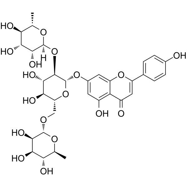
Ligustroflavone is an orally active flavonoid compound. Ligustroflavone can be extracted from Ligustrum lucidum. Ligustroflavone antagonizes the calcium-sensing receptor (CaSR), inhibits the RIPK1/RIPK3/MLKL pathway, and downregulates TGF-β/Smad signaling. Ligustroflavone regulates calcium metabolism, protects bone tissue, reduces cerebral ischemic injury, and inhibits liver fibrosis. Ligustroflavone can be used in the study of diabetic osteoporosis, ischemic stroke, and liver fibrosis.
Nos produits utilisent uniquement pour la recherche. Nous ne vendons pas aux patients.
- Pureté : 99.90%
- CAS No.: 260413-62-5
- Formule: C33H40O18
- Masse moléculaire:724.66
-
Stockage:
4°C, protect from light
* In solvent : -80°C, 6 months; -20°C, 1 month (protect from light)

To place orders, for customer services and technical support, please contact: MedChemExpress USA
Tel: 609-228-6898 E-mail: [email protected] [email protected]


-
 Activité biologique
-
Chemical Information
-
Solvant et solubilité
- Protocole
- Pureté et documentation
- Références
-
 Help & FAQs


-
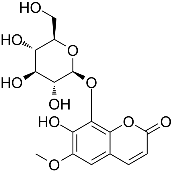
HY-N0579
FraxinPDE Inhibitor -
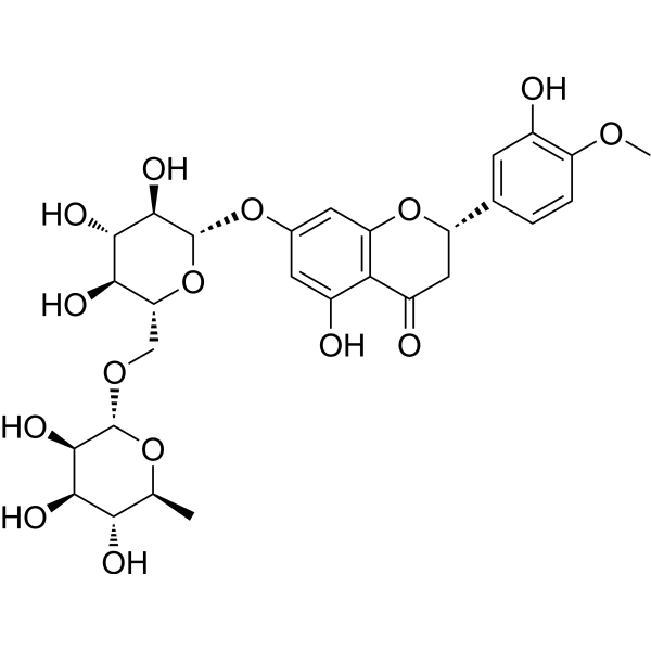
HY-15337
HesperidinAntioxidant -
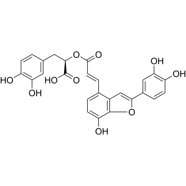
HY-N0319
Salvianolic acid CCytochrome Inhibitor -
HY-139201A
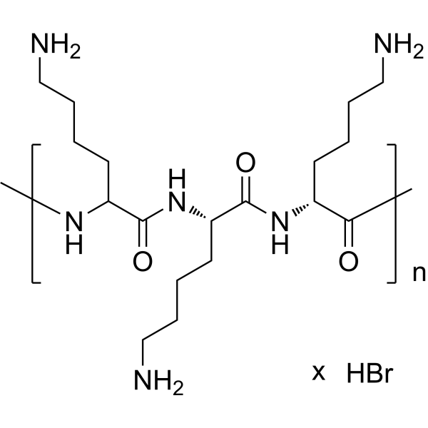
Poly-D-lysine hydrobromide (MW 70000-150000)CaSR Agonist -

HY-N0546R
Ligustroflavone (Standard)Standard Of Ligustroflavone -
HY-17397
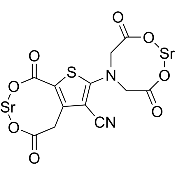
Strontium RanelateAntiosteoporotic Agent -
HY-P1955A
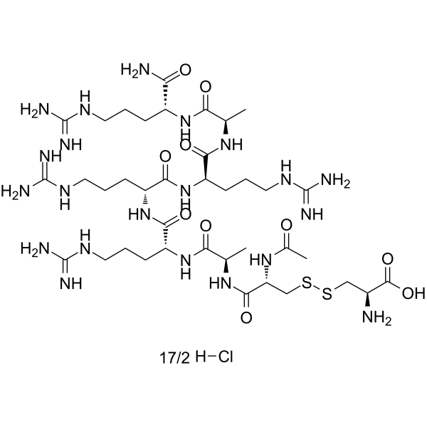
Etelcalcetide hydrochlorideCaSR Activator -
HY-70037
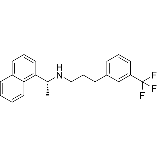
CinacalcetCaSR Agonist -
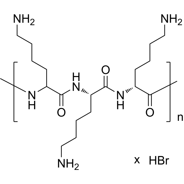
HY-139201
Poly-D-lysine hydrobromide (MW 30000-70000)CaSR Agonist -
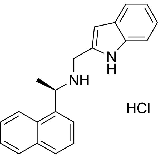
HY-122819
Calindol hydrochlorideCaSR PAM -
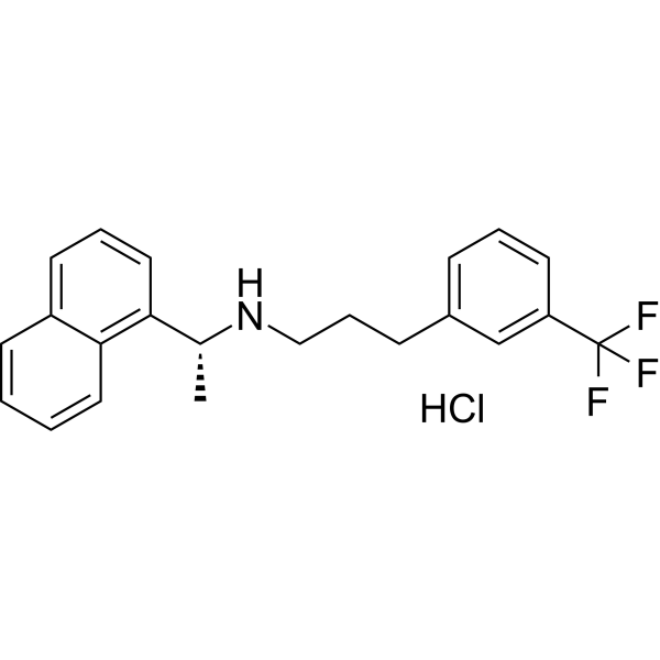
HY-70037A
Cinacalcet hydrochlorideCaSR Agonist -

HY-139201B
Poly-D-lysine hydrobromide (MW 84000)CaSR Agonist
Publications Citing Use of MedChemExpress (MCE) Ligustroflavone
More

Activité biologique
Description
|
RIPK1 |
RIPK3 |
In Vitro
Ligustroflavone (0.1 nM-0.1 mM; 2-24 h) increases PTH release in primary rat parathyroid gland cells in a dose- and time-dependent manner[1].
Ligustroflavone (25 μM) shows protective effects against hypoxia injury in PC12 cells, reducing LDH release[2].
Ligustroflavone (25 μM; 24 h) reduces α-SMA and COL 1A1 expression in TGF-β₁-stimulated human hepatic stellate LX-2 cells[3].
MedChemExpress (MCE) has not independently confirmed the accuracy of these methods. They are for reference only.
In Vivo
Ligustroflavone (30 mg/kg; i.g.; 15 min before ischemia) reduces necroptosis in rat brain after ischemic stroke through inhibiting RIPK1/RIPK3/MLKL pathway[2].
Ligustroflavone (5-20 mg/kg; i.p.; 3 times/week; 6 weeks) ameliorates CCl4 (HY-Y0298)-induced liver fibrosis in mice by reducing collagen deposition and inhibiting TGF-β/Smad signaling[3].
MedChemExpress (MCE) has not independently confirmed the accuracy of these methods. They are for reference only.
-
Animal Model:male Sprague-Dawley rats (250-300 g); MCAO-induced ischemic stroke model[2]
-
Dosage:30 mg/kg
-
Administration:Oral gavage (i.g.); 15 min before ischemia
-
Result:Improved neurological deficit scores, reduced cerebral infarct volume, and suppressed necroptosis-associated proteins RIPK3, MLKL, and p-MLKL in the brain.


Chemical Information
-
CAS No. 260413-62-5
-
Appearance Solid
-
Masse moléculaire 724.66
-
Formule C33H40O18
-
Color Light yellow to yellow
-
SMILES
O=C(C=C(C1=CC=C(O)C=C1)OC2=CC(O[C@H](O[C@H](CO[C@H](O[C@@H](C)[C@H](O)[C@H]3O)[C@@H]3O)[C@@H](O)[C@@H]4O)[C@@H]4O[C@@](O[C@@H](C)[C@H](O)[C@H]5O)([H])[C@@H]5O)=C6)C2=C6O
-
Synonyms
Nuezhenoside
-
Structure Classification
-
Initial Source
-
Livraison
Room temperature in continental US; may vary elsewhere.
-
Stockage
4°C, protect from light
* In solvent : -80°C, 6 months; -20°C, 1 month (protect from light)


Publications (2)
-
Journal Impact Factor
-
Most Recent
-
Food Chem
Effects of sun drying combined with baking processes on the flavor quality of Chongqing Tuocha raw tea. [Abstract]2025 Dec 30:497:146992. PMID: 41285060 -
Food Chem
Flavonoid-mediated metabolic underpinning quality variation in red bud-sport pear mutants. [Abstract]2025 Oct 15:489:144992. PMID: 40466530


Solvant et solubilité
In Vitro:
DMSO : 125 mg/mL (172.49 mM; Need ultrasonic; Hygroscopic DMSO has a significant impact on the solubility of product, please use newly opened DMSO)
Please refer to the solubility information to select the appropriate solvent. Once prepared, please aliquot and store the solution to prevent product inactivation from repeated freeze-thaw cycles.
Storage method and period of stock solution: -80°C, 6 months; -20°C, 1 month (protect from light). When stored at -80°C, please use it within 6 months. When stored at -20°C, please use it within 1 month.
Please refer to the solubility information to select the appropriate solvent. Once prepared, please aliquot and store the solution to prevent product inactivation from repeated freeze-thaw cycles.
Storage method and period of stock solution: -80°C, 6 months; -20°C, 1 month (protect from light). When stored at -80°C, please use it within 6 months. When stored at -20°C, please use it within 1 month.
Concentration (start) × Volume (start) = Concentration (final) × Volume (final)
In Vivo:
Select the appropriate dissolution method based on your experimental animal and administration route.
- For the following dissolution methods, please ensure to first prepare a clear stock solution using an In Vitro approach and then sequentially add co-solvents:
- To ensure reliable experimental results, the clarified stock solution can be appropriately stored based on storage conditions. As for the working solution for In Vivo experiments, it is recommended to prepare freshly and use it on the same day.
- The percentages shown for the solvents indicate their volumetric ratio in the final prepared solution. If precipitation or phase separation occurs during preparation, heat and/or sonication can be used to aid dissolution.
Add each solvent one by one: 10% DMSO 40% PEG300 5% Tween-80 45% Saline
Solubility: ≥ 2.08 mg/mL (2.87 mM); Clear solution
This protocol yields a clear solution of ≥ 2.08 mg/mL (saturation unknown).
Taking 1 mL working solution as an example, add 100 μL DMSO stock solution (20.8 mg/mL) to 400 μL PEG300, and mix evenly; then add 50 μL Tween-80 and mix evenly; then add 450 μL Saline to adjust the volume to 1 mL.
Preparation of Saline: Dissolve 0.9 g sodium chloride in ddH₂O and dilute to 100 mL to obtain a clear Saline solution.
Add each solvent one by one: 10% DMSO 90% (20% SBE-β-CD in Saline)
Solubility: ≥ 2.08 mg/mL (2.87 mM); Clear solution
This protocol yields a clear solution of ≥ 2.08 mg/mL (saturation unknown).
Taking 1 mL working solution as an example, add 100 μL DMSO stock solution (20.8 mg/mL) to 900 μL 20% SBE-β-CD in Saline, and mix evenly.
Preparation of 20% SBE-β-CD in Saline (4°C, storage for one week): 2 g SBE-β-CD powder is dissolved in 10 mL Saline, completely dissolve until clear.
In Vivo Dissolution Calculator
Please enter the basic information of animal experiments:
-
-
-
-
Recommended: Prepare an additional quantity of animals to account for potential losses during experiments.
Please enter your animal formula composition:
-
%DMSO +
Recommended: Keep the proportion of DMSO in working solution below 2% if your animal is weak.
-
%+
-
+%Tween-80 + +
-
%Saline +
The co-solvents required include: DMSO, . All of co-solvents are available by MedChemExpress (MCE). , Tween 80. All of co-solvents are available by MedChemExpress (MCE).
Working solution concentration: 0.22 mg/mL
Method for preparing stock solution: mg drug dissolved in μL DMSO. Stock solution concentration: mg/mL. * In solvent : -80°C, 6 months; -20°C, 1 month (protect from light)
1. Take μL DMSO stock solution;
2. Add μL .
μL , mix evenly;
3. Then add μL Tween 80, mix evenly;
4. Then add μL
Please ensure that the stock solution in the first step is dissolved to a clear state, and add co-solvents in sequence. You can use ultrasonic heating (ultrasonic cleaner, recommended frequency 20-40 kHz), vortexing, etc. to assist dissolution.


Pureté et documentation
-
Fiche technique (281 KB)
-
SDS (393 KB)
- English - EN (393 KB)
- Français - FR (393 KB)
- Deutsch - DE (393 KB)
- Norwegian - NO (393 KB)
- Español - ES (393 KB)
- Swedish - SV (393 KB)
- Italian - IT (393 KB)
- Korean - KR (393 KB)
- Portuguese - PT (393 KB)
-
Instruction de manipulation (2659 KB)
Références
[1]. Feng R, et al. Protective Effects of Ligustroflavone, an Active Compound from Ligustrum lucidum, on Diabetes-Induced Osteoporosis in Mice: A Potential Candidate as Calcium-Sensing Receptor Antagonist. Am J Chin Med. 2019;47(2):457-476. [Content Brief]
[2]. Zhang YY, et al. Ligustroflavone reduces necroptosis in rat brain after ischemic stroke through targeting RIPK1/RIPK3/MLKL pathway. Naunyn Schmiedebergs Arch Pharmacol. 2019 Sep;392(9):1085-1095. [Content Brief]
[3]. Kang R, et al. Ligustroflavone ameliorates CCl4-induced liver fibrosis through down-regulating the TGF-β/Smad signaling pathway. Chin J Nat Med. 2021 Mar;19(3):170-180. [Content Brief]


Complete Stock Solution Preparation Table
Please refer to the solubility information to select the appropriate solvent. Once prepared, please aliquot and store the solution to prevent product inactivation from repeated freeze-thaw cycles.
Storage method and period of stock solution: -80°C, 6 months; -20°C, 1 month (protect from light). When stored at -80°C, please use it within 6 months. When stored at -20°C, please use it within 1 month.
| Optional Solvent | Concentration Solvent Mass | 1 mg | 5 mg | 10 mg | 25 mg |
|---|---|---|---|---|---|
| DMSO | 1 mM | 1.3800 mL | 6.8998 mL | 13.7996 mL | 34.4989 mL |
| 5 mM | 0.2760 mL | 1.3800 mL | 2.7599 mL | 6.8998 mL | |
| 10 mM | 0.1380 mL | 0.6900 mL | 1.3800 mL | 3.4499 mL | |
| 15 mM | 0.0920 mL | 0.4600 mL | 0.9200 mL | 2.2999 mL | |
| 20 mM | 0.0690 mL | 0.3450 mL | 0.6900 mL | 1.7249 mL | |
| 25 mM | 0.0552 mL | 0.2760 mL | 0.5520 mL | 1.3800 mL | |
| 30 mM | 0.0460 mL | 0.2300 mL | 0.4600 mL | 1.1500 mL | |
| 40 mM | 0.0345 mL | 0.1725 mL | 0.3450 mL | 0.8625 mL | |
| 50 mM | 0.0276 mL | 0.1380 mL | 0.2760 mL | 0.6900 mL | |
| 60 mM | 0.0230 mL | 0.1150 mL | 0.2300 mL | 0.5750 mL | |
| 80 mM | 0.0172 mL | 0.0862 mL | 0.1725 mL | 0.4312 mL | |
| 100 mM | 0.0138 mL | 0.0690 mL | 0.1380 mL | 0.3450 mL |


Ligustroflavone Related Classifications
HY-N0546 Related Classifications
 Powered by Bioz