



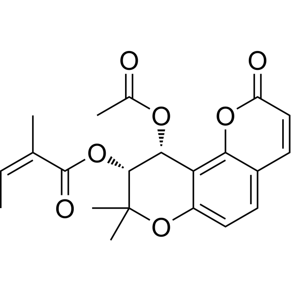
(-)-Praeruptorin A
(-)-Praeruptorin A is a nature product that could be isolated from the roots of Peucedanum praeruptorum Dunn. (-)-Praeruptorin A relaxes ileum and tracheal smooth muscles by activating NO/cGMP signaling pathway. (-)-Praeruptorin A has dramatically therapeutic effects on hypertension mainly through acting as a Ca2+-influx blocker.
For research use only. We do not sell to patients.
- CAS No.: 14017-71-1
- Formula: C21H22O7
- Molecular Weight:386.40
-
Storage:
Please store the product under the recommended conditions in the Certificate of Analysis.

To place orders, for customer services and technical support, please contact: MedChemExpress USA
Tel: 609-228-6898 E-mail: [email protected] [email protected]


-
 Biological Activity
-
Chemical Information
- Protocols
- Purity & Documentation
- References
-
 Help & FAQs


All Calcium Channel Isoforms
More

Biological Activity
Description


Chemical Information
-
CAS No. 14017-71-1
-
Molecular Weight 386.40
-
Formula C21H22O7
-
SMILES
C/C=C(C)\C(O[C@@H]1[C@H](OC(C)=O)C2=C3C(C=CC(O3)=O)=CC=C2OC1(C)C)=O
-
Structure Classification
-
Shipping
Room temperature in continental US; may vary elsewhere.
-
Storage
Please store the product under the recommended conditions in the Certificate of Analysis.

Protocols
-
Research Protocol for Cardiovascular Diseases
Cardiovascular disease can be modeled as maladaptive cardiac remodeling, where ischemic injury or pressure overload activates inflammatory signaling, fibroblast activation, extracellular-matrix deposition, cardiomyocyte hypertrophy, vascular remodeling, and progressive ventricular dysfunction. The TGF-β/SMAD axis is a central profibrotic pathway after myocardial injury and pressure overload, while innate immune and cytokine pathways regulate leukocyte recruitment, scar formation, and adverse remodeling. Key unresolved questions include which inflammatory signals are reparative versus harmful, when fibrosis is protective versus maladaptive, and whether pathway inhibition improves function without weakening necessary infarct healing or compensatory remodeling.
-
Ca2+ Staining Technique
Ca2+ staining is an experimental technique that utilizes specific fluorescent probes (such as Fluo-4 AM, Fura-2, etc.) to qualitatively or quantitatively detect dynamic changes in intracellular Ca2+ concentrations; this is achieved by monitoring the changes in fluorescent signals generated when these probes bind to free intracellular calcium ions. The underlying principle relies primarily on the presence of chelating groups within the probe's molecular structure that possess high affinity for calcium ions.


Purity & Documentation
References

Calculators
Concentration (start) × Volume (start) = Concentration (final) × Volume (final)


(-)-Praeruptorin A Related Classifications
HY-N0081A Related Classifications
 Powered by Bioz