



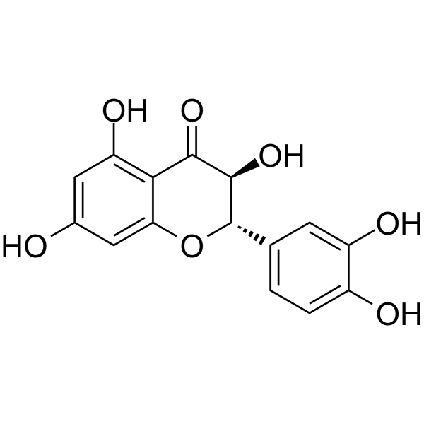
(-)-Taxifolin

Based on 1 Customer Validation
(-)-Taxifolin is the low-activity isomer of Taxifolin. Taxifolin is an orally effective collagenase inhibitor with an IC50 of 193.3 μM. Taxifolin dose-dependently reduces the levels of Smad-2, α-SMA, CTGF, type I collagen, NF-κB and FN in renal tissues, while decreasing serum levels of IL-1β, TNF-α, MDA, Scr and BUN, increasing SOD levels and reversing 27 serum metabolic biomarkers. Taxifolin inhibits the growth of a variety of pathogenic bacteria. Taxifolin can be used in studies related to renal fibrosis.
For research use only. We do not sell to patients.
- Purity : 98.98%
- CAS No.: 111003-33-9
- Formula: C15H12O7
- Molecular Weight:304.25
-
Storage:
-20°C, protect from light
* In solvent : -80°C, 6 months; -20°C, 1 month (protect from light)

To place orders, for customer services and technical support, please contact: MedChemExpress USA
Tel: 609-228-6898 E-mail: [email protected] [email protected]


-
 Biological Activity
-
Chemical Information
- Protocols
- Purity & Documentation
- References
-
 Help & FAQs




Biological Activity
Description
In Vitro
(-)-Taxifolin is the low-activity isomer of Taxifolin. Compared with the common (+)-Taxifolin (HY-N0136), (-)-Taxifolin shows lower or no obvious activity in some systems, which is mainly due to the difference in spatial configuration between the two isomers, leading to distinct interaction modes with target proteins.
MedChemExpress (MCE) has not independently confirmed the accuracy of these methods. They are for reference only. Further protocols information, click here.
In Vivo
-
Animal Model:Male SD rats (8 weeks old, 180-200 g) were anesthetized with 5% chloral hydrate, and the left ureter was exposed via laparotomy, doubly ligated with 4-0 silk suture at the upper-middle segment, and then transected between the two ligatures. Penicillin was administered intramuscularly after surgery, and oral administration of the drug was initiated before the surgery and continued daily thereafter[1]
-
Dosage:8 mg/kg; 16 mg/kg
-
Administration:p.o.; daily; 28 days
-
Result:Reduced renal interstitial inflammatory cell infiltration, tubular epithelial cell swelling/degeneration/necrosis, and interstitial fibrosis area, with the 16 mg/kg dose showing greater improvement.
Significantly reduced IL-1β, MDA, Scr, and BUN levels, and increased SOD levels compared to the model group; the 16 mg/kg dose additionally reduced TNF-α levels, while the 8 mg/kg dose had little effect on TNF-α.
Reduced renal tissue levels of TGF-β1, Smad-2, α-SMA, CTGF, collagen type I, NF-κB, and FN in a dose-dependent manner, with the 16 mg/kg dose showing greater reductions for most markers.
Regulated 16 serum metabolites at 8 mg/kg and 27 serum metabolites at 16 mg/kg, shifting the metabolic profile toward the healthy control group in a dose-dependent manner; the 16 mg/kg dose normalized levels of metabolites involved in 8 key pathways, while the 8 mg/kg dose regulated metabolites involved in 5 pathways.


Chemical Information
-
CAS No. 111003-33-9
-
Appearance Solid
-
Molecular Weight 304.25
-
Formula C15H12O7
-
Color White to off-white
-
SMILES
O=C1[C@@H](O)[C@H](C2=CC=C(O)C(O)=C2)OC3=CC(O)=CC(O)=C13
-
Synonyms
(-)-Dihydroquercetin
-
Structure Classification
-
Initial Source
-
Shipping
Room temperature in continental US; may vary elsewhere.
-
Storage
-20°C, protect from light
* In solvent : -80°C, 6 months; -20°C, 1 month (protect from light)

Protocols
-
Fibrosis/Collagen Morphometry
Fibrosis and collagen morphometry is based on the quantitative visualization of fibrillar collagen deposition in tissue sections using histochemical stains such as Sirius Red (Picrosirius Red) or Masson's trichrome, followed by image-based or polarization-enhanced analysis to estimate collagen proportional area as a surrogate of extracellular matrix accumulation during fibrotic remodeling. Sirius Red combined with polarized light microscopy enhances detection of collagen fibers due to birefringence properties, enabling more specific visualization of collagen type I and III fibrils compared to conventional bright-field histology, while whole-section or region-restricted digital morphometry reduces field-selection bias in fibrosis assessment. Alternative quantitative approaches include second harmonic generation (SHG) and two-photon excited fluorescence microscopy, which enable label-free detection of fibrillar collagen and have been validated against histological staining and biochemica
-
Collagen: Sirius Red Staining
Sirius Red or picrosirius red staining is a histochemical method for visualizing collagen-rich extracellular matrix in tissue sections, and collagen fibers are detected as red-stained structures under bright-field microscopy with enhanced birefringence under polarized light. Picrosirius red is useful for assessing total collagen organization, distribution, and fibrosis burden, but polarized color should not be interpreted as a definitive collagen type I versus type III readout because color is affected by fiber orientation, thickness, and packing.
-
Cell Cytotoxicity Assay
Cytotoxicity assays are usually based on the assessment of cell membrane damage, which can also be indirectly detected by measuring cell viability. Detection methods include MTT assay, CKK-8 assay, LDH assay and ATP assay, etc.
-
Connective Tissue: Masson's Trichrome/Collagen Trichrome Staining
Masson’s Trichrome (collagen/trichrome staining) is a histological technique that differentially stains tissue compartments using sequential acidic dyes to distinguish collagen from muscle and cytoplasmic components based on dye affinity and tissue permeability differences, enabling visualization of fibrosis and connective tissue architecture in histological sections. The classical formulation typically uses Weigert's iron hematoxylin for nuclear staining, Biebrich scarlet-acid fuchsin for cytoplasm and muscle, and aniline blue (or light green variants) for collagen, producing a characteristic blue/green collagen signal contrasted against red cytoplasm and dark nuclei. The staining principle relies on selective displacement of smaller dye molecules by larger anionic dyes in collagen-rich regions under controlled acidified conditions, which enhances collagen-specific dye retention. This property makes the method widely used for fibrosis assessment in organs such as heart, liver, lung, a
-
Research Protocol for Inflammation-related Diseases
The NLRP3 inflammasome is a cytosolic innate immune signaling platform that integrates priming signals and danger-signal activation to promote caspase-1 activation, maturation of IL-1β and IL-18, and gasdermin D-mediated pyroptotic cell death. The core experimental logic is to determine whether inflammatory disease phenotypes are driven by increased NLRP3 expression, ASC-containing inflammasome assembly, caspase-1 cleavage, GSDMD cleavage, and extracellular release of IL-1β/IL-18 rather than by nonspecific cell injury alone. The pathway is strongly linked to inflammation-related disease phenotypes because monosodium urate crystals activate NALP3/NLRP3 inflammasome signaling in gout-like crystal inflammation, cholesterol crystals activate NLRP3 inflammasomes in atherogenesis models, and DSS-induced intestinal inflammation has been reported to involve NLRP3 inflammasome activity. However, experimental colitis studies also show context-dependent protective effects of NLRP3 inflammasome co
-
Pyroptosis Solutions
Pyroptosis is a lytic inflammatory cell-death pathway executed by gasdermin pores, most classically through inflammasome-mediated activation of caspase-1, cleavage of gasdermin D, membrane pore formation, LDH release, and secretion of IL-1β and IL-18. The canonical pathway is commonly modeled by priming cells with an inflammatory signal such as LPS to induce pro-IL-1β and inflammasome components, followed by an activation signal such as ATP or nigericin to activate NLRP3, ASC speck formation, caspase-1 cleavage, GSDMD cleavage, cytokine release, and pyroptotic membrane rupture. The non-canonical pathway is triggered when cytosolic LPS activates mouse caspase-11 or human caspase-4/5, leading to GSDMD cleavage and pyroptosis, and this can secondarily activate NLRP3-dependent IL-1β release. Pyroptosis is linked to inflammatory injury, infection, cancer, liver disease, ocular disease, placental inflammation, and other disease phenotypes, but unresolved questions include which gasdermin fam


Purity & Documentation
-
Data Sheet (270 KB)
-
SDS (251 KB)
- English - EN (251 KB)
- Français - FR (251 KB)
- Deutsch - DE (251 KB)
- Norwegian - NO (251 KB)
- Español - ES (251 KB)
- Swedish - SV (251 KB)
- Italian - IT (251 KB)
- Korean - KR (251 KB)
- Portuguese - PT (251 KB)
-
Handling Instructions (2659 KB)
References
[1]. Ren L, et al. Dissecting Efficacy and Metabolic Characteristic Mechanism of on Renal Fibrosis by Multivariate Approach and Ultra-Performance Liquid Chromatography Coupled With Mass Spectrometry-Based Metabolomics Strategy. Frontiers in pharmacology. 2020;11:608511. [Content Brief]
[2]. Angelis A, et al. Bio-Guided Isolation of Methanol-Soluble Metabolites of Common Spruce (Picea abies) Bark by-Products and Investigation of Their Dermo-Cosmetic Properties. Molecules (Basel, Switzerland). 2016 Nov 21;21(11):1586. [Content Brief]

Calculators
Concentration (start) × Volume (start) = Concentration (final) × Volume (final)


(-)-Taxifolin Related Classifications
HY-N0136B Related Classifications
 Powered by Bioz