



MRL-494
Based on 2 publication(s) in Google Scholar
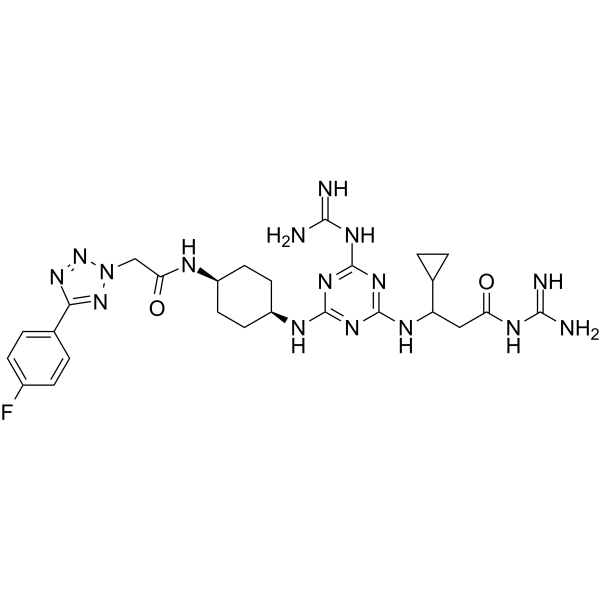
MRL-494, an antibacterial agent, is a inhibitor of β-barrel assembly machine A (BamA) impervious to efflux and the outer membrane permeability barrier. MRL-494 can inhibits Gram-positive (MIC of 12.5 μM for Staphylococcus aureus COL) and Gram-negative (MIC of 25 μM for E. coli JCM158) bacterias.
Nur für Forschungszwecke. Wir verkaufen nicht an Patienten.
- CAS. Nr.: 2434898-43-6
- Formel: C26H35FN16O2
- Molecular Weight:622.66
-
Speicherung:
Please store the product under the recommended conditions in the Certificate of Analysis.

To place orders, for customer services and technical support, please contact: MedChemExpress USA
Tel: 609-228-6898 E-mail: [email protected] [email protected]


-
 Biologische Aktivität
-
Chemical Information
- Protokoll
- Reinheit & Dokumentation
- Verweise
-
 Help & FAQs


Publications Citing Use of MedChemExpress (MCE) MRL-494
More

Biologische Aktivität
Beschreibung
IC50 & Target
β-barrel assembly machine A (BamA)[1]
In Vitro
MRL-494 lethally disrupts the cytoplasmic membrane. MRL-494 inhibits OM proteins (OMPs) biogenesis from outside the outer membrane (OM) by targeting BamA. MRL-494 exhibits strong anti-microbial properties against both Gram-positive and Gram-negative bacteria. The MIC values of MRL-494 against E. coli (WT), E. coli (ΔtolC), E. coli (ΔtolC envA101), K. pneumonia, A. baumannii (WT), A. baumannii (ΔlpxC) , P. aeruginosa (efflux deficient), P. aeruginosa (WT), Staphylococcus aureus (methicillin-resistant) and Bacillus subtilis rpoB18 are 25 μM, 25 μM, 25 μM, 100 μM, 200 μM, 200 μM, 100 μM, 100 μM, 12.5 μM and 25 μM, respectively[1].
MedChemExpress (MCE) has not independently confirmed the accuracy of these methods. They are for reference only. Further protocols information, click here.


Chemical Information
-
CAS. Nr. 2434898-43-6
-
Molecular Weight 622.66
-
Formel C26H35FN16O2
-
SMILES
FC1=CC=C(C2=NN(CC(N[C@@H]3CC[C@H](NC4=NC(NC(C5CC5)CC(NC(N)=N)=O)=NC(NC(N)=N)=N4)CC3)=O)N=N2)C=C1
-
Versand
Room temperature in continental US; may vary elsewhere.
-
Speicherung
Please store the product under the recommended conditions in the Certificate of Analysis.


Publications (2)
-
Journal Impact Factor
-
Most Recent
-
-
J Biol Chem
Mutations in the essential outer membrane protein BamA contribute to Escherichia coli resistance to the antimicrobial peptide TAT-RasGAP317-326. [Abstract]2025 Jan;301(1):108018. PMID: 39608713

Protokoll
-
Research Protocol for Infectious Diseases
Infectious-disease experiments test how pathogens interact with host barriers, innate immune receptors, inflammatory signaling, pathogen replication, and tissue injury; pattern-recognition receptors such as TLRs, RIG-I-like receptors, NOD-like receptors, and inflammasomes detect microbial molecules and activate NF-κB, interferon, and cytokine responses. The central hypothesis is that infection severity reflects the balance between pathogen burden and host response: protective inflammation restricts pathogen growth, whereas excessive or mislocalized inflammation contributes to tissue damage and disease phenotype. Unresolved questions include which host pathways are protective versus pathogenic, why some infection models fail to translate to human disease, and which combined readouts best predict clinically relevant infection outcomes.
-
Transepithelial/transendothelial electrical resistance assay
TEER measures electrical resistance across epithelial or endothelial monolayers cultured on permeable supports, and the readout reflects ionic conductance through the cell barrier, especially the paracellular pathway regulated by junctional integrity. TEER can be measured without destroying the monolayer and is commonly used before or during transport, permeability, barrier-disruption, and barrier-maturation experiments. TEER values are influenced by biological maturation and technical conditions; reported factors include temperature, medium formulation, passage number, electrode geometry, membrane properties, and junctional length during early monolayer maturation. Therefore, TEER should be interpreted with blank-insert subtraction, area normalization, repeated readings, and, when possible, orthogonal barrier readouts such as FITC-dextran flux or tight-junction staining.
-
Gram Staining of Tissue Sections
Gram staining of tissue sections is a histochemical technique used to differentiate Gram-positive and Gram-negative bacteria within histological specimens based on differences in bacterial cell wall structure and dye retention, adapted from classical bacteriological Gram staining into tissue-compatible “histological Gram stain” variants. In tissue applications, modifications of the Brown-Hopps and Brown-Brenn methods are commonly used to improve differentiation of microorganisms embedded within host connective tissue and to reduce overstaining or loss of Gram-negative signal, which are known limitations of earlier approaches. The principle relies on crystal violet-iodine complex retention in Gram-positive organisms and subsequent decolorization and counterstaining steps that allow contrast visualization of Gram-negative organisms against tissue background.


Reinheit & Dokumentation
Verweise

Calculators
Konzentration (Stammlösung) × Volumen (Stammlösung) = Konzentration (Ziellösung) × Volumen (Ziellösung)


MRL-494 Related Classifications
HY-128773 Related Classifications
 Powered by Bioz