



NADH
Based on 11 publication(s) in Google Scholar
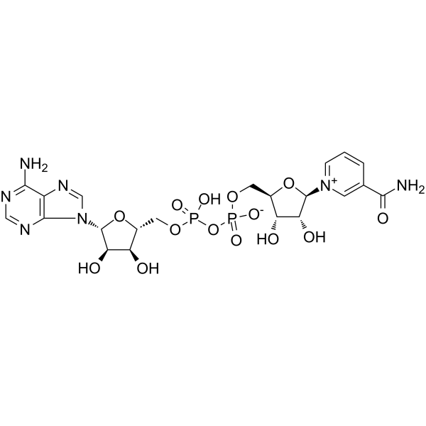
NADH is an orally active dehydrogenase coenzyme that acts as a crucial electron carrier in cellular respiration and participates in ATP production. NADH promotes metabolism, supports brain function, and counteracts oxidative stress by transferring electrons to the electron transport chain. As a signaling molecule, NADH regulates multiple biological processes, including anti-apoptosis, synaptic plasticity, gene expression, and calcium homeostasis. Redox imbalance of NADH/NAD⁺ is one of the key pathological mechanisms of various diseases, such as diabetic nephropathy, neurodegenerative diseases, and ischemia-reperfusion injury.
Para uso exclusivo en investigación. No vendemos a pacientes.
- No. CAS: 58-68-4
- Fòrmula: C21H29N7O14P2
- Peso molecular:665.44
-
Almacenamiento:
Please store the product under the recommended conditions in the Certificate of Analysis.


To place orders, for customer services and technical support, please contact: MedChemExpress USA
Tel: 609-228-6898 E-mail: [email protected] [email protected]


-
 Actividad biológica
-
Chemical Information
- Protocolo
- Pureza y Documentación
- Referencias
-
 Help & FAQs



Publications Citing Use of MedChemExpress (MCE) NADH
More- Cell Mol Immunol. 2024 Jun;21(6):561-574. [Abstract]
- Food Chem. 2023 Oct 15:423:136274. [Abstract]
- Biomater Res. 2026 Apr 29.
- Free Radic Biol Med. 2025 Jan:226:56-69. [Abstract]
- J Ginseng Res. 2025 Sep;49(5):541-552. [Abstract]
- Mol Plant Pathol. 2025 Dec;26(12):e70196. [Abstract]
- Microorganisms. 2026 May 9;14(5):1070. [Abstract]
- ACS Chem Biol. 2026 Jul 2.
- Biochemistry. 2023 Dec 5;62(23):3396-3410. [Abstract]
- Chemrxiv. 2025 Aug 28.
- Research Square Preprint. 2023 Sep 15.
Ver todos los productos específicos de isoformas Endogenous Metabolite
More

Actividad biológica
Descripciòn
IC50 & Target
|
Human Endogenous Metabolite |
In Vitro
Ensayo clínico
| NCT Number | Sponsor | Condition | Start Date |
Phase

|
|---|---|---|---|---|
| NCT01329991 | 2011-05 | PHASE1 |


Chemical Information
-
No. CAS 58-68-4
-
Peso molecular 665.44
-
Fòrmula C21H29N7O14P2
-
SMILES
O[C@H]1[C@@H](O)[C@H](N2C=CCC(C(N)=O)=C2)O[C@@H]1COP(O)(OP(OC[C@@H]3[C@@H](O)[C@@H](O)[C@H](N4C5=NC=NC(N)=C5N=C4)O3)(O)=O)=O
-
Structure Classification
-
Initial Source
-
Envío
Room temperature in continental US; may vary elsewhere.
-
Almacenamiento
Please store the product under the recommended conditions in the Certificate of Analysis.


Publications (11)
-
Journal Impact Factor
-
Most Recent
-
Cell Mol Immunol
2024 Jun;21(6):561-574. PMID: 38570588 -
Food Chem
Improvement of catalytic activity of sorbose dehydrogenase for deoxynivalenol degradation by rational design. [Abstract]2023 Oct 15:423:136274. PMID: 37159968 -
-
Free Radic Biol Med
TSPO exacerbates sepsis-induced cardiac dysfunction by inhibiting p62-Mediated autophagic flux via the ROS-RIP1/RIP3-exosome axis. [Abstract]2025 Jan:226:56-69. PMID: 39542185 -
J Ginseng Res
Total ginsenosides and ginsenoside Rb2 delay hepatocyte senescence by regulating NAD+ metabolism and promoting IDO2/QPRT expression. [Abstract]2025 Sep;49(5):541-552. PMID: 40843012 -
Mol Plant Pathol
Negative Immune Regulator CAD7 Functions as a Small-Molecule Aldehyde Reductase and Increases Histamine Accumulation in Arabidopsis. [Abstract]2025 Dec;26(12):e70196. PMID: 41456913 -
Microorganisms
Characterization of RmlABCD Enzymes from Marine Bacteria and Efficient Synthesis of dTDP-L-Rhamnose. [Abstract]2026 May 9;14(5):1070. PMID: 42197457 -
-
Biochemistry
Full-Length NAD+-I Riboswitches Bind a Single Cofactor but Cannot Discriminate against Adenosine Triphosphate. [Abstract]2023 Dec 5;62(23):3396-3410. PMID: 37947391 -
-


Pureza y Documentación
Referencias
[1].
Ying W. NAD+ and NADH in cellular functions and cell death. Front Biosci. 2006 Sep 1;11:3129-48.
[Content Brief]
[2].
Chen Z, et al. Therapeutic potential of NADH: in neurodegenerative diseases characterizde by mitochondrial dysfunction. Lin Chuang Er Bi Yan Hou Tou Jing Wai Ke Za Zhi. 2024 Jan;38(1):57-62.
[Content Brief]
[3]. Yan LJ, et al. NADH/NAD+ Redox Imbalance and Diabetic Kidney Disease. Biomolecules. 2021;11(5):730. Published 2021 May 14. [Content Brief]

Calculators
Concentration (start) × Volume (start) = Concentration (final) × Volume (final)


NADH Related Classifications
HY-113355 Related Classifications
 Powered by Bioz