



Nampt-IN-8
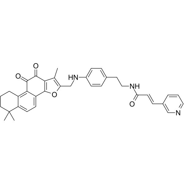
Nampt-IN-8 (Compound 10d) is an NAMPT inhibitor with an IC50 of 0.183 μM. Nampt-IN-8 is also a relatively good NQO1 substrate. Nampt-IN-8 induces cell apoptosis and ROS.
For research use only. We do not sell to patients.
- CAS No.: 2453183-75-8
- Formula: C36H35N3O4
- Molecular Weight:573.68
-
Storage:
Please store the product under the recommended conditions in the Certificate of Analysis.

To place orders, for customer services and technical support, please contact: MedChemExpress USA
Tel: 609-228-6898 E-mail: [email protected] [email protected]


-
 Biological Activity
-
Chemical Information
- Protocols
- Purity & Documentation
- References
-
 Help & FAQs




Biological Activity
Description
IC50 & Target
IC50: 0.183 μM (NAMPT)[1]
Cellular Effect
|
Cell Line

|
Type | Value | Description | References |
|---|---|---|---|---|
| A549 | IC50 |
1.15 μM
Compound: 10d
|
Antiproliferative activity against human A549 cells assessed as inhibition of cell growth incubated for 72 hrs by cell titer glo luminescent assay
Antiproliferative activity against human A549 cells assessed as inhibition of cell growth incubated for 72 hrs by cell titer glo luminescent assay
|
[PMID: 35640078] |
| A549/TR | IC50 |
11.82 μM
Compound: 10d
|
Antiproliferative activity against human A549/Taxol cells assessed as inhibition of cell growth pretreated with nicotinamide mononucleotide followed by compound addition and measured after 72 hrs by cell titer glo luminescent assay
Antiproliferative activity against human A549/Taxol cells assessed as inhibition of cell growth pretreated with nicotinamide mononucleotide followed by compound addition and measured after 72 hrs by cell titer glo luminescent assay
|
[PMID: 35640078] |
| A549/TR | IC50 |
124.9 μM
Compound: 10d
|
Antiproliferative activity against human A549/Taxol cells assessed as inhibition of cell growth pretreated with nicotinamide mononucleotide, dicumarol, N-acetyl-L-cysteine followed by compound addition and measured after 72 hrs by cell titer glo luminesce
Antiproliferative activity against human A549/Taxol cells assessed as inhibition of cell growth pretreated with nicotinamide mononucleotide, dicumarol, N-acetyl-L-cysteine followed by compound addition and measured after 72 hrs by cell titer glo luminesce
|
[PMID: 35640078] |
| A549/TR | IC50 |
14.48 μM
Compound: 10d
|
Antiproliferative activity against human A549/Taxol cells assessed as inhibition of cell growth pretreated with NQO1 inhibitor, dicumarol followed by compound addition and measured after for 72 hrs by cell titer glo luminescent assay
Antiproliferative activity against human A549/Taxol cells assessed as inhibition of cell growth pretreated with NQO1 inhibitor, dicumarol followed by compound addition and measured after for 72 hrs by cell titer glo luminescent assay
|
[PMID: 35640078] |
| A549/TR | IC50 |
4.1 μM
Compound: 10d
|
Antiproliferative activity against human A549/Taxol cells assessed as inhibition of cell growth measured after 72 hrs by cell titer glo luminescent assay
Antiproliferative activity against human A549/Taxol cells assessed as inhibition of cell growth measured after 72 hrs by cell titer glo luminescent assay
|
[PMID: 35640078] |
| A549/TR | IC50 |
45.29 μM
Compound: 10d
|
Antiproliferative activity against human A549/Taxol cells assessed as inhibition of cell growth pretreated with N-acetyl-L-cysteine followed by compound addition and measured after 72 hrs by cell titer glo luminescent assay
Antiproliferative activity against human A549/Taxol cells assessed as inhibition of cell growth pretreated with N-acetyl-L-cysteine followed by compound addition and measured after 72 hrs by cell titer glo luminescent assay
|
[PMID: 35640078] |
| L02 | IC50 |
120.4 μM
Compound: 10d
|
Antiproliferative activity against human L02 cells assessed as inhibition of cell growth measured after 72 hrs by cell titer glo luminescent assay
Antiproliferative activity against human L02 cells assessed as inhibition of cell growth measured after 72 hrs by cell titer glo luminescent assay
|
[PMID: 35640078] |
In Vitro
Nampt-IN-8 (Compound 10d) shows antiproliferative activity with an IC50 of 1.15 μM[1].
MedChemExpress (MCE) has not independently confirmed the accuracy of these methods. They are for reference only.
In Vivo


Chemical Information
-
CAS No. 2453183-75-8
-
Molecular Weight 573.68
-
Formula C36H35N3O4
-
SMILES
O=C1C2=C(CCCC3(C)C)C3=CC=C2C4=C(C1=O)C(C)=C(O4)CNC5=CC=C(C=C5)CCNC(/C=C/C6=CN=CC=C6)=O
-
Shipping
Room temperature in continental US; may vary elsewhere.
-
Storage
Please store the product under the recommended conditions in the Certificate of Analysis.


Purity & Documentation
References

Calculators
Concentration (start) × Volume (start) = Concentration (final) × Volume (final)


Nampt-IN-8 Related Classifications
HY-147795 Related Classifications
 Powered by Bioz