



Neoprzewaquinone A

Based on 1 Customer Validation
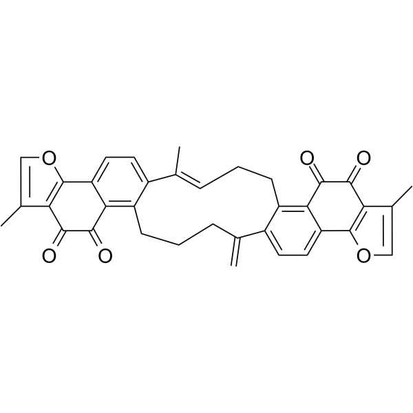
Neoprzewaquinone (NEO) is an orally active phenanthraquinone derivative isolated from Salvia miltiorrhiza Bunge. Neoprzewaquinone A inhibits IL-15Rα, blocks the ROCK2/STAT3 pathway to impair the migration and invasion abilities of triple-negative breast cancer cells; induces apoptosis of hepatocellular carcinoma cells by inhibiting the PI3K-AKT pathway; suppresses LPS-induced inflammation via binding to TLR4/MD2, and inhibits IL-15-induced proliferation, migration and phagocytosis of microglial cells; protects osteoblasts from mitochondrial dysfunction and apoptosis in diabetic osteoporosis by activating PRKD2 and the ELF5/PRKD2/PI3K/AKT/mTOR axis. Neoprzewaquinone A can be used in the research of diseases including rheumatoid arthritis, hepatocellular carcinoma, diabetic osteoporosis and triple-negative breast cancer.
Nur für Forschungszwecke. Wir verkaufen nicht an Patienten.
- Reinheit : 99.26%
- CAS. Nr.: 630057-39-5
- Formel: C36H28O6
- Molecular Weight:556.60
-
Speicherung:
4°C, sealed storage, away from moisture and light
* In solvent : -80°C, 6 months; -20°C, 1 month (sealed storage, away from moisture and light)

To place orders, for customer services and technical support, please contact: MedChemExpress USA
Tel: 609-228-6898 E-mail: [email protected] [email protected]


-
 Biologische Aktivität
-
Chemical Information
-
Lösungsmittel & Löslichkeit
- Protokoll
- Reinheit & Dokumentation
- Verweise
-
 Help & FAQs




Biologische Aktivität
Beschreibung
Cellular Effect
|
Cell Line

|
Type | Value | Description | References |
|---|---|---|---|---|
| MDA-MB-231 | IC50 |
11.14 μM
|
Cell viability inhibition against human MDA-MB-231 triple-negative breast cancer cells incubated for 24 hrs by MTT assay.
Cell viability inhibition against human MDA-MB-231 triple-negative breast cancer cells incubated for 24 hrs by MTT assay.
|
36982538 |
| MDA-MB-231 | IC50 |
7.11 μM
|
Cell viability inhibition against human MDA-MB-231 triple-negative breast cancer cells incubated for 48 hrs by MTT assay.
Cell viability inhibition against human MDA-MB-231 triple-negative breast cancer cells incubated for 48 hrs by MTT assay.
|
36982538 |
| MDA-MB-231 | IC50 |
4.69 μM
|
Cell viability inhibition against human MDA-MB-231 triple-negative breast cancer cells incubated for 72 hrs by MTT assay.
Cell viability inhibition against human MDA-MB-231 triple-negative breast cancer cells incubated for 72 hrs by MTT assay.
|
36982538 |
| HepG2 | IC50 |
6.08 μM
|
Cell viability inhibition against human HEPG-2 liver cancer cells by MTT assay.
Cell viability inhibition against human HEPG-2 liver cancer cells by MTT assay.
|
36982538 |
| NCI-H929 | IC50 |
7.25 μM
|
Cell viability inhibition against human NCI-H929 myeloma cells by MTT assay.
Cell viability inhibition against human NCI-H929 myeloma cells by MTT assay.
|
36982538 |
| AGS | IC50 |
9.06 μM
|
Cell viability inhibition against human AGS gastric adenocarcinoma cells by MTT assay.
Cell viability inhibition against human AGS gastric adenocarcinoma cells by MTT assay.
|
36982538 |
| MCF7 | IC50 |
9.51 μM
|
Cell viability inhibition against human MCF-7 breast cancer cells by MTT assay.
Cell viability inhibition against human MCF-7 breast cancer cells by MTT assay.
|
36982538 |
| NCI-H460 | IC50 |
13.46 μM
|
Cell viability inhibition against human H460 large cell lung cancer cells by MTT assay.
Cell viability inhibition against human H460 large cell lung cancer cells by MTT assay.
|
36982538 |
| ES-2 | IC50 |
14.54 μM
|
Cell viability inhibition against human ES-2 ovarian clear cell carcinoma cells by MTT assay.
Cell viability inhibition against human ES-2 ovarian clear cell carcinoma cells by MTT assay.
|
36982538 |
| A549 | IC50 |
48.28 μM
|
Cell viability inhibition against human A549 non-small cell lung cancer cells by MTT assay.
Cell viability inhibition against human A549 non-small cell lung cancer cells by MTT assay.
|
36982538 |
| MCF-10A | IC50 |
6.18 μM
|
Cell viability inhibition against human MCF-10A normal mammary epithelial cells by MTT assay.
Cell viability inhibition against human MCF-10A normal mammary epithelial cells by MTT assay.
|
36982538 |
| SH-SY5Y | IC50 |
12.61 μM
|
Cell viability inhibition against human SH-SY5Y neuroblastoma cells by MTT assay.
Cell viability inhibition against human SH-SY5Y neuroblastoma cells by MTT assay.
|
36982538 |
In Vitro
Neoprzewaquinone A (NEO) forms stable complexes with purified IL-1β, IL-6, and TNF-α proteins[1].
Neoprzewaquinone A (1-80000 nM) dose-dependently inhibits the in vitro binding of IL-1β to IL-1R1, IL-6 to IL-6R, and TNF-α to TNFR1[1].
Neoprzewaquinone A (5-20 μM; 24 h) dose-dependently inhibits the viability, migration, invasion capacity and cytotoxicity of MH7A rheumatoid arthritis fibroblast-like synoviocytes (RA-FLS)[1].
Neoprzewaquinone A (5-20 μM; 24 h) dose-dependently downregulates the expressions of IL-1β, IL-6, and TNF-α in MH7A rheumatoid arthritis fibroblast-like synoviocytes (RA-FLS)[1].
Neoprzewaquinone A (5-20 μM; 24 h) does not alter the cell cycle distribution of MH7A rheumatoid arthritis fibroblast-like synoviocytes (RA-FLS)[1].
Neoprzewaquinone A (5-20 μM; 24 h) dose-dependently inhibits the activation of the MAPK pathway in MH7A rheumatoid arthritis fibroblast-like synoviocytes (RA-FLS)[1].
Neoprzewaquinone A (20 μM; 24 h) modulates gene expression in primary RA-FLS[1].
Neoprzewaquinone A (5-20 μM; 24 h) dose-dependently inhibits the viability of primary RA-FLS and increases the cytotoxicity of primary RA-FLS[1].
Neoprzewaquinone A (0.3125-10 μM; 150 s) binds to purified PRKD2 protein, with a KD value of 6.39 × 10⁻⁶ M[3].
Neoprzewaquinone A (0.012-6.250 μM; 60 s binding, 60 s dissociation) specifically binds to recombinant IL-15Rα protein, with a dissociation constant (KD) of 0.62 μM[5].
Neoprzewaquinone A binds to the IL-15/IL-15Rα protein complex with a KD value of 0.6203 μM[6].
Neoprzewaquinone A (5-20 μM; 24 h) dose-dependently induces apoptosis of MH7A rheumatoid arthritis fibroblast-like synoviocytes (RA-FLS)[1].
Neoprzewaquinone A (1-20 μM) inhibits the viability of HepG2, Huh7 and Hepa1-6 hepatocellular carcinoma cells in a dose-dependent manner[2].
Neoprzewaquinone A (5-20 μM; 24 h) inhibits the migration of HepG2 and Huh7 hepatocellular carcinoma cells in a dose-dependent manner[2].
Neoprzewaquinone A (5-20 μM; 3 days followed by 2 weeks of normal culture) inhibits colony formation of HepG2 hepatocellular carcinoma cells in a dose-dependent manner[2].
Neoprzewaquinone A (0.3-10 μM; 24-72 h) potently inhibits the viability of MDA-MB-231 triple-negative breast cancer cells, with an IC50 of 4.69 μM at 72 h[4].
Neoprzewaquinone A (0.5-1 μM; 7 days) inhibits the colony-forming ability of MDA-MB-231 triple-negative breast cancer cells[4].
Neoprzewaquinone A potently and selectively inhibits purified PIM1 kinase with an IC50 of 0.56 μM[4].
Neoprzewaquinone A (10 μM) directly binds to PIM1 in MDA-MB-231 triple-negative breast cancer cells[4].
Neoprzewaquinone A (5-20 μM; 20 h) inhibits the PIM1/ROCK2/STAT3 signaling pathway in MDA-MB-231 triple-negative breast cancer cells, reduces the levels of phosphorylated and total ROCK pathway proteins, but does not alter the total PIM1 level[4].
Neoprzewaquinone A (5-20 μM; 20 h) dose-dependently inhibits the EMT process in MDA-MB-231 triple-negative breast cancer cells, upregulates E-cadherin levels and downregulates Vimentin levels[4].
Neoprzewaquinone A (10-20 μM) protects MC3T3-E1 mouse preosteoblasts against HGPA-induced mitochondrial dysfunction and apoptosis by restoring the activation of the ELF5/PRKD2 and PI3K/AKT/mTOR pathways, reducing ROS, and improving mitochondrial function[3].
Neoprzewaquinone A (1-3 μM; 24 h) inhibits the migration of MDA-MB-231 triple-negative breast cancer cells[4].
Neoprzewaquinone A (1-3 μM; 24 h) inhibits the invasion of MDA-MB-231 triple-negative breast cancer cells[4].
Neoprzewaquinone A (5-20 μM; 24 h) induces G0/G1 phase arrest, apoptosis and autophagy in triple-negative breast cancer MDA-MB-231 cells[4].
Neoprzewaquinone A inhibits the proliferation of IL-15-stimulated Mo7e cells, with an IC50 value of 1.075 μM[6].
Neoprzewaquinone A (5-20 μM; 24 h) regulates the expression of autophagy-related proteins in MDA-MB-231 triple-negative breast cancer cells, upregulating Beclin1 and LC3B while downregulating ATG5[4].
Neoprzewaquinone A (0.18-5.40 μM; 72 h) inhibits IL-15-induced proliferation of the human megakaryocytic leukemia cell line Mo7e, with an IC50 value of 1.075 μM[5].
Neoprzewaquinone A (0.3 μg/mL; 24-72 h) inhibits basal proliferation and migration, as well as IL-15-induced proliferation and migration, of human microglial cells HMC3[6].
Neoprzewaquinone A (0.3 μg/mL; 3-48 h) inhibits both basal phagocytosis and IL-15-induced phagocytosis in human microglial HMC3 cells[6].
Neoprzewaquinone A inhibits the binding of lipopolysaccharide (LPS) to the TLR4/MD2 receptor on the cell membrane[7].
Neoprzewaquinone A inhibits LPS-mediated dimerization of TLR4 and MD2 in cells[7].
Neoprzewaquinone A inhibits the phosphorylation of NF-κB p65 in LPS-stimulated cells[7].
Neoprzewaquinone A (0.3 μg/mL) inhibits multiple parameters of glycolysis and mitochondrial respiration in human microglial HMC3 cells, and partially reduces the non-mitochondrial respiration level and respiratory capacity of HMC3 cells treated with IL-15[6].
Neoprzewaquinone A inhibits the secretion of NO, TNF-α, IL-6 and IL-1β in LPS-stimulated cells[7].
MedChemExpress (MCE) has not independently confirmed the accuracy of these methods. They are for reference only.
-
Cell Line:MH7A RA fibroblast-like synoviocytes (RA-FLS)
-
Concentration:5, 10, 20 μM
-
Incubation Time:24 h
-
Result:Reduced cell viability.
-
Cell Line:MH7A RA fibroblast-like synoviocytes (RA-FLS)
-
Concentration:5, 10, 20 μM
-
Incubation Time:24 h
-
Result:Increased cytotoxicity.
-
Cell Line:MH7A RA fibroblast-like synoviocytes (RA-FLS)
-
Concentration:5, 10, 20 μM
-
Incubation Time:24 h
-
Result:Downregulated relative expression levels of IL-1β, IL-6 and TNF-α.
-
Cell Line:MH7A RA fibroblast-like synoviocytes (RA-FLS)
-
Concentration:5, 10, 20 μM
-
Incubation Time:24 h
-
Result:Enhanced apoptosis.
-
Cell Line:MH7A RA fibroblast-like synoviocytes (RA-FLS)
-
Concentration:5, 10, 20 μM
-
Incubation Time:24 h
-
Result:Had no effect on cell cycle distribution in MH7A cells.
-
Cell Line:MH7A RA fibroblast-like synoviocytes (RA-FLS)
-
Concentration:5, 10, 20 μM
-
Incubation Time:24 h
-
Result:Reduced migrated cell counts.
-
Cell Line:MH7A RA fibroblast-like synoviocytes (RA-FLS)
-
Concentration:10, 20 μM
-
Incubation Time:24 h
-
Result:Reduced invaded cell counts.
-
Cell Line:MH7A RA fibroblast-like synoviocytes (RA-FLS)
-
Concentration:5, 10, 20 μM
-
Incubation Time:24 h
-
Result:Decreased phosphorylation levels of MEK and ERK.
-
Cell Line:primary RA-FLS
-
Concentration:5, 10, 20 μM
-
Incubation Time:24 h
-
Result:Reduced cell viability in primary RA-FLS, following a similar trend to observations in MH7A cells.
-
Cell Line:primary RA-FLS
-
Concentration:5, 10, 20 μM
-
Incubation Time:24 h
-
Result:Increased cytotoxicity in primary RA-FLS, following a similar trend to observations in MH7A cells.
Reached cytotoxicity levels comparable to those seen in MH7A cells at matching concentrations.
-
Cell Line:HepG2 human hepatoma cells, Huh7 human hepatoma cells
-
Concentration:5, 10, 20 μM
-
Incubation Time:24 h
-
Result:Reduced the number of migrating HepG2 and Huh7 cells in a concentration-dependent manner.
Induced significant decreases in migrating cell number at all tested concentrations relative to untreated controls.
Exhibited greater inhibitory efficacy at 20 μM than the positive control sorafenib.
-
Cell Line:MDA-MB-231
-
Concentration:1, 2, 3 μM
-
Incubation Time:24 h
-
Result:Significantly reduced the wound closure rate of MDA-MB-231 cells in a concentration-dependent manner compared to the control group.
-
Cell Line:MDA-MB-231
-
Concentration:5, 10, 20 μM
-
Incubation Time:24 h
-
Result:Significantly reduced the number of invasive MDA-MB-231 cells in a concentration-dependent manner compared to the control group.
-
Cell Line:MDA-MB-231
-
Concentration:5, 10, 20 μM
-
Incubation Time:20 h
-
Result:Suppressed the expression of ROCK1, ROCK2, p-MYPT1, p-mTOR, and p-STAT3 in a concentration-dependent manner.
Left total protein levels of PIM1, BAD, MYPT1, mTOR, and STAT3 unchanged.\nDose-dependently upregulated the expression of the epithelial marker E-cadherin.
Downregulated the expression of the mesenchymal marker Vimentin compared to the control group.
-
Cell Line:MDA-MB-231
-
Concentration:5, 10, 20 μM
-
Incubation Time:24 h
-
Result:Induced G0/G1 phase arrest in a concentration-dependent manner.
Increased the proportion of cells in G0/G1 phase.
Decreased the proportion of cells in S phase.
Downregulated cyclin B1 and upregulated cyclin D1 in a concentration-dependent manner.
-
Cell Line:MDA-MB-231
-
Concentration:5, 10, 20 μM
-
Incubation Time:24 h
-
Result:Induced apoptosis in a concentration-dependent manner.
Increased nuclear shrinkage and bright blue fluorescence detected at higher concentrations.
Increased the apoptotic cell proportion.
-
Cell Line:MDA-MB-231
-
Concentration:5, 10, 20 μM
-
Incubation Time:24 h
-
Result:Induced autophagy in MDA-MB-231 cells.
-
Cell Line:MDA-MB-231
-
Concentration:5, 10, 20 μM
-
Incubation Time:24 h
-
Result:Upregulated the expression of Beclin1 and LC3B in a concentration-dependent manner.
Downregulated ATG5 expression in a concentration-dependent manner.
-
Cell Line:human microglial HMC3 cells
-
Concentration:0.3 μg/mL
-
Incubation Time:24, 48, 72 h
-
Result:Significantly reduced HMC3 cell proliferation.
Significantly reversed the IL-15-induced increase in HMC3 cell proliferation, reducing proliferation rates to levels below the IL-15-only group at all three time points.
-
Cell Line:human microglial HMC3 cells
-
Concentration:0.3 μg/mL
-
Incubation Time:24, 48 h
-
Result:Significantly reduced HMC3 cell migration compared to the control group.
Significantly reversed the IL-15-induced increase in HMC3 cell migration, reducing migration rates to levels below the IL-15-only group.
In Vivo
Neoprzewaquinone A (0.1875-0.75 mg/kg; intravenous injection; once daily for 13 consecutive days) inhibits hepatocellular carcinoma progression in a dose-dependent manner in BALB/c nude mice[2].
Neoprzewaquinone A (2.3-3.8 mg/kg; p.o.; once daily for 4 consecutive weeks of observation) alleviates diabetic bone loss in male C57BL/6 mice, and improves bone mineral density, trabecular bone structure and mechanical strength[3].
Neoprzewaquinone A (0.1-1.0%; single oral administration) reduces intraocular pressure in normotensive NZW rabbits in a dose-dependent manner[4].
MedChemExpress (MCE) has not independently confirmed the accuracy of these methods. They are for reference only.
-
Animal Model:DBA/1 (male, 6-8 weeks old, collagen-induced rheumatoid arthritis model)[1]
-
Dosage:0.1875, 0.375, 0.75 mg/kg
-
Administration:i.v.; once daily for 35 days
-
Result:Reduced arthritis index scores.
Normalized spleen index.
Reduced synovial hyperplasia scores.
Reduced inflammatory infiltration scores.
Reduced cartilage damage score.
Decreased joint tissue IL-1β, IL-6, TNF-α mean density.
Decreased serum IL-1β, IL-6, TNF-α concentrations.
Reduced Th1/Th17 cell population.
Reduced Th1/Th2 ratio.
Increased Treg cell population.
Reduced Th17/Treg ratio.
Reduced joint tissue MEK and ERK phosphorylation levels.
Caused no significant changes in body weight, serum ALT, AST, or Cr levels, or liver/kidney histopathology compared to control groups.
-
Animal Model:BALB/c nude (male, 5 weeks old, hepatocellular carcinoma model via HepG2 cell subcutaneous injection)[2]
-
Dosage:0.1875, 0.375, 0.75 mg/kg
-
Administration:i.v.; once daily for 13 days
-
Result:Inhibited tumor progression in a dose-dependent manner.
Reduced final tumor volume and tumor weight to the lowest levels at 0.75 mg/kg dose.
Did not affect mouse body weight.
Caused no significant changes in serum ALT, AST, BUN, or creatinine levels.
Showed no notable architectural changes in liver, kidney, or lung tissues.
-
Animal Model:C57BL/6 (male, 8-10 weeks, 16-20 g, induced by 6-week high-fat diet followed by 3 consecutive daily intraperitoneal injections of streptozotocin at 35 mg/kg)[3]
-
Dosage:2.3, 3.8 mg/kg
-
Administration:i.g.; once daily for 4 weeks
-
Result:Improved bone mineral density, trabecular architecture, and mechanical strength in diabetic osteoporosis mice.
-
Animal Model:New Zealand White (NZW)[4]
-
Dosage:0.1 %, 0.3 %, 1.0 %
-
Administration:o.a.; single dose
-
Result:Reduced IOP levels starting 15 minutes post-administration, with effects lasting 3-4 hours.
Achieved a maximum IOP reduction of 2.67 mmHg at 60 minutes post-administration of 0.3% formulation.
Reduced IOP from a baseline of 12.67 mmHg to 8.34 mmHg at 240 minutes post-administration of 1.0% formulation, with a maximum IOP reduction of 4.33 mmHg.
- Annexin V plus membrane-impermeant dye apoptosis staining
- Cell migration
- Liver Cancer Modeling
- Research Protocol for Inflammation-related Diseases
- Breast Cancer Modeling
- Cell Viability Determination by MTT Colorimetric Assay
- Phagocytosis Functional Assay
- Collagen-Induced Arthritis
- Detection of Apoptosis
- TUNEL staining for apoptotic DNA fragmentation
- LPS-Induced Endotoxemia/Systemic Inflammation
- Mitochondrial membrane-potential and mitochondrial mass staining
- Apoptosis Solutions
- Cell invasion


Chemical Information
-
CAS. Nr. 630057-39-5
-
Appearance Solid
-
Molecular Weight 556.60
-
Formel C36H28O6
-
Color Light brown to brown
-
SMILES
O=C1C2=C(CC/C=C(C3=CC=C(C4=C(C(C)=CO4)C5=O)C(C5=O)=C3CCCC6=C)\C)C6=CC=C2C7=C(C(C)=CO7)C1=O
-
Structure Classification
-
Versand
Room temperature in continental US; may vary elsewhere.
-
Speicherung
4°C, sealed storage, away from moisture and light
* In solvent : -80°C, 6 months; -20°C, 1 month (sealed storage, away from moisture and light)


Lösungsmittel & Löslichkeit
In Vitro:
DMSO : 4.55 mg/mL (8.17 mM; ultrasonic and warming and heat to 60°C; Hygroscopic DMSO has a significant impact on the solubility of product, please use newly opened DMSO)
Please refer to the solubility information to select the appropriate solvent. Once prepared, please aliquot and store the solution to prevent product inactivation from repeated freeze-thaw cycles.
Storage method and period of stock solution: -80°C, 6 months; -20°C, 1 month (sealed storage, away from moisture and light). When stored at -80°C, please use it within 6 months. When stored at -20°C, please use it within 1 month.
Please refer to the solubility information to select the appropriate solvent. Once prepared, please aliquot and store the solution to prevent product inactivation from repeated freeze-thaw cycles.
Storage method and period of stock solution: -80°C, 6 months; -20°C, 1 month (sealed storage, away from moisture and light). When stored at -80°C, please use it within 6 months. When stored at -20°C, please use it within 1 month.
Konzentration (Stammlösung) × Volumen (Stammlösung) = Konzentration (Ziellösung) × Volumen (Ziellösung)


Reinheit & Dokumentation
-
Data Sheet (314 KB)
-
SDS (393 KB)
- English - EN (393 KB)
- Français - FR (393 KB)
- Deutsch - DE (393 KB)
- Norwegian - NO (393 KB)
- Español - ES (393 KB)
- Swedish - SV (393 KB)
- Italian - IT (393 KB)
- Korean - KR (393 KB)
- Portuguese - PT (393 KB)
-
Handling Instructions (2659 KB)
Verweise
[2]. Zhang C, et al. Neoprzewaquinone A suppresses hepatocellular carcinoma through promoting the ubiquitin-related degradation of EGFR and inhibiting PI3K-AKT pathway. European journal of pharmacology. 2025 Nov 15;1007:178274. [Content Brief]
[5]. 贺毅,王海霞,刘敏,等。基于分子对接和表面等离子体共振技术筛选 IL‑15Rα 小分子抑制剂 [J]. 生理学报,2023,75 (5):623‑628.
[7]. van der Meer JW, et al. Abstract!. The Netherlands journal of medicine. 2002 Dec;60(11):418. [Content Brief]


Complete Stock Solution Preparation Table
Please refer to the solubility information to select the appropriate solvent. Once prepared, please aliquot and store the solution to prevent product inactivation from repeated freeze-thaw cycles.
Storage method and period of stock solution: -80°C, 6 months; -20°C, 1 month (sealed storage, away from moisture and light). When stored at -80°C, please use it within 6 months. When stored at -20°C, please use it within 1 month.
| Optional Solvent | Concentration Solvent Mass | 1 mg | 5 mg | 10 mg | 25 mg |
|---|---|---|---|---|---|
| DMSO | 1 mM | 1.7966 mL | 8.9831 mL | 17.9662 mL | 44.9156 mL |
| 5 mM | 0.3593 mL | 1.7966 mL | 3.5932 mL | 8.9831 mL |


Neoprzewaquinone A Related Classifications
HY-N3201 Related Classifications
 Powered by Bioz