



HIOC
Based on 1 publication(s) in Google Scholar
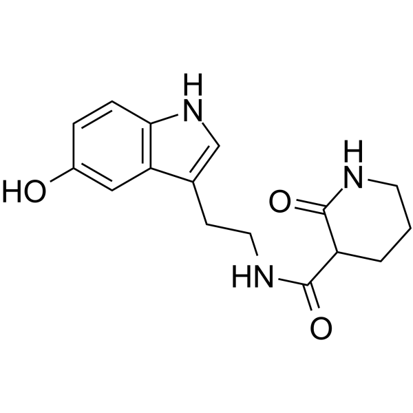
HIOC is a potent and selective activator of TrkB (tropomyosin related kinase B) receptor. HIOC can pass the blood-brain and blood-retinal barriers.HIOC activates TrkB/ERK pathway and decreases neuronal cell apoptosis. HIOC attenuates early brain injury after SAH (subarachnoid hemorrhage). HIOC shows protective activity in an animal model for light-induced retinal degeneration.
For research use only. We do not sell to patients.
- Purity : 99.94%
- CAS No.: 314054-36-9
- Formula: C16H19N3O3
- Molecular Weight:301.34
-
Storage:
4°C, protect from light
* In solvent : -80°C, 6 months; -20°C, 1 month (protect from light)

To place orders, for customer services and technical support, please contact: MedChemExpress USA
Tel: 609-228-6898 E-mail: [email protected] [email protected]


-
 Biological Activity
-
Chemical Information
-
Solvent & Solubility
- Protocols
- Purity & Documentation
- References
-
 Help & FAQs



Publications Citing Use of MedChemExpress (MCE) HIOC
More

Biological Activity
Description
IC50 & Target
|
TrkB |
ERK |
In Vivo


Chemical Information
-
CAS No. 314054-36-9
-
Appearance Solid
-
Molecular Weight 301.34
-
Formula C16H19N3O3
-
Color White to off-white
-
SMILES
O=C(C1C(NCCC1)=O)NCCC2=CNC3=C2C=C(O)C=C3
-
Shipping
Room temperature in continental US; may vary elsewhere.
-
Storage
4°C, protect from light
* In solvent : -80°C, 6 months; -20°C, 1 month (protect from light)


Publications (1)
-
Journal Impact Factor
-
Most Recent
-
J Biol Chem
Stress-induced glucocorticoid signaling impairs enteric neurotrophin BDNF-TrkB pathway and drives gastrointestinal dysmotility. [Abstract]2026 Apr 27;302(7):113095. PMID: 42055341


Solvent & Solubility
In Vitro:
DMSO : 100 mg/mL (331.85 mM; Need ultrasonic; Hygroscopic DMSO has a significant impact on the solubility of product, please use newly opened DMSO)
Please refer to the solubility information to select the appropriate solvent. Once prepared, please aliquot and store the solution to prevent product inactivation from repeated freeze-thaw cycles.
Storage method and period of stock solution: -80°C, 6 months; -20°C, 1 month (protect from light). When stored at -80°C, please use it within 6 months. When stored at -20°C, please use it within 1 month.
Please refer to the solubility information to select the appropriate solvent. Once prepared, please aliquot and store the solution to prevent product inactivation from repeated freeze-thaw cycles.
Storage method and period of stock solution: -80°C, 6 months; -20°C, 1 month (protect from light). When stored at -80°C, please use it within 6 months. When stored at -20°C, please use it within 1 month.
Concentration (start) × Volume (start) = Concentration (final) × Volume (final)

Protocols
-
Apoptosis
Apoptosis, also called programmed cell death, is generally characterized by distinct morphological characteristics.
-
TUNEL staining for apoptotic DNA fragmentation
TUNEL staining detects DNA strand breaks by using terminal deoxynucleotidyl transferase to add labeled nucleotides to exposed 3′-OH DNA termini, generating either microscopic staining in fixed cells or tissue sections, or fluorescence/cytometric signal in cell suspensions. TUNEL positivity reflects DNA fragmentation but should not be interpreted alone as definitive apoptosis, because TUNEL can also label necrotic, autolytic, mechanically damaged, or DNA-repair-associated DNA breaks.
-
Annexin V plus membrane-impermeant dye apoptosis staining
Annexin V-based apoptosis assays rely on the detection of phosphatidylserine (PS) externalization from the inner leaflet of the plasma membrane to the outer leaflet, an early biochemical hallmark of apoptosis. Fluorescently labeled Annexin V binds PS in a calcium-dependent manner, enabling identification of early apoptotic cells by flow cytometry or fluorescence microscopy. When combined with a membrane-impermeant DNA-binding dye (e. g. , propidium iodide), this approach allows discrimination between viable (Annexin V−/dye−), early apoptotic (Annexin V+/dye−), and late apoptotic or necrotic (Annexin V+/dye+) cell populations by assessing membrane integrity and PS exposure.
-
Apoptosis Solutions
Apoptosis is a regulated, generally non-lytic cell-death pathway that removes unwanted, damaged, infected, or abnormal cells through coordinated morphological changes, caspase activation, DNA fragmentation, and membrane remodeling. The intrinsic apoptosis pathway is controlled mainly by mitochondrial outer membrane permeabilization, BCL-2 family proteins, cytochrome c release, apoptosome formation, caspase-9 activation, and downstream executioner caspase-3/7 activation. The extrinsic apoptosis pathway is initiated by death receptors such as Fas, TNFR, and TRAIL receptors, which recruit adaptor proteins and activate caspase-8 before engaging executioner caspases or mitochondrial amplification through BID cleavage. Apoptosis is linked to many phenotypes, including cancer cell killing, tissue homeostasis, immune regulation, neurodegeneration, infection response, and treatment-induced cytotoxicity; unresolved questions include how apoptosis interacts with necroptosis, pyroptosis, ferroptos
-
How to Choose the Right Model Animal
Choosing the right model animal is a validity-driven decision in which the species, strain, sex, age, genetic background, disease-induction method, outcome measures, and welfare burden must match the scientific question rather than laboratory tradition or convenience. A model should be selected by judging face validity, construct validity, and predictive validity: whether it resembles the human phenotype, whether it reproduces relevant mechanisms, and whether results are likely to predict human biology or treatment response. Animal studies often fail to translate because of species differences, weak disease resemblance, poor experimental design, inadequate reporting, publication bias, and underuse of randomization, blinding, and sample-size justification. Unresolved questions include how to rank competing models objectively, how much human-disease complexity must be reproduced for a given objective, and when non-animal systems such as organoids, ex vivo tissue, or computational models


Purity & Documentation
-
Data Sheet (274 KB)
-
SDS (251 KB)
- English - EN (251 KB)
- Français - FR (251 KB)
- Deutsch - DE (251 KB)
- Norwegian - NO (251 KB)
- Español - ES (251 KB)
- Swedish - SV (251 KB)
- Italian - IT (251 KB)
- Korean - KR (251 KB)
- Portuguese - PT (251 KB)
-
Handling Instructions (2659 KB)
References


Complete Stock Solution Preparation Table
Please refer to the solubility information to select the appropriate solvent. Once prepared, please aliquot and store the solution to prevent product inactivation from repeated freeze-thaw cycles.
Storage method and period of stock solution: -80°C, 6 months; -20°C, 1 month (protect from light). When stored at -80°C, please use it within 6 months. When stored at -20°C, please use it within 1 month.
| Optional Solvent | Concentration Solvent Mass | 1 mg | 5 mg | 10 mg | 25 mg |
|---|---|---|---|---|---|
| DMSO | 1 mM | 3.3185 mL | 16.5926 mL | 33.1851 mL | 82.9628 mL |
| 5 mM | 0.6637 mL | 3.3185 mL | 6.6370 mL | 16.5926 mL | |
| 10 mM | 0.3319 mL | 1.6593 mL | 3.3185 mL | 8.2963 mL | |
| 15 mM | 0.2212 mL | 1.1062 mL | 2.2123 mL | 5.5309 mL | |
| 20 mM | 0.1659 mL | 0.8296 mL | 1.6593 mL | 4.1481 mL | |
| 25 mM | 0.1327 mL | 0.6637 mL | 1.3274 mL | 3.3185 mL | |
| 30 mM | 0.1106 mL | 0.5531 mL | 1.1062 mL | 2.7654 mL | |
| 40 mM | 0.0830 mL | 0.4148 mL | 0.8296 mL | 2.0741 mL | |
| 50 mM | 0.0664 mL | 0.3319 mL | 0.6637 mL | 1.6593 mL | |
| 60 mM | 0.0553 mL | 0.2765 mL | 0.5531 mL | 1.3827 mL | |
| 80 mM | 0.0415 mL | 0.2074 mL | 0.4148 mL | 1.0370 mL | |
| 100 mM | 0.0332 mL | 0.1659 mL | 0.3319 mL | 0.8296 mL |


HIOC Related Classifications
HY-101446 Related Classifications
 Powered by Bioz