



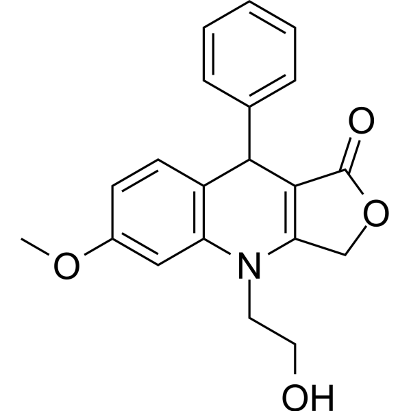
NSC756093

Based on 1 Customer Validation
NSC756093 (SU093) is a GBP1:PIM1 interaction inhibitor. NSC756093 binds to GBP1-PIM1 with a Kd of 38 nM. NSC756093 suppresses proliferation, reduces migration, induces G1 phase cell-cycle arrest, and increases apoptotic cell death in ovarian cancer cells. NSC756093 reduces cellular proteasomal activity, induces accumulation of ubiquitinated proteins, and restrains tumor progression and lung metastasis in murine ovarian cancer xenograft models. NSC756093 increases sensitivity of prostate cancer cells to Docetaxel (HY-B0011) and sensitizes GBP1-overexpressing ovarian cancer cells to Paclitaxel (HY-B0015). NSC756093 can be used for the research of prostate cancer and ovarian cancer.
For research use only. We do not sell to patients.
- Purity : 99.58%
- CAS No.: 1629908-92-4
- Formula: C20H19NO4
- Molecular Weight:337.37
-
Storage:Powder -20°C, 3 years , 4°C, 2 years
* The compound is unstable in solutions, freshly prepared is recommended.

To place orders, for customer services and technical support, please contact: MedChemExpress USA
Tel: 609-228-6898 E-mail: [email protected] [email protected]


-
 Biological Activity
-
Chemical Information
-
Solvent & Solubility
- Protocols
- Purity & Documentation
- References
-
 Help & FAQs




Biological Activity
Description
Cellular Effect
|
Cell Line

|
Type | Value | Description | References |
|---|---|---|---|---|
| FaDu | IC50 |
0.496 μM
Compound: SU093
|
Synergistic antiproliferative activity against human FADU cells assessed as reduction in colony formation unit followed by irradiation with 5 Gy IR and measured after 10 to 14 days by crystal violet staining based assay
Synergistic antiproliferative activity against human FADU cells assessed as reduction in colony formation unit followed by irradiation with 5 Gy IR and measured after 10 to 14 days by crystal violet staining based assay
|
[PMID: 31531203] |
| FaDu | IC50 |
6.5 μM
Compound: SU093
|
Inhibition of colony formation in human FADU cells incubated for 10 to 14 days by crystal violet staining based assay
Inhibition of colony formation in human FADU cells incubated for 10 to 14 days by crystal violet staining based assay
|
[PMID: 31531203] |
In Vitro
NSC756093 (0.1-100 nM) potently and dose-dependently inhibits the recombinant GBP1:PIM1 protein-protein interaction in an SPR-based assay, achieving 65% inhibition at 100 nM[1].
NSC756093 (100 nM; 3 h) inhibits the endogenous GBP1:PIM1 interaction in SKOV3 ovarian cancer cells[1].
NSC756093 (10 nM-100 μM) inhibits growth of NCI-60 human cancer cell lines, with increased sensitivity in cell lines expressing higher levels of both GBP1 and PIM1[1].
NSC756093 (2-8 μM; up to 144 h) potently inhibits the proliferation of parental DU145 and PC3 prostate cancer cells, but has no effect on GBP1 knockout DU145 and PC3 cells, indicating its anti-proliferative activity is dependent on GBP1 expression[2].
NSC756093 (4 μM; 24 h) potently inhibits the migration of DU145 and PC3 prostate cancer cells[2].
NSC756093 (48 h) inhibits viability of multiple ovarian cancer cell lines, demonstrating IC50 values of 1.18 μM (OVCAR3), 13.69 μM (OVCAR4), 14.96 μM (OVCAR5), 2.03 μM (OVCAR8), 1.9 μM (SKOV3), 6.95 μM (ID8) after 48 h treatment[3].
NSC756093 (0.5-1 μM; 7 days) dose-dependently inhibits colony formation of OVCAR8, SKOV3, and ID8 ovarian cancer cells when treated for 7 days, with significant effects at 0.5 and 1 μM[3].
NSC756093 (1-5 μM; 12 h) causes dose-dependent cell cycle arrest in sub-G1 and G1 phases of OVCAR8, SKOV3, and ID8 ovarian cancer cells after 12 h treatment at 1, 2.5, and 5 μM[3].
NSC756093 (2.5-5 μM; 24 h) dose-dependently induces apoptotic cell death in OVCAR8, SKOV3, and ID8 ovarian cancer cells after 24 h treatment at 2.5 and 5 μM[3].
NSC756093 (1 μM; 12 h pre-treatment, 16 h migration) inhibits migration of OVCAR8, SKOV3, and ID8 ovarian cancer cells by 40-46% in a Boyden chamber assay[3].
NSC756093 (SU093) significantly inhibits proteasomal activity in ovarian cancer cells[4].
MedChemExpress (MCE) has not independently confirmed the accuracy of these methods. They are for reference only. Further protocols information, click here.
-
Cell Line:Human prostate cancer DU145 cells, human prostate cancer PC3 cells, GBP1 knockout DU145 cells, GBP1 knockout PC3 cells
-
Concentration:2 μM, 4 μM, 8 μM (preliminary dose optimization); 4 μM (growth monitoring)
-
Incubation Time:Up to 96 h (DU145 growth monitoring); up to 144 h (PC3 growth monitoring)
-
Result:Significantly reduced the growth rate of parental DU145 and PC3 cells.
Had no apparent growth-inhibitory effect on GBP1 knockout DU145 and PC3 cells.
Caused more severe growth inhibition of parental cells at 8 μM, while induced milder effect at 2 μM.
-
Cell Line:Human prostate cancer DU145 cells, human prostate cancer PC3 cells
-
Concentration:4 μM
-
Incubation Time:24 h
-
Result:Significantly reduced the number of migrated DU145 and PC3 cells per field compared to untreated control cells.
-
Cell Line:OVCAR8, SKOV3, ID8
-
Concentration:1 μM, 2.5 μM, 5 μM
-
Incubation Time:12 h
-
Result:Induced significant, dose-dependent arrest in sub-G1 and G1 phases, with concomitant decreases in S or G2/M phase cells across all three cell lines.
-
Cell Line:OVCAR8, SKOV3, ID8
-
Concentration:2.5 μM, 5 μM
-
Incubation Time:24 h
-
Result:Significantly increased the proportion of apoptotic cells at both doses in all three cell lines.
Showed higher apoptosis observed at 5 μM compared to 2.5 μM.
In Vivo
MedChemExpress (MCE) has not independently confirmed the accuracy of these methods. They are for reference only.
-
Animal Model:C57BL/6 (female, 6-7 weeks old, syngeneic xenograft model via subcutaneous implantation of luciferase-expressing ID8 cells)[3]
-
Dosage:20 mg/kg
-
Administration:i.p.; daily; 42 days
-
Result:Significantly delayed tumor progression compared to vehicle control.
Reduced tumor weight by 1.5-fold relative to vehicle controls at study termination.
Reduced the number of lung metastatic nodules relative to vehicle controls.
Caused no significant changes in liver toxicity parameters (aspartate aminotransferase, alanine aminotransferase, alkaline phosphatase).


Chemical Information
-
CAS No. 1629908-92-4
-
Appearance Solid
-
Molecular Weight 337.37
-
Formula C20H19NO4
-
Color White to off-white
-
SMILES
O=C1OCC(N2CCO)=C1C(C3=CC=CC=C3)C4=C2C=C(OC)C=C4
-
Synonyms
SU093
-
Shipping
Room temperature in continental US; may vary elsewhere.
-
Storage
Powder -20°C 3 years 4°C 2 years * The compound is unstable in solutions, freshly prepared is recommended.


Solvent & Solubility
In Vitro:
DMSO : 62.5 mg/mL (185.26 mM; Need ultrasonic; Hygroscopic DMSO has a significant impact on the solubility of product, please use newly opened DMSO)
Please refer to the solubility information to select the appropriate solvent. The compound is unstable in solutions, freshly prepared is recommended.
Please refer to the solubility information to select the appropriate solvent. The compound is unstable in solutions, freshly prepared is recommended.
Concentration (start) × Volume (start) = Concentration (final) × Volume (final)

Protocols
-
Apoptosis
Apoptosis, also called programmed cell death, is generally characterized by distinct morphological characteristics.
-
Cell Cytotoxicity Assay
Cytotoxicity assays are usually based on the assessment of cell membrane damage, which can also be indirectly detected by measuring cell viability. Detection methods include MTT assay, CKK-8 assay, LDH assay and ATP assay, etc.
-
Mammalian live/dead viability and cytotoxicity staining
Live/dead viability and cytotoxicity staining assays are based on the simultaneous detection of intracellular esterase activity in metabolically active (viable) cells and membrane integrity loss in non-viable cells. In commonly used dual-staining approaches, membrane-permeant fluorogenic substrates are converted by intracellular esterases into fluorescent products in live cells, while impermeant DNA-binding dyes selectively enter cells with compromised plasma membranes and label nucleic acids in dead or dying cells, enabling discrimination between viable and non-viable populations by fluorescence microscopy or flow cytometry.
-
Cell migration
Cell migration is a method that plays an important role in wound healing, cell differentiation, embryonic development, etc.
-
Tail-Vein Experimental Metastasis Xenograft
Tail-vein experimental metastasis xenograft models assess the ability of injected tumor cells to survive circulation, arrest in vascular beds, extravasate, and colonize distant organs, most commonly lung after lateral tail-vein injection; this model bypasses primary-tumor formation, local invasion, and intravasation, so the readout reflects late metastatic colonization rather than the full metastatic cascade. The main readouts are metastatic burden measured by bioluminescence imaging, gross metastatic nodules, histology, organ weight, survival, or ex vivo tumor-cell quantification; luciferase-labeled tumor cells permit longitudinal noninvasive monitoring, while histology confirms organ colonization and tissue localization.
-
Patient-Derived Xenograft (PDX)
Patient-derived xenograft (PDX) models are generated by engrafting primary human tumor tissue directly into immunodeficient mice, allowing in vivo propagation of patient tumor biology without initial in vitro adaptation. These models are used to preserve key histopathological and molecular characteristics of the original tumor and enable assessment of tumor growth dynamics and therapeutic response in a living organism. The biological readout is tumor engraftment and subsequent growth in the murine host, which reflects the ability of human tumor cells to survive, vascularize, and expand in an immunocompromised microenvironment.
-
Apoptosis Solutions
Apoptosis is a regulated, generally non-lytic cell-death pathway that removes unwanted, damaged, infected, or abnormal cells through coordinated morphological changes, caspase activation, DNA fragmentation, and membrane remodeling. The intrinsic apoptosis pathway is controlled mainly by mitochondrial outer membrane permeabilization, BCL-2 family proteins, cytochrome c release, apoptosome formation, caspase-9 activation, and downstream executioner caspase-3/7 activation. The extrinsic apoptosis pathway is initiated by death receptors such as Fas, TNFR, and TRAIL receptors, which recruit adaptor proteins and activate caspase-8 before engaging executioner caspases or mitochondrial amplification through BID cleavage. Apoptosis is linked to many phenotypes, including cancer cell killing, tissue homeostasis, immune regulation, neurodegeneration, infection response, and treatment-induced cytotoxicity; unresolved questions include how apoptosis interacts with necroptosis, pyroptosis, ferroptos
-
Splenic/Portal-Vein Liver Metastasis Xenograft
Splenic and portal-vein liver metastasis xenograft models deliver tumor cells into the portal circulation so that cells reach the liver first and form hepatic metastatic lesions; splenic injection uses the spleen as an access route to the portal system, while direct portal-vein injection introduces cells into the portal vein without requiring splenectomy. The assay detects liver colonization, intrahepatic tumor growth, tumor distribution, treatment response, survival, and liver-metastasis microenvironment changes; readouts include bioluminescence or fluorescence imaging, gross liver nodule counts, liver weight or tumor burden, histology, and survival.
-
Subcutaneous Cell-Line-Derived Xenograft
Subcutaneous cell-line-derived xenograft (CDX) models are established by implanting cultured human cancer cell lines into immunodeficient mice, where the injected cells form localized tumors that can be monitored in vivo as a measure of tumorigenic potential, growth kinetics, and treatment response. These models are widely used in oncology research because they allow reproducible tumor formation and enable comparative assessment of tumor growth between different cell lines or genetic manipulations in a controlled in vivo microenvironment. Subcutaneous implantation of cancer cells in immunodeficient mice is a standard approach for evaluating tumor growth behavior and therapeutic response across multiple cancer types, including prostate, esophageal, pancreatic, and colon cancer models.
-
Orthotopic Cell-Line Xenograft
Orthotopic cell-line xenograft models involve implantation of human cancer cell lines into the anatomically corresponding organ of immunodeficient mice to reproduce tumor growth within a native microenvironment, enabling more clinically relevant tumor behavior compared with subcutaneous models. These models are widely used because orthotopic placement better recapitulates tumor progression, including invasion and metastatic spread, which are often underrepresented in heterotopic implantation systems. Compared with conventional xenografts, orthotopic implantation is described as more technically complex but provides improved simulation of tumor-microenvironment interactions and metastatic behavior, making it particularly valuable for translational oncology research. Surgical orthotopic implantation approaches have been emphasized as enabling faithful reproduction of clinical cancer features, including metastasis and disease progression patterns that align with the tumor’s organ of origi
-
Cell Viability Determination by MTT Colorimetric Assay
The following protocol uses the MTT colorimetric assay as a classic literature-established method for assessing cell viability/metabolic activity in cultured mammalian cells. MTT[3-(4,5-dimethylthiazol-2-yl)-2,5-diphenyltetrazolium bromide] is reduced by metabolically active cells to a colored formazan product; the amount of formazan is quantified spectrophotometrically and provides an indirect measure of metabolically active viable cells. Importantly, MTT reduction reflects cellular oxidoreductase/metabolic activity rather than an absolute direct count of living cells, so changes in cellular metabolism can alter the signal independently of cell number.
-
Intraperitoneal/Peritoneal Dissemination Xenograft
Intraperitoneal (IP) or peritoneal dissemination xenograft models are based on the introduction of human cancer cells into the peritoneal cavity of immunodeficient mice, where they attach to peritoneal surfaces, form multicellular aggregates or spheroids, and progressively generate disseminated tumor nodules that mimic advanced peritoneal metastatic disease. These models are widely used to study ovarian cancer progression, tumor-microenvironment interactions, and intraperitoneal therapeutic responses, often incorporating bioluminescence or fluorescence imaging to longitudinally monitor tumor burden in vivo. The biological principle relies on the capacity of tumor cells such as SKOV3 or related ovarian carcinoma lines to survive in suspension, aggregate within ascites-like fluid, adhere to mesothelial surfaces, and invade peritoneal organs, thereby recapitulating human peritoneal carcinomatosis patterns observed in advanced disease.


Purity & Documentation
-
Data Sheet (284 KB)
-
SDS (536 KB)
- English - EN (536 KB)
- Français - FR (536 KB)
- Deutsch - DE (536 KB)
- Norwegian - NO (536 KB)
- Español - ES (536 KB)
- Swedish - SV (536 KB)
- Italian - IT (536 KB)
- Korean - KR (536 KB)
- Portuguese - PT (536 KB)
-
Handling Instructions (2659 KB)
References
[1]. Andreoli M, et al. Identification of the first inhibitor of the GBP1:PIM1 interaction. Implications for the development of a new class of anticancer agents against paclitaxel resistant cancer cells. J Med Chem. 2014;57(19):7916-7932. [Content Brief]
[2]. Zhao J, et al. Oncogenic Role of Guanylate Binding Protein 1 in Human Prostate Cancer. Front Oncol. 2020 Jan 10;9:1494. [Content Brief]
[3]. Tailor D, et al. Y box binding protein 1 inhibition as a targeted therapy for ovarian cancer. Cell Chem Biol. 2021;28(8):1206-1220.e6. [Content Brief]
[4]. Tailor D, et al. Guanylate-binding protein 1 modulates proteasomal machinery in ovarian cancer. iScience. 2023;26(11):108292. Published 2023 Oct 24. [Content Brief]


Complete Stock Solution Preparation Table
Please refer to the solubility information to select the appropriate solvent. The compound is unstable in solutions, freshly prepared is recommended.
| Optional Solvent | Concentration Solvent Mass | 1 mg | 5 mg | 10 mg | 25 mg |
|---|---|---|---|---|---|
| DMSO | 1 mM | 2.9641 mL | 14.8205 mL | 29.6410 mL | 74.1026 mL |
| 5 mM | 0.5928 mL | 2.9641 mL | 5.9282 mL | 14.8205 mL | |
| 10 mM | 0.2964 mL | 1.4821 mL | 2.9641 mL | 7.4103 mL | |
| 15 mM | 0.1976 mL | 0.9880 mL | 1.9761 mL | 4.9402 mL | |
| 20 mM | 0.1482 mL | 0.7410 mL | 1.4821 mL | 3.7051 mL | |
| 25 mM | 0.1186 mL | 0.5928 mL | 1.1856 mL | 2.9641 mL | |
| 30 mM | 0.0988 mL | 0.4940 mL | 0.9880 mL | 2.4701 mL | |
| 40 mM | 0.0741 mL | 0.3705 mL | 0.7410 mL | 1.8526 mL | |
| 50 mM | 0.0593 mL | 0.2964 mL | 0.5928 mL | 1.4821 mL | |
| 60 mM | 0.0494 mL | 0.2470 mL | 0.4940 mL | 1.2350 mL | |
| 80 mM | 0.0371 mL | 0.1853 mL | 0.3705 mL | 0.9263 mL | |
| 100 mM | 0.0296 mL | 0.1482 mL | 0.2964 mL | 0.7410 mL |


NSC756093 Related Classifications
HY-12929 Related Classifications
 Powered by Bioz
Keywords