



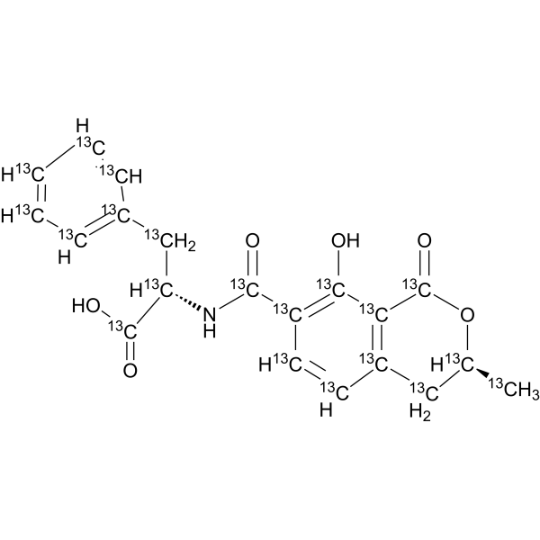
Ochratoxin B13C20
Ochratoxin B-13C20 is 13C-labeled Ochratoxin B (HY-N6786). Ochratoxin B is an orally active secondary metabolite of Aspergillus ochraceus and non-chlorinated analog of the mycotoxin Ochratoxin A. Ochratoxin B reduces the toxic effects of Ochratoxin A (HY-N6788). Ochratoxin B inhibits cell division. Ochratoxin B causes craniofacial malformations in Xenopus laevis embryos.
For research use only. We do not sell to patients.
- Formula: 13C20H19NO6
- Molecular Weight:389.22
-
Storage:
Please store the product under the recommended conditions in the Certificate of Analysis.

To place orders, for customer services and technical support, please contact: MedChemExpress USA
Tel: 609-228-6898 E-mail: [email protected] [email protected]


-
 Biological Activity
-
Chemical Information
- Protocols
- Purity & Documentation
- References
-
 Help & FAQs


All Endogenous Metabolite Isoforms
More

Biological Activity
Description
In Vitro
Stable heavy isotopes of hydrogen, carbon, and other elements have been incorporated into drug molecules, largely as tracers for quantitation during the drug development process. Deuteration has gained attention because of its potential to affect the pharmacokinetic and metabolic profiles of drugs[1].
MedChemExpress (MCE) has not independently confirmed the accuracy of these methods. They are for reference only. Further protocols information, click here.
Application
1. This compound can be used as a tracer
2. This compound can be used as an internal standard for quantitative analysis by NMR, GC-MS, or LC-MS.


Chemical Information
-
Unlabeled CAS 4825-86-9
-
Molecular Weight 389.22
-
Formula 13C20H19NO6
-
SMILES
O=[13C]1O[13C@@H]([13CH3])[13CH2][13C]2=[13C]1[13C](O)=[13C]([13C](N[13C@H]([13C](O)=O)[13CH2][13C]3=[13CH][13CH]=[13CH][13CH]=[13CH]3)=O)[13CH]=[13CH]2
-
Shipping
Room temperature in continental US; may vary elsewhere.
-
Storage
Please store the product under the recommended conditions in the Certificate of Analysis.

Protocols
-
CFSE Dye Dilution Proliferation Assay
The CFSE (carboxyfluorescein diacetate succinimidyl ester) dye dilution proliferation assay is based on the covalent labeling of intracellular proteins by a cell-permeant fluorescent dye that becomes fluorescent upon intracellular ester cleavage and then is stably retained within cells. As labeled cells divide, the dye is partitioned equally between daughter cells, resulting in a stepwise halving of fluorescence intensity that can be quantified by flow cytometry to determine the number of cell divisions undergone by each cell population. This fluorescence dilution approach enables quantitative tracking of lymphocyte proliferation at the single-cell level over multiple rounds of division. CFSE-based proliferation analysis has been widely applied to measure antigen-driven lymphocyte expansion in vitro, where discrete fluorescence peaks correspond to successive cell divisions and allow reconstruction of proliferative history within heterogeneous populations.
-
Dye-dilution cell tracking and proliferation staining
Dye-dilution cell tracking assays quantify cell proliferation by covalently labeling intracellular proteins with a stable fluorescent dye that is equally partitioned between daughter cells during mitosis, resulting in stepwise halving of fluorescence intensity with each cell division as measured by flow cytometry histograms. Carboxyfluorescein diacetate succinimidyl ester (CFSE) is a prototypical dye that diffuses into cells, is enzymatically converted into a fluorescent compound, and then covalently binds intracellular amine groups, producing long-lived fluorescence suitable for tracking multiple rounds of division in vitro and in vivo. Successive generations of dividing cells form discrete peaks of decreasing fluorescence intensity, enabling estimation of proliferation history, precursor frequency, and division index within heterogeneous populations. Alternative dyes such as CellTrace Violet (CTV) and far-red membrane dyes (e. g. , PKH26) follow the same dilution principle but differ


Purity & Documentation
References
[1]. Russak EM, et al. Impact of Deuterium Substitution on the Pharmacokinetics of Pharmaceuticals. Ann Pharmacother. 2019 Feb;53(2):211-220. [Content Brief]
[2]. Knasmüller S, et al. Structurally related mycotoxins ochratoxin A, ochratoxin B, and citrinin differ in their genotoxic activities and in their mode of action in human-derived liver (HepG2) cells: implications for risk assessment. Nutr Cancer. 2004;50(2):190-7. [Content Brief]
[3]. O'Brien E, et al. Investigation of the teratogenic potential of ochratoxin A and B using the FETAX system. Birth Defects Res B Dev Reprod Toxicol. 2005 Oct;74(5):417-23. [Content Brief]
[4]. Dietrich DR, et al. Species- and sex-specific renal cytotoxicity of ochratoxin A and B in vitro. Exp Toxicol Pathol. 2001 Jun;53(2-3):215-25. [Content Brief]
[5]. Csenki Z, et al. The individual and combined effects of ochratoxin A with citrinin and their metabolites (ochratoxin B, ochratoxin C, and dihydrocitrinone) on 2D/3D cell cultures, and zebrafish embryo models. Food Chem Toxicol. 2021 Dec;158:112674. [Content Brief]
[6]. Mally A, et al. Biotransformation and nephrotoxicity of ochratoxin B in rats. Toxicol Appl Pharmacol. 2005 Aug 1;206(1):43-53. [Content Brief]
[7]. Størmer FC, et al. Metabolism of ochratoxin B and its possible effects upon the metabolism and toxicity of ochratoxin A in rats. Appl Environ Microbiol. 1985 May;49(5):1108-12. [Content Brief]

Calculators
Concentration (start) × Volume (start) = Concentration (final) × Volume (final)


Ochratoxin B13C20 Related Classifications
HY-N6786S Related Classifications
 Powered by Bioz