



PAPA NONOate
Based on 2 publication(s) in Google Scholar
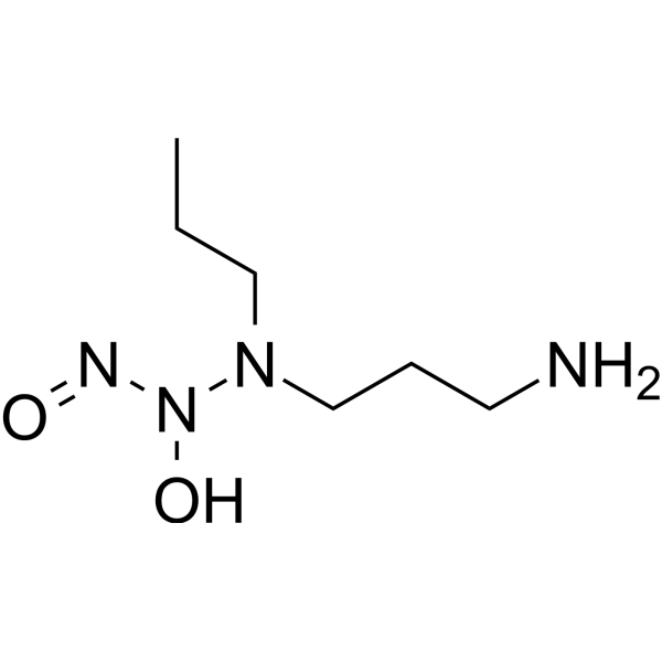
PAPA NONOate is a NO donor with a NO release half-life of 77 min (22-25°C). PAPA NONOate may represent a potential research for impaired wound healing in diabetes by increasing the rate of collagen synthesis at the wound site.
For research use only. We do not sell to patients.
- Purity : 98.0%
- CAS No.: 146672-58-4
- Formula: C6H16N4O2
- Molecular Weight:176.22
-
Storage:
Please store the product under the recommended conditions in the Certificate of Analysis.

To place orders, for customer services and technical support, please contact: MedChemExpress USA
Tel: 609-228-6898 E-mail: [email protected] [email protected]


-
 Biological Activity
-
Chemical Information
-
Solvent & Solubility
- Protocols
- Purity & Documentation
- References
-
 Help & FAQs




Publications Citing Use of MedChemExpress (MCE) PAPA NONOate
MoreAll Endogenous Metabolite Isoforms
More

Biological Activity
Description
In Vitro
PAPA NONOate (50 μM; 12 h) significantly reduces 70% caspase-3-like activity in caspase-3-overexpressing HUVEC and abolishs the induction of cell death by caspase-3[1].
MedChemExpress (MCE) has not independently confirmed the accuracy of these methods. They are for reference only. Further protocols information, click here.
In Vivo
MedChemExpress (MCE) has not independently confirmed the accuracy of these methods. They are for reference only.
-
Animal Model:Anesthetized male Sprague-Dawley rats with streptozotocin-induced diabetes and full thickness dermal wound[2]
-
Dosage:100 μM
-
Administration:Wounds received 100 μM PAPA NONOate in phosphate buffer
-
Result:Increased the wound healing rate in test group.


Chemical Information
-
CAS No. 146672-58-4
-
Appearance Solid
-
Molecular Weight 176.22
-
Formula C6H16N4O2
-
Color White to light yellow
-
SMILES
NCCCN(CCC)N(O)N=O
-
Structure Classification
-
Initial Source
-
Shipping
Shipping with dry ice.
-
Storage
Please store the product under the recommended conditions in the Certificate of Analysis.


Publications (2)
-
Journal Impact Factor
-
Most Recent
-
ACS Nano
Triple-Cascade Responsive Pneumatic Nanomotor Enhance Cancer Immunotherapy by Oncolytic Virus-Triggered Pyroptosis. [Abstract]2026 Mar 31;20(12):9784-9801. PMID: 41842752 -
Neurosci Bull
2025 Jun 28. PMID: 40580390


Solvent & Solubility
In Vitro:
H2O : 50 mg/mL (283.74 mM; ultrasonic and warming and heat to 60°C)
Please refer to the solubility information to select the appropriate solvent. The compound is unstable in solutions, freshly prepared is recommended.
* Note: If you choose water as the stock solution, please dilute it to the working solution, then filter and sterilize it with a 0.22 μm filter before use.
Please refer to the solubility information to select the appropriate solvent. The compound is unstable in solutions, freshly prepared is recommended.
* Note: If you choose water as the stock solution, please dilute it to the working solution, then filter and sterilize it with a 0.22 μm filter before use.
Concentration (start) × Volume (start) = Concentration (final) × Volume (final)

Protocols
-
Cell migration
Cell migration is a method that plays an important role in wound healing, cell differentiation, embryonic development, etc.
-
Protocol for Pharmacokinetic Study
Pharmacokinetic studies quantify how an organism handles a drug over time through absorption, distribution, metabolism, and excretion, and the core experimental readout is the concentration-time profile of parent drug and, when relevant, metabolites in biological matrices such as plasma, whole blood, urine, bile, or tissue. Pharmacokinetic analysis links dose, route, exposure, clearance, half-life, distribution, bioavailability, and systemic exposure to drug efficacy and toxicity hypotheses rather than measuring a signaling pathway directly. The literature links pharmacokinetics to drug-development phenotypes by showing that drug metabolism and pharmacokinetics influence compound progression, exposure-response interpretation, safety margins, dosing strategy, and failure risk during discovery and development. DMPK science contributes to compound optimization by integrating physicochemical properties, in vitro metabolism, transporter behavior, in vivo exposure, and pharmacodynamic contex
-
Cell-Exclusion Zone Migration Assay
The Cell-Exclusion Zone (CEZ) migration assay is an in vitro 2D cell migration method in which a defined cell-free area is created using removable physical barriers such as silicone stoppers, allowing cells to be seeded around the barrier and subsequently migrate into the cleared zone after barrier removal. This approach enables quantification of collective cell migration by monitoring repopulation of the initially cell-free region over time using microscopy-based imaging. Compared with scratch-based wound healing assays, barrier-based exclusion methods are designed to avoid mechanical damage to the extracellular matrix and reduce injury-induced effects on boundary cells, thereby improving interpretability of migration behavior in vitro. The assay readout is typically the progressive reduction in the cell-free area or the number of cells invading the exclusion zone, reflecting coordinated cell motility relevant to physiological processes such as wound healing, epithelial repair, and ca
-
Research Protocol for Metabolic Diseases
AMP-activated protein kinase, AMPK, is a conserved cellular energy sensor that responds to reduced cellular energy status and coordinates metabolism by increasing ATP-generating catabolic pathways while suppressing ATP-consuming anabolic processes. In metabolic disease research, the AMPK pathway is experimentally relevant because it regulates hepatic lipid synthesis, fatty acid oxidation, glucose production, skeletal-muscle glucose disposal, mTORC1-linked biosynthesis, autophagy, mitochondrial homeostasis, and whole-body energy balance. The central pathway logic is that energy stress, metformin, exercise-like stimulation, or direct AMPK activators increase AMPKα Thr172 phosphorylation and downstream substrate phosphorylation, including ACC and RAPTOR. Phosphorylation of ACC suppresses lipogenesis and supports fatty acid oxidation, whereas phosphorylation of RAPTOR suppresses mTORC1 signaling and links cellular energy status to growth and protein synthesis control. The pathway is linked


Purity & Documentation
-
Data Sheet (271 KB)
-
SDS (392 KB)
- English - EN (392 KB)
- Français - FR (392 KB)
- Deutsch - DE (392 KB)
- Norwegian - NO (392 KB)
- Español - ES (392 KB)
- Swedish - SV (392 KB)
- Italian - IT (392 KB)
- Korean - KR (392 KB)
- Portuguese - PT (392 KB)
-
Handling Instructions (2659 KB)
References
[1]. Rössig L, et al. Nitric oxide inhibits caspase-3 by S-nitrosation in vivo. J Biol Chem. 1999 Mar 12;274(11):6823-6. [Content Brief]
[3]. Keefer LK, et al. "NONOates" (1-substituted diazen-1-ium-1,2-diolates) as nitric oxide donors: convenient nitric oxide dosage forms. Methods Enzymol. 1996;268:281-93. [Content Brief]


Complete Stock Solution Preparation Table
Please refer to the solubility information to select the appropriate solvent. The compound is unstable in solutions, freshly prepared is recommended.
| Optional Solvent | Concentration Solvent Mass | 1 mg | 5 mg | 10 mg | 25 mg |
|---|---|---|---|---|---|
| H2O | 1 mM | 5.6747 mL | 28.3736 mL | 56.7472 mL | 141.8681 mL |
| 5 mM | 1.1349 mL | 5.6747 mL | 11.3494 mL | 28.3736 mL | |
| 10 mM | 0.5675 mL | 2.8374 mL | 5.6747 mL | 14.1868 mL | |
| 15 mM | 0.3783 mL | 1.8916 mL | 3.7831 mL | 9.4579 mL | |
| 20 mM | 0.2837 mL | 1.4187 mL | 2.8374 mL | 7.0934 mL | |
| 25 mM | 0.2270 mL | 1.1349 mL | 2.2699 mL | 5.6747 mL | |
| 30 mM | 0.1892 mL | 0.9458 mL | 1.8916 mL | 4.7289 mL | |
| 40 mM | 0.1419 mL | 0.7093 mL | 1.4187 mL | 3.5467 mL | |
| 50 mM | 0.1135 mL | 0.5675 mL | 1.1349 mL | 2.8374 mL | |
| 60 mM | 0.0946 mL | 0.4729 mL | 0.9458 mL | 2.3645 mL | |
| 80 mM | 0.0709 mL | 0.3547 mL | 0.7093 mL | 1.7734 mL | |
| 100 mM | 0.0567 mL | 0.2837 mL | 0.5675 mL | 1.4187 mL |
* Note: If you choose water as the stock solution, please dilute it to the working solution, then filter and sterilize it with a 0.22 μm filter before use.


PAPA NONOate Related Classifications
HY-134636 Related Classifications
 Powered by Bioz