



Parishin B
Based on 1 publication(s) in Google Scholar
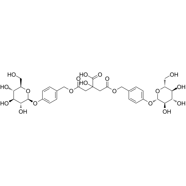
Parishin B is a multi-target biological agent with anti-epileptic, anti-fascioliasis and anti-breast cancer lung metastasis activities. Parishin B regulates the gene/protein activities of ACLY, unc13c, γ-aminobutynergic system, pro-inflammatory system, antioxidant system, monoaminergic system, ferroptosis-related pathways, as well as GST, CatL, Trx and TRIB3. Parishin B modulates energy-lipid metabolism, synaptic function, neurotransmitter balance, neuroinflammation, oxidative stress, parasite detoxification/invasion processes, as well as apoptosis, cell cycle, proliferation and metastasis of cancer cells[1][2][3]. Parishin B can be used for related research of epilepsy, fascioliasis and breast cancer lung metastasis[3].
For research use only. We do not sell to patients.
- Purity : 99.42%
- CAS No.: 174972-79-3
- Formula: C32H40O19
- Molecular Weight:728.65
-
Storage:
4°C, protect from light
* In solvent : -80°C, 6 months; -20°C, 1 month (protect from light)

To place orders, for customer services and technical support, please contact: MedChemExpress USA
Tel: 609-228-6898 E-mail: [email protected] [email protected]


-
 Biological Activity
-
Chemical Information
-
Solvent & Solubility
- Protocols
- Purity & Documentation
- References
-
 Help & FAQs


-
HY-N2125
Parishin C5-HT1A Receptor Agonist -
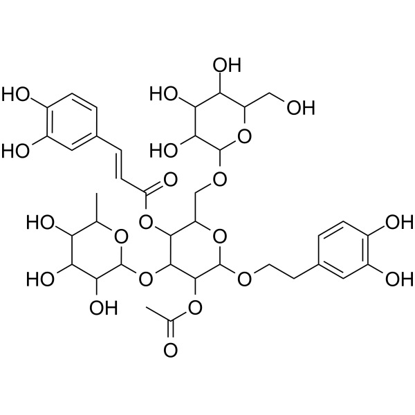
HY-N2155
Tubuloside AHepatoprotective Agent -
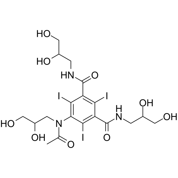
HY-B0594
IohexolContrast Agent -
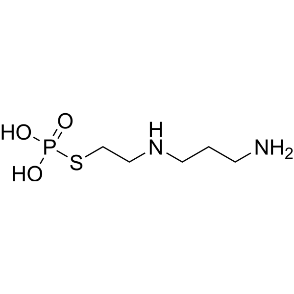
HY-B0639
AmifostineCytoprotective Agent -
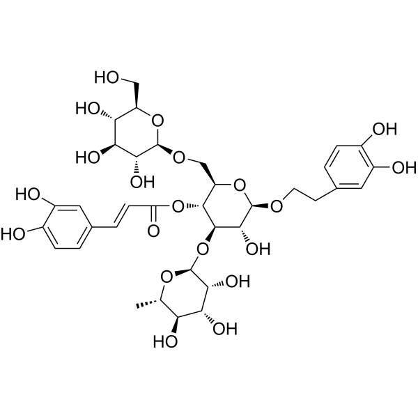
HY-N0020
EchinacosideWnt/β-catenin Signaling Inhibitor -
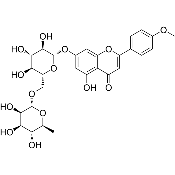
HY-N0528
LinarinAChE Inhibitor -
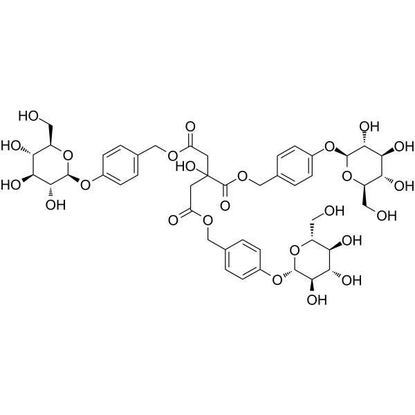
HY-N2031
ParishinSir2/Uth1/TOR Modulator -
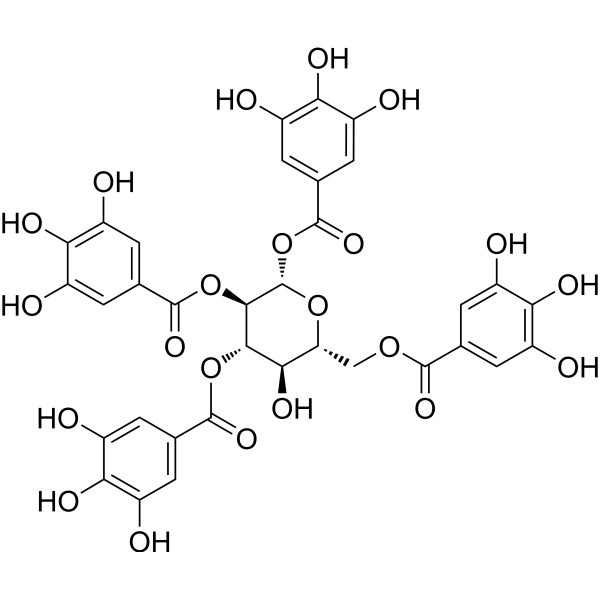
HY-111832
1,2,3,6-TetragalloylglucoseUGT1A1 Inhibitor -
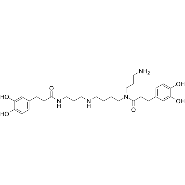
HY-N2393
Kukoamine BActive Compound -
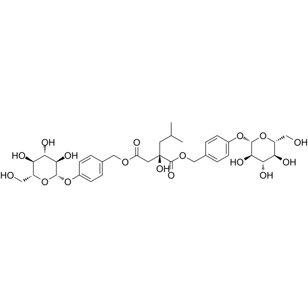
HY-122308
MilitarineAnti-inflammatory Agent -
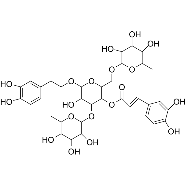
HY-N0033
PoliumosideAldose Reductase Inhibitor -
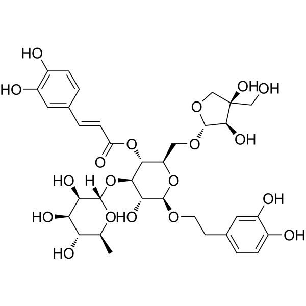
HY-N0029
Forsythoside BTNF Inhibitor
Publications Citing Use of MedChemExpress (MCE) Parishin B
More

Biological Activity
Description
In Vitro
Parishin B (10 μM; 1 h) increases the thermal stability of TRIB3 in MDA-MB-231 cell lysate, confirming direct binding to the protein[3].
Parishin B (20 μM; 24 h) blocks the TRIB3-AKT1 protein-protein interaction in MDA-MB-231 breast cancer cells[3].
Parishin B (0-120 μM; 48 h) selectively reduces viability in MDA-MB-231, SK-BR-3, MCF-7, HCC1954, and T-47D breast cancer cells with minimal toxicity to MCF-10A normal mammary epithelial cells[3].
Parishin B (5-20 μM; 12 day) suppresses colony formation in MDA-MB-231 breast cancer cells, reducing colony numbers by ~50% at 20 μM[3].
Parishin B (2.5-5 μM; 24 h) significantly reduces the migratory and invasive capabilities of MDA-MB-231 breast cancer cells, with ~80% inhibition of both processes at 5 μM[3].
Parishin B (5-20 μM; 24 h) induces concentration-dependent apoptosis in MDA-MB-231 breast cancer cells, with a ~40% apoptosis rate at 20 μM[3].
Parishin B (20 μM; 24 h) alters the gene expression profile of MDA-MB-231 breast cancer cells, with significant enrichment of DEGs in the cell cycle signaling pathway[3].
Parishin B (5-20 μM; 24 h) induces concentration-dependent G2/M phase arrest in MDA-MB-231 breast cancer cells, with ~50% of cells in G2/M at 20 μM[3].
Parishin B (5-20 μM; 24 h) upregulates CDK1 and Cyclin B1 expression, and increases PI3K and AKT1 phosphorylation levels, in MDA-MB-231 breast cancer cells[3].
MedChemExpress (MCE) has not independently confirmed the accuracy of these methods. They are for reference only. Further protocols information, click here.
-
Cell Line:MDA-MB-231, SK-BR-3, MCF-7, HCC1954, T-47D breast cancer cells, MCF-10A normal mammary epithelial cells
-
Concentration:1.25 μM; 2.5 μM; 5 μM; 10 μM; 20 μM; 40 μM; 80 μM;120 μM
-
Incubation Time:24 h; 48 h
-
Result:Reduced viability in all tested breast cancer cell lines, with the strongest effects observed in MDA-MB-231 and MCF-7 cells.
Showed minimal toxicity to MCF-10A normal mammary epithelial cells at concentrations up to 120 μM, indicating selective cytotoxicity toward cancer cells.
-
Cell Line:MDA-MB-231 breast cancer cells
-
Concentration:5-20 μM
-
Incubation Time:24 h
-
Result:Induced apoptosis in MDA-MB-231 cells in a concentration-dependent manner, with apoptosis rates reaching ~15% at 5 μM, ~30% at 10 μM, and ~40% at 20 μM.
-
Cell Line:MDA-MB-231 breast cancer cells
-
Concentration:5-20 μM
-
Incubation Time:12 day
-
Result:Significantly suppressed colony formation in MDA-MB-231 cells, reducing colony numbers by ~20% at 5 μM, ~20% at 10 μM, and ~50% at 20 μM compared to control.
-
Cell Line:MDA-MB-231 breast cancer cells
-
Concentration:2.5-5 μM
-
Incubation Time:24 h
-
Result:Significantly reduced the migratory capacity of MDA-MB-231 cells, reducing relative migration by ~50% at 2.5 μM and ~80% at 5 μM.
Significantly reduced invasive capacity, reducing relative invasion by ~50% at 2.5 μM and ~80% at 5 μM.
-
Cell Line:MDA-MB-231 breast cancer cells
-
Concentration:5-20 μM
-
Incubation Time:24 h
-
Result:Induced a concentration-dependent G2/M phase arrest in MDA-MB-231 cells, with the percentage of cells in G2/M increasing from ~15% in control to ~20% at 5 μM, ~30% at 10 μM, and ~50% at 20 μM.
-
Cell Line:MDA-MB-231 breast cancer cells
-
Concentration:5-20 μM
-
Incubation Time:24 h
-
Result:Increased the expression of CDK1 and Cyclin B1 in a concentration-dependent manner, with significant upregulation observed at 10 μM and 20 μM.
Increased the phosphorylation levels of PI3K (Tyr458) and AKT1 (Ser473) in a concentration-dependent manner, with significant increases observed at 10 μM and 20 μM.
In Vivo
Parishin B (0.0625-0.25 mg/mL; immersion; 5 days) causes no apparent developmental toxicity or behavioral impairment in zebrafish embryos[1].
Parishin B (4-8 mg/kg; i.p.; once a day; 6 weeks) inhibits breast cancer lung metastasis in female BALB/c nude mice in vivo in a dose-dependent manner, with the 8 mg/kg daily intraperitoneal dose reducing average lung tumor nodule number by ~55% and lung fluorescence flux by ~67%, while upregulating AKT/PI3K phosphorylation and Cyclin B1/CDK1 expression in lung tissues without causing detectable toxicity[3].
MedChemExpress (MCE) has not independently confirmed the accuracy of these methods. They are for reference only.
-
Animal Model:wild-type AB strain (7 days post-fertilization larvae)[1]
-
Dosage:0.0625 mg/mL; 0.125 mg/mL; 0.25 mg/mL
-
Administration:immersion; 60 min
-
Result:Significantly reduced PTZ-induced hyperlocomotion, including total swimming distance and average swimming speed (p < 0.05, p < 0.01) compared to the PTZ-only group.
Significantly increased GABA (p < 0.05, p < 0.001), dopamine (p < 0.001), and norepinephrine (p < 0.01, p < 0.001, p < 0.0001) levels, while reducing elevated 5-HT levels (p < 0.05, p < 0.001) compared to the PTZ-only group; did not significantly alter glutamate levels.
Exerted dose-dependent anti-inflammatory effects: at 0.125 mg/mL, reduced IL-1β levels more effectively than the positive control VPA (p < 0.0001), partially restored PTZ-reduced IL-6 levels (p < 0.001, p < 0.0001), and suppressed PTZ-increased TNF-α levels.
Significantly increased superoxide dismutase (SOD) activity at all tested doses (p < 0.0001), with effects superior to VPA.
Partially normalized PTZ-induced global metabolic alterations at 0.125 mg/mL, restoring key energy metabolism metabolites (e.g., inositol, Alpha-D-glucose) and reversing lipid accumulation.
Reversed PTZ-induced proteomic dysregulation at 0.125 mg/mL, stabilizing cytoskeleton proteins, restoring synaptic protein (unc-13) homeostasis, and upregulating ATP-citrate lyase (ACLY).
Upregulated GABAergic signaling genes (gad2, slc32a1, gabra1), synaptic priming gene unc13c, and metabolic regulatory gene aclya (p < 0.001, p < 0.0001); suppressed pro-inflammatory genes (il1b, tnfa, nfkb1) (p < 0.05, p < 0.001, p < 0.0001); upregulated oxidative stress and mitochondrial function genes (sod1, gpx1a, ndufs1, ucp2) (p < 0.001, p < 0.0001); and suppressed monoaminergic neurotransmitter metabolism genes (th, dbh, tph1a, tph2) (p < 0.05, p < 0.001, p < 0.0001).
-
Animal Model:BALB/c nude (female, 4-5 weeks old, breast cancer lung metastasis model via tail vein injection of MDA-MB-231-LUC cells)[3]
-
Dosage:4 mg/kg; 8 mg/kg
-
Administration:i.p.; once a day; 6 weeks
-
Result:Reduced average lung tumor nodule number from ~11 to ~7 (4 mg/kg) and from ~11 to ~5 (8 mg/kg).
Reduced lung fluorescence flux from ~6 × 10⁶ p/s to ~3 × 10⁶ p/s (4 mg/kg) and from ~6 × 10⁶ p/s to ~2 × 10⁶ p/s (8 mg/kg).
Increased relative phosphorylation levels of AKT by 1.8-fold (4 mg/kg) and 2.0-fold (8 mg/kg) relative to controls.
Increased relative phosphorylation levels of PI3K by 1.4-fold (4 mg/kg) and 1.8-fold (8 mg/kg) relative to controls.
Increased relative expression of Cyclin B1 by 1.7-fold (4 mg/kg) and 1.9-fold (8 mg/kg) relative to controls.
Increased relative expression of CDK1 by 1.9-fold (4 mg/kg) and 2.8-fold (8 mg/kg) relative to controls.
Showed marked improvement in pulmonary metastatic lesions via H&E staining in both treated groups compared to controls.
Caused no significant drug toxicity, with all organ indices remaining within normal ranges.


Chemical Information
-
CAS No. 174972-79-3
-
Appearance Solid
-
Molecular Weight 728.65
-
Formula C32H40O19
-
Color White to off-white
-
SMILES
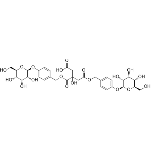
O=C(OCC1=CC=C(O[C@H]2[C@@H]([C@H]([C@@H]([C@@H](CO)O2)O)O)O)C=C1)CC(O)(CC(O)=O)C(OCC3=CC=C(O[C@H]4[C@@H]([C@H]([C@@H]([C@@H](CO)O4)O)O)O)C=C3)=O
-
Structure Classification
-
Initial Source
-
Shipping
Room temperature in continental US; may vary elsewhere.
-
Storage
4°C, protect from light
* In solvent : -80°C, 6 months; -20°C, 1 month (protect from light)


Publications (1)
-
Journal Impact Factor
-
Most Recent


Solvent & Solubility
In Vitro:
DMSO : 50 mg/mL (68.62 mM; Need ultrasonic; Hygroscopic DMSO has a significant impact on the solubility of product, please use newly opened DMSO)
H2O : 50 mg/mL (68.62 mM; Need ultrasonic)
Please refer to the solubility information to select the appropriate solvent. Once prepared, please aliquot and store the solution to prevent product inactivation from repeated freeze-thaw cycles.
Storage method and period of stock solution: -80°C, 6 months; -20°C, 1 month (protect from light). When stored at -80°C, please use it within 6 months. When stored at -20°C, please use it within 1 month.
* Note: If you choose water as the stock solution, please dilute it to the working solution, then filter and sterilize it with a 0.22 μm filter before use.
Please refer to the solubility information to select the appropriate solvent. Once prepared, please aliquot and store the solution to prevent product inactivation from repeated freeze-thaw cycles.
Storage method and period of stock solution: -80°C, 6 months; -20°C, 1 month (protect from light). When stored at -80°C, please use it within 6 months. When stored at -20°C, please use it within 1 month.
* Note: If you choose water as the stock solution, please dilute it to the working solution, then filter and sterilize it with a 0.22 μm filter before use.
Concentration (start) × Volume (start) = Concentration (final) × Volume (final)
In Vivo:
Select the appropriate dissolution method based on your experimental animal and administration route.
- For the following dissolution methods, please ensure to first prepare a clear stock solution using an In Vitro approach and then sequentially add co-solvents:
- To ensure reliable experimental results, the clarified stock solution can be appropriately stored based on storage conditions. As for the working solution for In Vivo experiments, it is recommended to prepare freshly and use it on the same day.
- The percentages shown for the solvents indicate their volumetric ratio in the final prepared solution. If precipitation or phase separation occurs during preparation, heat and/or sonication can be used to aid dissolution.
Add each solvent one by one: 10% DMSO 40% PEG300 5% Tween-80 45% Saline
Solubility: ≥ 2.5 mg/mL (3.43 mM); Clear solution
This protocol yields a clear solution of ≥ 2.5 mg/mL (saturation unknown).
Taking 1 mL working solution as an example, add 100 μL DMSO stock solution (25.0 mg/mL) to 400 μL PEG300, and mix evenly; then add 50 μL Tween-80 and mix evenly; then add 450 μL Saline to adjust the volume to 1 mL.
Preparation of Saline: Dissolve 0.9 g sodium chloride in ddH₂O and dilute to 100 mL to obtain a clear Saline solution.
Add each solvent one by one: 10% DMSO 90% (20% SBE-β-CD in Saline)
Solubility: ≥ 2.5 mg/mL (3.43 mM); Clear solution
This protocol yields a clear solution of ≥ 2.5 mg/mL (saturation unknown).
Taking 1 mL working solution as an example, add 100 μL DMSO stock solution (25.0 mg/mL) to 900 μL 20% SBE-β-CD in Saline, and mix evenly.
Preparation of 20% SBE-β-CD in Saline (4°C, storage for one week): 2 g SBE-β-CD powder is dissolved in 10 mL Saline, completely dissolve until clear.
In Vivo Dissolution Calculator
Please enter the basic information of animal experiments:
-
-
-
-
Recommended: Prepare an additional quantity of animals to account for potential losses during experiments.
Working solution concentration: 0.22 mg/mL
This product has good water solubility, please refer to the measured solubility data in water/PBS/Saline for details.

Protocols
-
Annexin V plus membrane-impermeant dye apoptosis staining
Annexin V-based apoptosis assays rely on the detection of phosphatidylserine (PS) externalization from the inner leaflet of the plasma membrane to the outer leaflet, an early biochemical hallmark of apoptosis. Fluorescently labeled Annexin V binds PS in a calcium-dependent manner, enabling identification of early apoptotic cells by flow cytometry or fluorescence microscopy. When combined with a membrane-impermeant DNA-binding dye (e. g. , propidium iodide), this approach allows discrimination between viable (Annexin V−/dye−), early apoptotic (Annexin V+/dye−), and late apoptotic or necrotic (Annexin V+/dye+) cell populations by assessing membrane integrity and PS exposure.
-
ROS/oxidative-stress fluorescent staining
ROS/oxidative-stress fluorescent staining uses cell-permeant fluorogenic probes that become fluorescent after oxidation inside cells or tissues; commonly used examples include DCFH-DA/DCFDA for broad cellular oxidant detection, DHE for superoxide-related signal detection, MitoSOX for mitochondrial superoxide-related signal detection, and CellROX probes for oxidative-stress-associated fluorescence readouts. The assay detects probe oxidation rather than a single ROS species unless the probe and analysis method have been chemically validated for that species. DCFH-DA enters cells, is deacetylated by intracellular esterases to DCFH, and produces fluorescent DCF after oxidation, so the readout is used as an operational measure of total cellular oxidative stress rather than a species-specific ROS measurement. DHE and MitoSOX can report superoxide-related oxidation, but red fluorescence alone can include non-specific ethidium-like oxidation products; HPLC or optimized spectral approaches are
-
BrdU Incorporation Assay
Bromodeoxyuridine (BrdU) incorporation assay is based on the principle that BrdU, a thymidine analog, is incorporated into newly synthesized DNA during the S phase of the cell cycle, thereby serving as a marker of DNA replication and cellular proliferation. Incorporated BrdU can be detected using anti-BrdU antibodies following DNA denaturation, enabling visualization or quantification of proliferating cells through immunochemical detection methods such as immunofluorescence or immunohistochemistry.
-
Protocol for Cell Cycle
Cell-cycle analysis by flow cytometry measures DNA content in single cells to estimate the fraction of cells in G0/G1, S, and G2/M phases. Propidium iodide intercalates into DNA, and after RNA removal with RNase, fluorescence intensity reflects cellular DNA content: 2N cells are assigned to G0/G1, cells between 2N and 4N to S phase, and 4N cells to G2/M. DNA-content analysis alone cannot reliably separate G0 from G1 or G2 from M. Ki-67 can distinguish quiescent G0 cells from cycling cells, EdU or BrdU incorporation marks active DNA synthesis in S phase, and phospho-histone H3 staining identifies mitotic cells within the 4N population.
-
Research Protocol for Inflammation-related Diseases
The NLRP3 inflammasome is a cytosolic innate immune signaling platform that integrates priming signals and danger-signal activation to promote caspase-1 activation, maturation of IL-1β and IL-18, and gasdermin D-mediated pyroptotic cell death. The core experimental logic is to determine whether inflammatory disease phenotypes are driven by increased NLRP3 expression, ASC-containing inflammasome assembly, caspase-1 cleavage, GSDMD cleavage, and extracellular release of IL-1β/IL-18 rather than by nonspecific cell injury alone. The pathway is strongly linked to inflammation-related disease phenotypes because monosodium urate crystals activate NALP3/NLRP3 inflammasome signaling in gout-like crystal inflammation, cholesterol crystals activate NLRP3 inflammasomes in atherogenesis models, and DSS-induced intestinal inflammation has been reported to involve NLRP3 inflammasome activity. However, experimental colitis studies also show context-dependent protective effects of NLRP3 inflammasome co
-
Research Protocol for Neurological Diseases
PINK1/Parkin-mediated mitophagy pathway is a mitochondrial quality-control signaling axis in which mitochondrial depolarization stabilizes PINK1 on damaged mitochondria, activates Parkin recruitment and E3 ubiquitin ligase activity, promotes ubiquitination of outer mitochondrial membrane proteins, recruits selective autophagy adaptors, and drives lysosomal degradation of damaged mitochondria. In neurological disease research, this pathway is experimentally important because neurons, especially dopaminergic neurons, are highly dependent on mitochondrial integrity, and defective mitochondrial turnover can lead to mitochondrial dysfunction, oxidative stress, impaired neuronal survival, α-synuclein accumulation, and neuroinflammatory damage-associated signals. The genetic disease link is strongest in Parkinson’s disease because mutations in PRKN/parkin cause autosomal recessive juvenile parkinsonism, mutations in PINK1 cause hereditary early-onset Parkinson’s disease, and Drosophila studie
-
Breast Cancer Modeling
Breast cancer is a heterogeneous cancer, and it has been distinguished into four subtypes: luminal A, luminal B, HER2-positive and basal-like. Molecular mutations, epigenetic alterations, hormone exposure and immune microenvironment are related to the progression of breast cancer.
-
Cell Viability Determination by MTT Colorimetric Assay
The following protocol uses the MTT colorimetric assay as a classic literature-established method for assessing cell viability/metabolic activity in cultured mammalian cells. MTT[3-(4,5-dimethylthiazol-2-yl)-2,5-diphenyltetrazolium bromide] is reduced by metabolically active cells to a colored formazan product; the amount of formazan is quantified spectrophotometrically and provides an indirect measure of metabolically active viable cells. Importantly, MTT reduction reflects cellular oxidoreductase/metabolic activity rather than an absolute direct count of living cells, so changes in cellular metabolism can alter the signal independently of cell number.
-
Apoptosis
Apoptosis, also called programmed cell death, is generally characterized by distinct morphological characteristics.
-
TUNEL staining for apoptotic DNA fragmentation
TUNEL staining detects DNA strand breaks by using terminal deoxynucleotidyl transferase to add labeled nucleotides to exposed 3′-OH DNA termini, generating either microscopic staining in fixed cells or tissue sections, or fluorescence/cytometric signal in cell suspensions. TUNEL positivity reflects DNA fragmentation but should not be interpreted alone as definitive apoptosis, because TUNEL can also label necrotic, autolytic, mechanically damaged, or DNA-repair-associated DNA breaks.
-
Flow cytometric DNA-content cell-cycle staining
Flow cytometric DNA-content cell-cycle staining measures the fluorescence intensity of DNA-bound fluorochromes in single cells or nuclei to estimate DNA content distributions, allowing assignment of populations to G0/G1, S, and G2/M phases by DNA histogram deconvolution. Propidium iodide (PI) intercalates into DNA, and PI fluorescence is proportional to cellular DNA content when staining is performed under conditions that make DNA accessible and minimize non-DNA signal. Cells with G2/M DNA content are expected to show approximately twice the fluorescence intensity of G0/G1 cells, while S-phase cells occupy intermediate fluorescence values. PI-based DNA-content analysis can also detect cells with fractional DNA content, often reported as sub-G1, when DNA fragmentation and extraction during staining reduce retained DNA signal in apoptotic cells. DAPI is an alternative DNA fluorochrome for univariate DNA-content analysis, while bivariate approaches combining DNA content with proliferation
-
Tail-Vein Experimental Metastasis Xenograft
Tail-vein experimental metastasis xenograft models assess the ability of injected tumor cells to survive circulation, arrest in vascular beds, extravasate, and colonize distant organs, most commonly lung after lateral tail-vein injection; this model bypasses primary-tumor formation, local invasion, and intravasation, so the readout reflects late metastatic colonization rather than the full metastatic cascade. The main readouts are metastatic burden measured by bioluminescence imaging, gross metastatic nodules, histology, organ weight, survival, or ex vivo tumor-cell quantification; luciferase-labeled tumor cells permit longitudinal noninvasive monitoring, while histology confirms organ colonization and tissue localization.
-
Apoptosis Solutions
Apoptosis is a regulated, generally non-lytic cell-death pathway that removes unwanted, damaged, infected, or abnormal cells through coordinated morphological changes, caspase activation, DNA fragmentation, and membrane remodeling. The intrinsic apoptosis pathway is controlled mainly by mitochondrial outer membrane permeabilization, BCL-2 family proteins, cytochrome c release, apoptosome formation, caspase-9 activation, and downstream executioner caspase-3/7 activation. The extrinsic apoptosis pathway is initiated by death receptors such as Fas, TNFR, and TRAIL receptors, which recruit adaptor proteins and activate caspase-8 before engaging executioner caspases or mitochondrial amplification through BID cleavage. Apoptosis is linked to many phenotypes, including cancer cell killing, tissue homeostasis, immune regulation, neurodegeneration, infection response, and treatment-induced cytotoxicity; unresolved questions include how apoptosis interacts with necroptosis, pyroptosis, ferroptos
-
Cell invasion
Cell invasion is the ability of cells to migrate from one area to another via the extracellular matrix. Cell invasion is the response of normal and cancer cells to chemical and mechanical stimuli. Before migrating to a new region, the extracellular matrix is degraded by proteases within the cell. Cell invasion often occurs during wound repair, vascularization and inflammation, abnormal tissue invasion, and tumor cell metastasis.
-
Ferroptosis Solutions
Ferroptosis is an iron-dependent, non-apoptotic form of regulated cell death characterized by lethal lipid peroxidation and sensitivity to suppression by iron chelators or lipophilic radical-trapping antioxidants. The core pathway links cystine uptake through system Xc−, glutathione availability, GPX4-dependent detoxification of phospholipid hydroperoxides, iron-dependent oxidative reactions, and polyunsaturated-phospholipid metabolism into a cell-death program that is biochemically and morphologically distinct from apoptosis, necrosis, and autophagy. The ferroptosis pathway is experimentally linked to phenotype through chemical and genetic perturbation. Erastin induces ferroptosis by inhibiting cystine uptake through system Xc− and weakening antioxidant defenses, while GPX4 inhibition or depletion causes lipid peroxide accumulation and ferroptotic cancer-cell death. ACSL4 and oxidizable arachidonoyl- or adrenoyl-containing phosphatidylethanolamines shape ferroptosis sensitivity by con
-
Orthotopic Cell-Line Xenograft
Orthotopic cell-line xenograft models involve implantation of human cancer cell lines into the anatomically corresponding organ of immunodeficient mice to reproduce tumor growth within a native microenvironment, enabling more clinically relevant tumor behavior compared with subcutaneous models. These models are widely used because orthotopic placement better recapitulates tumor progression, including invasion and metastatic spread, which are often underrepresented in heterotopic implantation systems. Compared with conventional xenografts, orthotopic implantation is described as more technically complex but provides improved simulation of tumor-microenvironment interactions and metastatic behavior, making it particularly valuable for translational oncology research. Surgical orthotopic implantation approaches have been emphasized as enabling faithful reproduction of clinical cancer features, including metastasis and disease progression patterns that align with the tumor’s organ of origi


Purity & Documentation
-
Data Sheet (288 KB)
-
SDS (393 KB)
- English - EN (393 KB)
- Français - FR (393 KB)
- Deutsch - DE (393 KB)
- Norwegian - NO (393 KB)
- Español - ES (393 KB)
- Swedish - SV (393 KB)
- Italian - IT (393 KB)
- Korean - KR (393 KB)
- Portuguese - PT (393 KB)
-
Handling Instructions (2659 KB)
References
[1]. Sun M, et al. Parishin B Attenuates PTZ-Induced Seizures in Zebrafish and Is Associated with Neurotransmitter Balance and ACLY-Related Metabolic Pathways. Metabolites. 2026 Apr 18;16(4):275. [Content Brief]
[2]. Zulqarnain M, et al. Integrated in vitro and in silico analysis of Artemisia absinthium identifies Parishin C and Parishin B as promising multitarget antifasciolic agents. Fitoterapia. 2026 Jan;188:107038. [Content Brief]
[3]. Cheng X, et al. Parishin B blocking TRIB3-AKT1 interaction inhibits breast cancer lung metastasis. Frontiers in pharmacology. 2024;15:1517708. [Content Brief]
[4]. Cheng X, et al. An Optimized and Sensitive Pharmacokinetic Quantitative Method of Investigating Gastrodin, Parishin, and Parishin B, C and E in Beagle Dog Plasma using LC-MS/MS after Intragastric Administration of Tall Gastrodia Capsules. Molecules. 2017 Nov 10;22(11):1938. [Content Brief]


Complete Stock Solution Preparation Table
Please refer to the solubility information to select the appropriate solvent. Once prepared, please aliquot and store the solution to prevent product inactivation from repeated freeze-thaw cycles.
Storage method and period of stock solution: -80°C, 6 months; -20°C, 1 month (protect from light). When stored at -80°C, please use it within 6 months. When stored at -20°C, please use it within 1 month.
| Optional Solvent | Concentration Solvent Mass | 1 mg | 5 mg | 10 mg | 25 mg |
|---|---|---|---|---|---|
| DMSO / H2O | 1 mM | 1.3724 mL | 6.8620 mL | 13.7240 mL | 34.3100 mL |
| 5 mM | 0.2745 mL | 1.3724 mL | 2.7448 mL | 6.8620 mL | |
| 10 mM | 0.1372 mL | 0.6862 mL | 1.3724 mL | 3.4310 mL | |
| 15 mM | 0.0915 mL | 0.4575 mL | 0.9149 mL | 2.2873 mL | |
| 20 mM | 0.0686 mL | 0.3431 mL | 0.6862 mL | 1.7155 mL | |
| 25 mM | 0.0549 mL | 0.2745 mL | 0.5490 mL | 1.3724 mL | |
| 30 mM | 0.0457 mL | 0.2287 mL | 0.4575 mL | 1.1437 mL | |
| 40 mM | 0.0343 mL | 0.1716 mL | 0.3431 mL | 0.8578 mL | |
| 50 mM | 0.0274 mL | 0.1372 mL | 0.2745 mL | 0.6862 mL | |
| 60 mM | 0.0229 mL | 0.1144 mL | 0.2287 mL | 0.5718 mL |
* Note: If you choose water as the stock solution, please dilute it to the working solution, then filter and sterilize it with a 0.22 μm filter before use.


Parishin B Related Classifications
HY-N2124 Related Classifications
 Powered by Bioz