



PBP10 TFA
Based on 1 publication(s) in Google Scholar
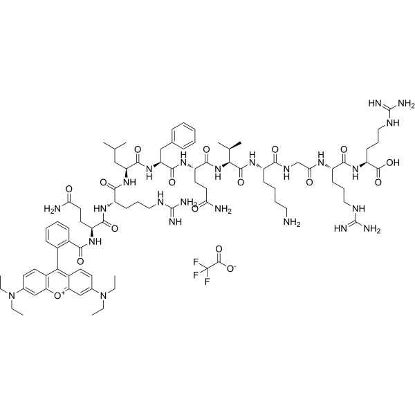
PBP10 TFA is a decapeptide. PBP10 TFA selectively binds to lipoteichoic acid (LTA), lipopolysaccharide (LPS) and phosphatidylinositol-4,5-bisphosphate (PIP2). PBP10 TFA penetrates cell membranes and possesses bactericidal, anti-inflammatory, cell motility-inhibiting and actin assembly-regulating activities. PBP10 TFA is applicable to relevant research on bacterial infections, microbe-induced inflammation, skin and soft tissue infections, as well as sepsis.
For research use only. We do not sell to patients.
- Purity : 98.22%
- Formula: C86H127F3N24O17
- Molecular Weight:1826.07
-
Storage:
Sealed storage, away from moisture.
Powder -80°C, 2 years , -20°C, 1 year* In solvent : -80°C, 6 months; -20°C, 1 month (sealed storage, away from moisture)

To place orders, for customer services and technical support, please contact: MedChemExpress USA
Tel: 609-228-6898 E-mail: [email protected] [email protected]


-
 Biological Activity
-
Chemical Information
-
Solvent & Solubility
- Protocols
- Purity & Documentation
- References
-
 Help & FAQs


Publications Citing Use of MedChemExpress (MCE) PBP10 TFA
More

Biological Activity
Description
IC50 & Target
IC50: formyl peptide receptor 2 (FPR2)[1]
In Vitro
PBP10 (2 μM; 15 min) TFA selectively binds to LTA, LPS, and PIP2, but does not bind to PE[1].
PBP10 (1-18 h) TFA exhibits bactericidal activity against E. coli SG13009 with an MIC of 12.5 μg/mL, and also shows bactericidal activity against B. subtilis ATCC 6051 with an MIC of 3.125 μg/mL. Its bactericidal activity is strongly inhibited by LPS and LTA[1].
PBP10 (0-50 μM) TFA inhibits the directional migration of NIH3T3 fibroblasts, human A7 melanoma cells and human neutrophils in a concentration-dependent manner[2].
PBP10 (1-50 μM) TFA potently inhibits thrombin-stimulated human platelet aggregation and platelet actin assembly in a concentration-dependent manner[2].
PBP10 (5-20 μM) TFA concentration-dependently inhibits the migration speed and actin assembly of human neutrophils stimulated by fMLP[2].
PBP10 (0-100 µg/mL; 24 h) TFA exhibits extremely low cytotoxicity against human keratinocyte HaCaT cells[3].
PBP10 (2-10 µg/mL; 1 h) TFA exhibits potent dose-dependent bactericidal activity against E. coli RS218 and S. aureus A1[3].
PBP10 (2-10 µg/mL; 24 h) TFA dose-dependently inhibits the production of NO and ROS in human keratinocyte HaCaT cells stimulated by LPS (HY-D1056), LTA, heat-inactivated E. coli or heat-inactivated S. aureus[3].
PBP10 (2-10 µg/mL; 24 h) TFA reduces IL-8 release in LPS- and LTA-stimulated human keratinocyte HaCaT cells in a dose-dependent manner[3].
MedChemExpress (MCE) has not independently confirmed the accuracy of these methods. They are for reference only.
-
Cell Line:HaCaT human keratinocytes (stimulated with LPS or LTA)
-
Concentration:2, 5, 10 µg/mL
-
Incubation Time:24 h (co-incubated with LPS or LTA)
-
Result:Reduced IL-8 release in LPS/LTA in a dose-dependent manner.


Chemical Information
-
Appearance Solid
-
Molecular Weight 1826.07
-
Formula C86H127F3N24O17
-
Color Purple to purplish red
-
Sequence
RhB-Gln-Arg-Leu-Phe-Gln-Val-Lys-Gly-Arg-Arg-OH
-
Sequence Shortening
RhB-QRLFQVKGRR-OH
-
Shipping
Room temperature in continental US; may vary elsewhere.
-
Storage
Sealed storage, away from moisture
Powder -80°C 2 years -20°C 1 year * In solvent : -80°C, 6 months; -20°C, 1 month (sealed storage, away from moisture)


Publications (1)
-
Journal Impact Factor
-
Most Recent
-
Cancer Res
DLGAP1-AS2-Mediated Phosphatidic Acid Synthesis Activates YAP Signaling and Confers Chemoresistance in Squamous Cell Carcinoma. [Abstract]2022 Aug 16;82(16):2887-2903. PMID: 35731019


Solvent & Solubility
In Vitro:
DMSO : 100 mg/mL (54.76 mM; Need ultrasonic; Hygroscopic DMSO has a significant impact on the solubility of product, please use newly opened DMSO)
Please refer to the solubility information to select the appropriate solvent. Once prepared, please aliquot and store the solution to prevent product inactivation from repeated freeze-thaw cycles.
Storage method and period of stock solution: -80°C, 6 months; -20°C, 1 month (sealed storage, away from moisture). When stored at -80°C, please use it within 6 months. When stored at -20°C, please use it within 1 month.
Please refer to the solubility information to select the appropriate solvent. Once prepared, please aliquot and store the solution to prevent product inactivation from repeated freeze-thaw cycles.
Storage method and period of stock solution: -80°C, 6 months; -20°C, 1 month (sealed storage, away from moisture). When stored at -80°C, please use it within 6 months. When stored at -20°C, please use it within 1 month.
Concentration (start) × Volume (start) = Concentration (final) × Volume (final)


Purity & Documentation
-
Data Sheet (277 KB)
-
SDS (252 KB)
- English - EN (252 KB)
- Français - FR (252 KB)
- Deutsch - DE (252 KB)
- Norwegian - NO (252 KB)
- Español - ES (252 KB)
- Swedish - SV (252 KB)
- Italian - IT (252 KB)
- Korean - KR (252 KB)
- Portuguese - PT (252 KB)
-
Handling Instructions (2659 KB)
References
[1]. Ewelina Piktel, et al. Inhibition of inflammatory response in human keratinocytes by magnetic nanoparticles functionalized with PBP10 peptide derived from the PIP2-binding site of human plasma gelsolin. J Nanobiotechnology. 2019 Feb 2;17(1):22. [Content Brief]
[2]. C C Cunningham, et al.Cell permeant polyphosphoinositide-binding peptides that block cell motility and actin assembly. J Biol Chem [Content Brief]


Complete Stock Solution Preparation Table
Please refer to the solubility information to select the appropriate solvent. Once prepared, please aliquot and store the solution to prevent product inactivation from repeated freeze-thaw cycles.
Storage method and period of stock solution: -80°C, 6 months; -20°C, 1 month (sealed storage, away from moisture). When stored at -80°C, please use it within 6 months. When stored at -20°C, please use it within 1 month.
| Optional Solvent | Concentration Solvent Mass | 1 mg | 5 mg | 10 mg | 25 mg |
|---|---|---|---|---|---|
| DMSO | 1 mM | 0.5476 mL | 2.7381 mL | 5.4762 mL | 13.6906 mL |
| 5 mM | 0.1095 mL | 0.5476 mL | 1.0952 mL | 2.7381 mL | |
| 10 mM | 0.0548 mL | 0.2738 mL | 0.5476 mL | 1.3691 mL | |
| 15 mM | 0.0365 mL | 0.1825 mL | 0.3651 mL | 0.9127 mL | |
| 20 mM | 0.0274 mL | 0.1369 mL | 0.2738 mL | 0.6845 mL | |
| 25 mM | 0.0219 mL | 0.1095 mL | 0.2190 mL | 0.5476 mL | |
| 30 mM | 0.0183 mL | 0.0913 mL | 0.1825 mL | 0.4564 mL | |
| 40 mM | 0.0137 mL | 0.0685 mL | 0.1369 mL | 0.3423 mL | |
| 50 mM | 0.0110 mL | 0.0548 mL | 0.1095 mL | 0.2738 mL |


PBP10 TFA Related Classifications
HY-P1116A Related Classifications
 Powered by Bioz