



PBP10
Based on 1 publication(s) in Google Scholar
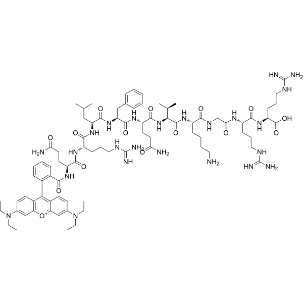
PBP10 is a decapeptide. PBP10 selectively binds to lipoteichoic acid (LTA), lipopolysaccharide (LPS) and phosphatidylinositol-4,5-bisphosphate (PIP2). PBP10 penetrates cell membranes and possesses bactericidal, anti-inflammatory, cell motility-inhibiting and actin assembly-regulating activities. PBP10 is applicable to relevant research on bacterial infections, microbe-induced inflammation, skin and soft tissue infections, as well as sepsis.
For research use only. We do not sell to patients.
- Purity : 96.75%
- CAS No.: 794466-43-6
- Formula: C84H127N24O15+
- Molecular Weight:1713.06
-
Storage:
Sealed storage, away from moisture.
Powder -80°C, 2 years , -20°C, 1 year* In solvent : -80°C, 6 months; -20°C, 1 month (sealed storage, away from moisture)

To place orders, for customer services and technical support, please contact: MedChemExpress USA
Tel: 609-228-6898 E-mail: [email protected] [email protected]


-
 Biological Activity
-
Chemical Information
- Protocols
- Purity & Documentation
- References
-
 Help & FAQs


-
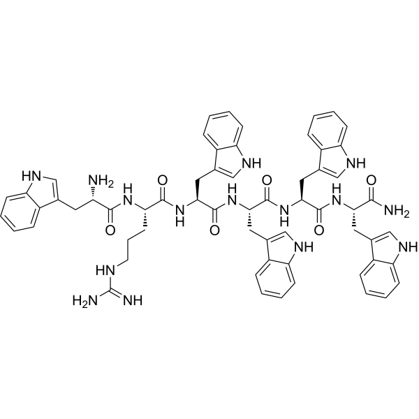
HY-P1119
WRW4FPRL1 Antagonist -
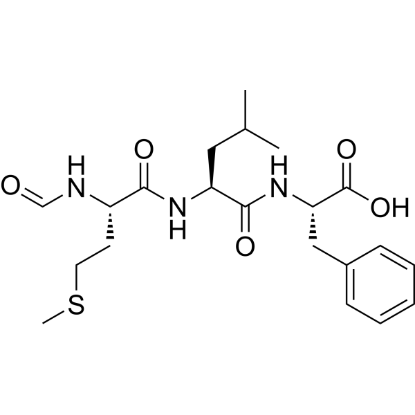
HY-P0224
N-Formyl-Met-Leu-PheTNF Receptor Inhibitor -

HY-P1117
MMK1FPRL-1 Agonist -
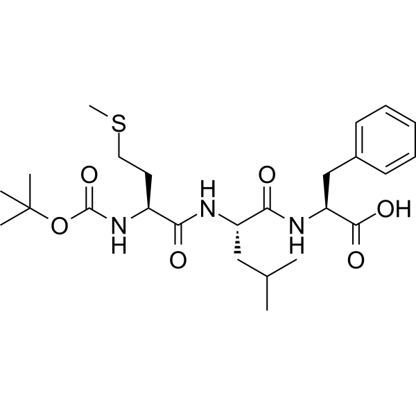
HY-103473
Boc-MLFFPR Antagonist -
HY-P1120
WKYMVmN-Formyl Peptide Receptor Agonist -

HY-P1118
Ac9-25FPR Agonist -

HY-P1121
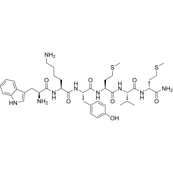
WKYMVM-NH2FPRL1/FPRL2 Recaptor Agonist -
HY-P1122
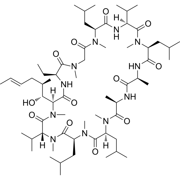
Cyclosporin HFPR-1 Inhibitor -
HY-103472
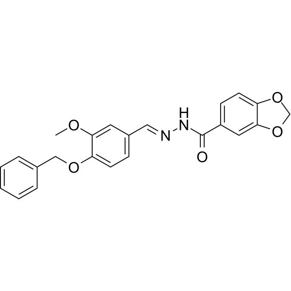
FPR-A14FPR Agonist -

HY-103472R
FPR-A14 (Standard)Reference Standard
Publications Citing Use of MedChemExpress (MCE) PBP10
More

Biological Activity
Description
IC50 & Target
IC50: formyl peptide receptor 2 (FPR2)[1]
In Vitro
PBP10 (2 μM; 15 min) selectively binds to LTA, LPS, and PIP2, but does not bind to PE[1].
PBP10 (1-18 h) exhibits bactericidal activity against E. coli SG13009 with an MIC of 12.5 μg/mL, and also shows bactericidal activity against B. subtilis ATCC 6051 with an MIC of 3.125 μg/mL. Its bactericidal activity is strongly inhibited by LPS and LTA[1].
PBP10 (0-50 μM) inhibits the directional migration of NIH3T3 fibroblasts, human A7 melanoma cells and human neutrophils in a concentration-dependent manner[2].
PBP10 (1-50 μM) potently inhibits thrombin-stimulated human platelet aggregation and platelet actin assembly in a concentration-dependent manner[2].
PBP10 (5-20 μM) concentration-dependently inhibits the migration speed and actin assembly of human neutrophils stimulated by fMLP[2].
PBP10 (0-100 µg/mL; 24 h) exhibits extremely low cytotoxicity against human keratinocyte HaCaT cells[3].
PBP10 (2-10 µg/mL; 1 h) exhibits potent dose-dependent bactericidal activity against E. coli RS218 and S. aureus A1[3].
PBP10 (2-10 µg/mL; 24 h) dose-dependently inhibits the production of NO and ROS in human keratinocyte HaCaT cells stimulated by LPS (HY-D1056), LTA, heat-inactivated E. coli or heat-inactivated S. aureus[3].
PBP10 (2-10 µg/mL; 24 h) reduces IL-8 release in LPS- and LTA-stimulated human keratinocyte HaCaT cells in a dose-dependent manner[3].
MedChemExpress (MCE) has not independently confirmed the accuracy of these methods. They are for reference only. Further protocols information, click here.
-
Cell Line:HaCaT human keratinocytes (stimulated with LPS or LTA)
-
Concentration:2, 5, 10 µg/mL
-
Incubation Time:24 h (co-incubated with LPS or LTA)
-
Result:Reduced IL-8 release in LPS/LTA in a dose-dependent manner.


Chemical Information
-
CAS No. 794466-43-6
-
Appearance Solid
-
Molecular Weight 1713.06
-
Formula C84H127N24O15+
-
Color White to off-white
-
Sequence
RhB-Gln-Arg-Leu-Phe-Gln-Val-Lys-Gly-Arg-Arg-OH
-
Sequence Shortening
RhB-QRLFQVKGRR-OH
-
Shipping
Room temperature in continental US; may vary elsewhere.
-
Storage
Sealed storage, away from moisture
Powder -80°C 2 years -20°C 1 year * In solvent : -80°C, 6 months; -20°C, 1 month (sealed storage, away from moisture)


Publications (1)
-
Journal Impact Factor
-
Most Recent
-
Cancer Res
DLGAP1-AS2-Mediated Phosphatidic Acid Synthesis Activates YAP Signaling and Confers Chemoresistance in Squamous Cell Carcinoma. [Abstract]2022 Aug 16;82(16):2887-2903. PMID: 35731019

Protocols
-
Scratch/Wound-Healing Migration Assay
The scratch/wound-healing migration assay measures collective migration of adherent cells into an experimentally created cell-free gap in a confluent monolayer. The readout is generated by imaging the gap immediately after scratching and at later time points, then quantifying reduction in wound area, wound width, or percentage closure as cells move into the denuded region. Gap closure reflects cell migration but may also include cell proliferation, so interpretation should distinguish migration-focused conditions from proliferation-driven closure when possible, such as by using short assay windows, serum-controlled conditions, cell counting, or proliferation controls reported in published protocols.
-
LPS-Induced Endotoxemia/Systemic Inflammation
Lipopolysaccharide (LPS)-induced endotoxemia is a widely used in vivo model of acute systemic inflammation in which LPS, a Gram-negative bacterial endotoxin, activates innate immune signaling primarily through TLR4, leading to rapid and transient induction of pro-inflammatory cytokines such as TNF-α, IL-6, and IL-1β in circulation and tissues. This cytokine surge is commonly used as a measurable readout of systemic inflammatory activation and immune dysregulation, and is typically assessed within hours after intraperitoneal LPS administration in mouse models of endotoxemia. The model captures key features of systemic inflammatory response syndrome, including cytokine release, immune cell activation, and downstream tissue responses, and has been used to evaluate anti-inflammatory interventions such as cytokine modulation, lipid mediators, and immune cell-targeting therapies.
-
Cell migration
Cell migration is a method that plays an important role in wound healing, cell differentiation, embryonic development, etc.
-
Under-Agarose Cell Migration Assay
The under-agarose cell migration assay is a classical in vitro chemotaxis method designed to evaluate directed cell movement through a semi-solid agarose matrix toward soluble chemoattractant gradients, enabling visualization and quantification of leukocyte or motile cell migration in a confined 2D-like environment. In this system, cells and chemoattractants are placed in separate wells cut into an agarose gel, allowing diffusion-driven gradient formation that guides directional migration, which is typically assessed by measuring migration distance, cell morphology changes, and accumulation toward the chemoattractant source. This assay has been widely used to study neutrophil and leukocyte chemotaxis as a simple alternative to filter-based migration systems and allows direct microscopic observation of migrating cells under near-physiological confinement conditions.
-
Transwell/Boyden Chamber Migration Assay
The Transwell/Boyden chamber migration assay measures cell movement through a porous membrane separating an upper and lower chamber, usually after a chemoattractant gradient is established by placing cells in the upper chamber and chemoattractant-containing medium in the lower chamber. The readout is generated by quantifying cells that traverse the membrane and appear on the lower membrane surface or in the lower chamber, depending on whether the cell type is adherent or non-adherent. This assay reflects chemotactic or haptotactic migration rather than matrix invasion unless an extracellular-matrix barrier is added to the membrane.
-
Research Protocol for Inflammation-related Diseases
The NLRP3 inflammasome is a cytosolic innate immune signaling platform that integrates priming signals and danger-signal activation to promote caspase-1 activation, maturation of IL-1β and IL-18, and gasdermin D-mediated pyroptotic cell death. The core experimental logic is to determine whether inflammatory disease phenotypes are driven by increased NLRP3 expression, ASC-containing inflammasome assembly, caspase-1 cleavage, GSDMD cleavage, and extracellular release of IL-1β/IL-18 rather than by nonspecific cell injury alone. The pathway is strongly linked to inflammation-related disease phenotypes because monosodium urate crystals activate NALP3/NLRP3 inflammasome signaling in gout-like crystal inflammation, cholesterol crystals activate NLRP3 inflammasomes in atherogenesis models, and DSS-induced intestinal inflammation has been reported to involve NLRP3 inflammasome activity. However, experimental colitis studies also show context-dependent protective effects of NLRP3 inflammasome co
-
Cell-Exclusion Zone Migration Assay
The Cell-Exclusion Zone (CEZ) migration assay is an in vitro 2D cell migration method in which a defined cell-free area is created using removable physical barriers such as silicone stoppers, allowing cells to be seeded around the barrier and subsequently migrate into the cleared zone after barrier removal. This approach enables quantification of collective cell migration by monitoring repopulation of the initially cell-free region over time using microscopy-based imaging. Compared with scratch-based wound healing assays, barrier-based exclusion methods are designed to avoid mechanical damage to the extracellular matrix and reduce injury-induced effects on boundary cells, thereby improving interpretability of migration behavior in vitro. The assay readout is typically the progressive reduction in the cell-free area or the number of cells invading the exclusion zone, reflecting coordinated cell motility relevant to physiological processes such as wound healing, epithelial repair, and ca


Purity & Documentation
-
Data Sheet (264 KB)
-
SDS (252 KB)
- English - EN (252 KB)
- Français - FR (252 KB)
- Deutsch - DE (252 KB)
- Norwegian - NO (252 KB)
- Español - ES (252 KB)
- Swedish - SV (252 KB)
- Italian - IT (252 KB)
- Korean - KR (252 KB)
- Portuguese - PT (252 KB)
-
Handling Instructions (2659 KB)
References
[1]. Bucki R, et al. Interaction of the gelsolin-derived antibacterial PBP 10 peptide with lipid bilayers and cell membranes. Antimicrob Agents Chemother. 2006;50(9):2932-2940. [Content Brief]
[2]. Cunningham CC, et al. Cell permeant polyphosphoinositide-binding peptides that block cell motility and actin assembly. J Biol Chem. 2001;276(46):43390-43399. [Content Brief]
[3]. Piktel E, et al. Inhibition of inflammatory response in human keratinocytes by magnetic nanoparticles functionalized with PBP10 peptide derived from the PIP2-binding site of human plasma gelsolin. J Nanobiotechnology. 2019;17(1):22. Published 2019 Feb 2. [Content Brief]

Calculators
Concentration (start) × Volume (start) = Concentration (final) × Volume (final)


PBP10 Related Classifications
HY-P1116 Related Classifications
 Powered by Bioz
Keywords
- PBP10
- 794466-43-6
- PBP 10
- PBP-10
- Bacterial
- Reactive Oxygen Species (ROS)
- Interleukin Related
- lipopolysaccharides
- lipoteichoic acid
- B. subtilis ATCC 6051
- human A7 melanoma cells
- phosphatidylinositol 3-kinase
- gelsolin
- NIH3T3 fibroblasts
- human neutrophils
- phosphatidylinositol-4
- 5-bisphosphate
- E. coli SG13009
- Inhibitor
- inhibitor
- inhibit