



Pladienolide B
Based on 6 publication(s) in Google Scholar
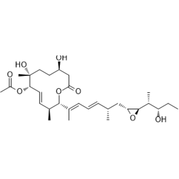
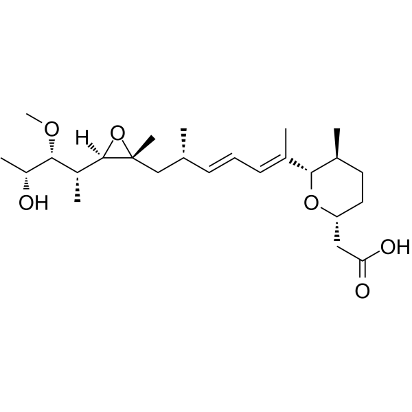
Pladienolide B is a potent cancer cell growth inhibitor that targets the SF3B1 subunit of the spliceosome. Pladienolide B exerts antitumor activities mediated through the inhibition of pre-mRNA splicing. Pladienolide B induces apoptosis.
For research use only. We do not sell to patients.
- Purity : 98.29%
- CAS No.: 445493-23-2
- Formula: C30H48O8
- Molecular Weight:536.70
-
Storage:Powder -20°C, 3 years ; In solvent -80°C, 6 months , -20°C, 1 month

To place orders, for customer services and technical support, please contact: MedChemExpress USA
Tel: 609-228-6898 E-mail: [email protected] [email protected]


-
 Biological Activity
-
Chemical Information
-
Solvent & Solubility
- Protocols
- Purity & Documentation
- References
-
 Help & FAQs


-
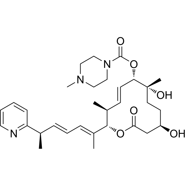
HY-111517
H3B-8800SF3B Aplicing Modulator -
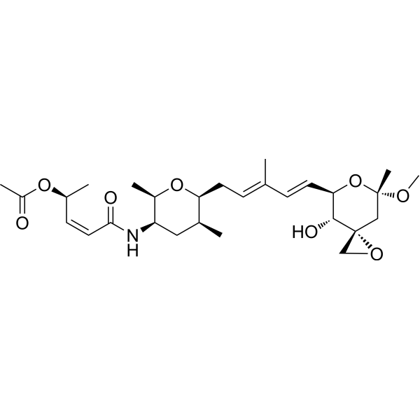
HY-16466
Spliceostatin ASF3b Inhibitor -
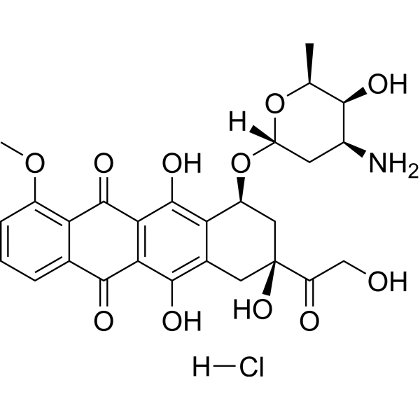
HY-15142
Doxorubicin hydrochlorideTopoisomerase Inhibitor -
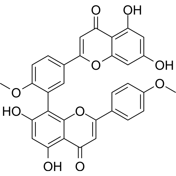
HY-N2117
Isoginkgetinpre-mRNA Splicing Inhibitor -
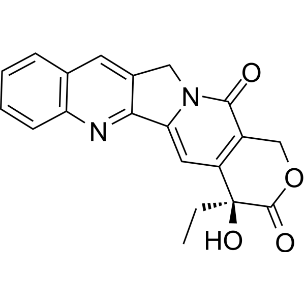
HY-16560
CamptothecinTopo I inhibitor -
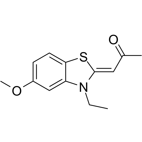
HY-15338
TG003CLK1 Inhibitor -
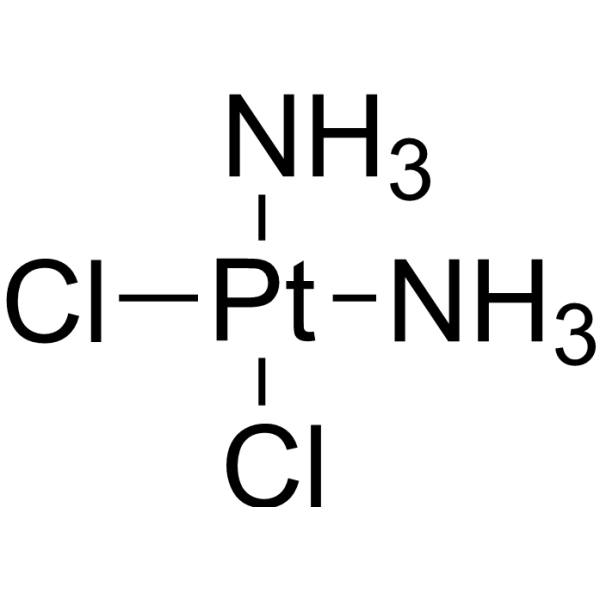
HY-17394
CisplatinDNA Alkylator -
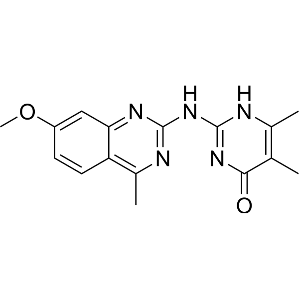
HY-100236
MadrasinSplicing Inhibitor -
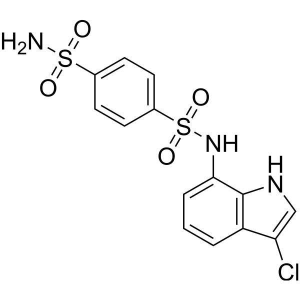
HY-13650
IndisulamCarbonic Anhydrase Inhibitor/RBM39 Molecular Glue Degrader -
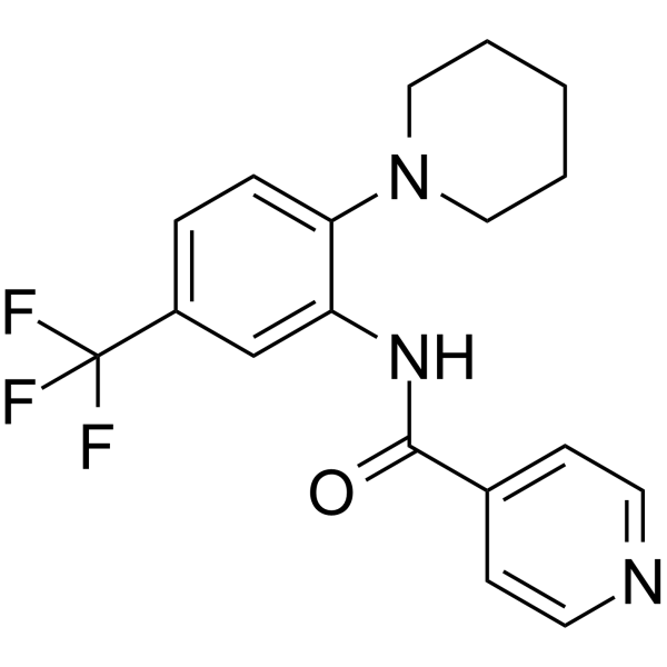
HY-13949
SRPIN340Virus Protease Inhibitor -
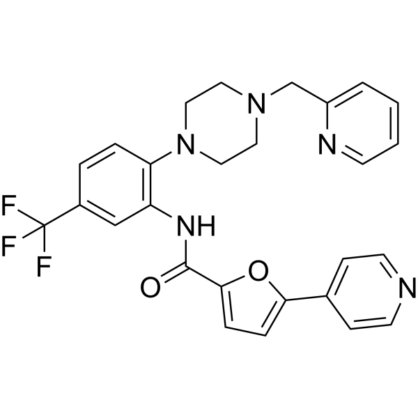
HY-117661
SPHINX31SRPK1 Inhibitor -
HY-19828
HerboxidienePre-mRNA Splicing Inhibitor
Publications Citing Use of MedChemExpress (MCE) Pladienolide B
More Customer Validation & Images

-
RT-PCR
-
WB
-
Bio/Physico-chemical Assay
-
Bio/Physico-chemical Assay
-
Bio/Physico-chemical Assay


Biological Activity
Description
In Vitro
Pladienolide B (0.1-2 nM; 24-72 hours) inhibits human cervical carcinoma cells viability[3].
Pladienolide B (0.1-2 nM; 24-48 hours) reduces SF3b1 expression in human cervical carcinoma cells[3].
Pladienolide B induces (0.1-2 nM; 24 hours) cell cycle arrest and apoptosis[3].
MedChemExpress (MCE) has not independently confirmed the accuracy of these methods. They are for reference only. Further protocols information, click here.
-
Cell Line:HeLa cells
-
Concentration:0.1, 0.5, 1, 1.5, 2 nM
-
Incubation Time:24, 48, 72 hours
-
Result:Significantly decreased cell viability, and the decrease was concentration- and time-dependent.
-
Cell Line:HeLa cells
-
Concentration:0.1, 0.5, and 2 nM
-
Incubation Time:24 hours
-
Result:The apoptotic cells were highly induced at 24 hours.
-
Cell Line:HeLa cells
-
Concentration:0.1, 0.5, and 2 nM
-
Incubation Time:24, 48 hours
-
Result:Induced a time- and concentration-dependent decrease in cellular SF3b1 proteins.
In Vivo
MedChemExpress (MCE) has not independently confirmed the accuracy of these methods. They are for reference only.
-
Animal Model:Female or male BALB/c nu/nu mice (7 weeks of age) (PC-3, OVCAR-3, DU-145, WiDr, and HCT-116, BSY-1 xenografts)[4]
-
Dosage:2.5, 5, and 10 mg/kg
-
Administration:I.v.; daily for 5 days
-
Result:Showed strong growth inhibitory or regressive activities against these xenografts.


Chemical Information
-
CAS No. 445493-23-2
-
Appearance Solid
-
Molecular Weight 536.70
-
Formula C30H48O8
-
Color White to off-white
-
SMILES
O=C1O[C@H](/C(C)=C/C=C/[C@@H](C)C[C@H]2O[C@@H]2[C@H](C)[C@@H](O)CC)[C@@H](C)/C=C/[C@H](OC(C)=O)[C@](C)(O)CC[C@@H](O)C1
-
Shipping
Room temperature in continental US; may vary elsewhere.
-
Storage
Powder -20°C 3 years In solvent -80°C 6 months -20°C 1 month


Publications (6)
-
Journal Impact Factor
-
Most Recent
-
Nat Commun
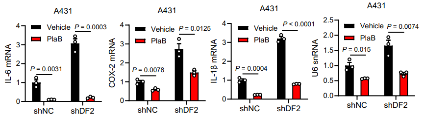
YTHDF2 regulates self non-coding RNA metabolism to control inflammation and tumorigenesis. [Abstract]2025 Nov 12;16(1):9946. PMID: 41224760
Pladienolide B purchased from MedChemExpress. Usage Cited in: Nat Commun. 2025 Nov 12;16(1):9946. [Abstract]
The splicing inhibitor Pladienolide B (500 nM, 3 h) reduced the expression of pro-inflammatory cytokines in control and YTHDF2 knockdown cells, and also reduced the level of U6 snRNA.
-
Mol Oncol
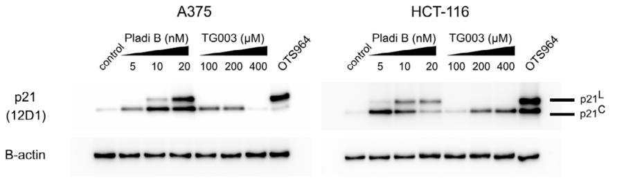
CDK11 inhibition induces cytoplasmic p21WAF1 splice variant by p53 stabilisation and SF3B1 inactivation. [Abstract]2025 Oct 17. PMID: 41105927
Pladienolide B purchased from MedChemExpress. Usage Cited in: Mol Oncol. 2025 Oct 17. [Abstract]
A375 and HCT-116 cells were treated with increasing concentrations of Pladienolide B (0-20 nM) and TG003 for 24 h and the levels of p21C and p21L were assessed with immunoblotting using 12D1 antibody; β-actin served as a loading control. OTS964 (200 nm) was used as a positive control. n = 3 biologically independent replicates.
-
J Proteomics
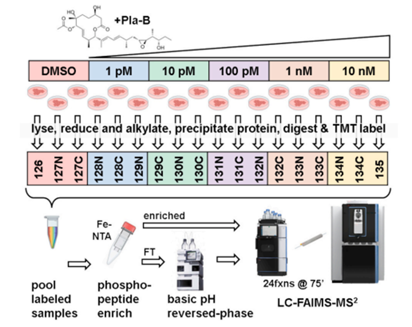
Decoding spliceosome inhibition: Isobaric tag-based proteomic profiling of pladienolide B treated human cell lines. [Abstract]2026 Jan 20:323:105565. PMID: 41224195
Pladienolide B purchased from MedChemExpress. Usage Cited in: J Proteomics. 2026 Jan 20:323:105565. [Abstract]
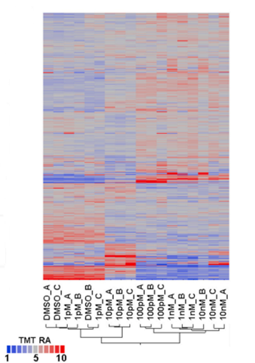
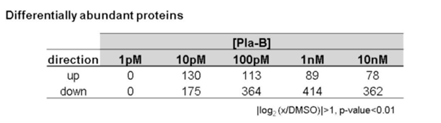
A protocol for quantitative proteomics analysis of SH-SY5Y cells treated with praldinolactone B (Pla-B) based on isotope labeling was developed to determine the dose-related effects of Pla-B on the cellular proteome. Cells were treated for 24 hours with increasing concentrations of Pla-B (1 pM to 10 nM) or DMSO (as a control).
Pladienolide B purchased from MedChemExpress. Usage Cited in: J Proteomics. 2026 Jan 20:323:105565. [Abstract]
The heatmap shows the changes in protein expression under different concentrations of Pla-B (1 pM to 10 nM) treatment.
Pladienolide B purchased from MedChemExpress. Usage Cited in: J Proteomics. 2026 Jan 20:323:105565. [Abstract]
The differential protein statistics table shows the number of proteins with significant changes in Pla-B concentration from 1 pM to 10 nM.
-
-
-


Solvent & Solubility
In Vitro:
DMSO : 100 mg/mL (186.32 mM; Need ultrasonic; Hygroscopic DMSO has a significant impact on the solubility of product, please use newly opened DMSO)
Please refer to the solubility information to select the appropriate solvent. Once prepared, please aliquot and store the solution to prevent product inactivation from repeated freeze-thaw cycles.
Storage method and period of stock solution: -80°C, 6 months; -20°C, 1 month. When stored at -80°C, please use it within 6 months. When stored at -20°C, please use it within 1 month.
Please refer to the solubility information to select the appropriate solvent. Once prepared, please aliquot and store the solution to prevent product inactivation from repeated freeze-thaw cycles.
Storage method and period of stock solution: -80°C, 6 months; -20°C, 1 month. When stored at -80°C, please use it within 6 months. When stored at -20°C, please use it within 1 month.
Concentration (start) × Volume (start) = Concentration (final) × Volume (final)
In Vivo:
Select the appropriate dissolution method based on your experimental animal and administration route.
- For the following dissolution methods, please ensure to first prepare a clear stock solution using an In Vitro approach and then sequentially add co-solvents:
- To ensure reliable experimental results, the clarified stock solution can be appropriately stored based on storage conditions. As for the working solution for In Vivo experiments, it is recommended to prepare freshly and use it on the same day.
- The percentages shown for the solvents indicate their volumetric ratio in the final prepared solution. If precipitation or phase separation occurs during preparation, heat and/or sonication can be used to aid dissolution.
Add each solvent one by one: 10% DMSO 40% PEG300 5% Tween-80 45% Saline
Solubility: ≥ 2.5 mg/mL (4.66 mM); Clear solution
This protocol yields a clear solution of ≥ 2.5 mg/mL (saturation unknown).
Taking 1 mL working solution as an example, add 100 μL DMSO stock solution (25.0 mg/mL) to 400 μL PEG300, and mix evenly; then add 50 μL Tween-80 and mix evenly; then add 450 μL Saline to adjust the volume to 1 mL.
Preparation of Saline: Dissolve 0.9 g sodium chloride in ddH₂O and dilute to 100 mL to obtain a clear Saline solution.
Add each solvent one by one: 10% DMSO 90% (20% SBE-β-CD in Saline)
Solubility: ≥ 2.5 mg/mL (4.66 mM); Clear solution
This protocol yields a clear solution of ≥ 2.5 mg/mL (saturation unknown).
Taking 1 mL working solution as an example, add 100 μL DMSO stock solution (25.0 mg/mL) to 900 μL 20% SBE-β-CD in Saline, and mix evenly.
Preparation of 20% SBE-β-CD in Saline (4°C, storage for one week): 2 g SBE-β-CD powder is dissolved in 10 mL Saline, completely dissolve until clear.
In Vivo Dissolution Calculator
Please enter the basic information of animal experiments:
-
-
-
-
Recommended: Prepare an additional quantity of animals to account for potential losses during experiments.
Please enter your animal formula composition:
-
%DMSO +
Recommended: Keep the proportion of DMSO in working solution below 2% if your animal is weak.
-
%+
-
+%Tween-80 + +
-
%Saline +
The co-solvents required include: DMSO, . All of co-solvents are available by MedChemExpress (MCE). , Tween 80. All of co-solvents are available by MedChemExpress (MCE).
Working solution concentration: 0.22 mg/mL
Method for preparing stock solution: mg drug dissolved in μL DMSO. Stock solution concentration: mg/mL.
1. Take μL DMSO stock solution;
2. Add μL .
μL , mix evenly;
3. Then add μL Tween 80, mix evenly;
4. Then add μL
Please ensure that the stock solution in the first step is dissolved to a clear state, and add co-solvents in sequence. You can use ultrasonic heating (ultrasonic cleaner, recommended frequency 20-40 kHz), vortexing, etc. to assist dissolution.

Protocols
-
RNA extraction experimental
By lysing cells, releasing RNA, and removing impurities such as proteins and DNA, high-purity RNA products are finally obtained. The commonly used traditional method is the guanidine isothiocyanate/phenol/chloroform method (Trizol), which is suitable for a variety of animal materials including animal tissues, microorganisms, cultured cells, etc., and most plant materials.
-
Apoptosis
Apoptosis, also called programmed cell death, is generally characterized by distinct morphological characteristics.
-
TUNEL staining for apoptotic DNA fragmentation
TUNEL staining detects DNA strand breaks by using terminal deoxynucleotidyl transferase to add labeled nucleotides to exposed 3′-OH DNA termini, generating either microscopic staining in fixed cells or tissue sections, or fluorescence/cytometric signal in cell suspensions. TUNEL positivity reflects DNA fragmentation but should not be interpreted alone as definitive apoptosis, because TUNEL can also label necrotic, autolytic, mechanically damaged, or DNA-repair-associated DNA breaks.
-
Annexin V plus membrane-impermeant dye apoptosis staining
Annexin V-based apoptosis assays rely on the detection of phosphatidylserine (PS) externalization from the inner leaflet of the plasma membrane to the outer leaflet, an early biochemical hallmark of apoptosis. Fluorescently labeled Annexin V binds PS in a calcium-dependent manner, enabling identification of early apoptotic cells by flow cytometry or fluorescence microscopy. When combined with a membrane-impermeant DNA-binding dye (e. g. , propidium iodide), this approach allows discrimination between viable (Annexin V−/dye−), early apoptotic (Annexin V+/dye−), and late apoptotic or necrotic (Annexin V+/dye+) cell populations by assessing membrane integrity and PS exposure.
-
Protocol for Cell Counting and Cell Density Analysis
Cell counting and cell-density analysis estimate the number of cells in a known volume or field area. Manual hemocytometer counting uses a chamber of defined geometry to convert counted cells into cells/mL, while automated counters and image-analysis workflows detect cell objects from optical, brightfield, fluorescence, impedance, or digital-image features. Trypan blue viability counting is based on dye exclusion: viable cells with intact membranes exclude dye, while non-viable cells with compromised membranes stain blue. The readout is total cell density, viable-cell density, dead-cell density, and percent viability. Cell density can also be estimated from microscopy images by counting objects per image area, from flow cytometry using calibrated volume or reference particles, or from in situ microscopy in bioreactors after calibration against reference methods such as hemocytometer or flow cytometry.
-
Somatic Cell Culture
A method of simulating the in vivo environment in vitro to maintain the cell growth, differentation and main functions.
-
Apoptosis Solutions
Apoptosis is a regulated, generally non-lytic cell-death pathway that removes unwanted, damaged, infected, or abnormal cells through coordinated morphological changes, caspase activation, DNA fragmentation, and membrane remodeling. The intrinsic apoptosis pathway is controlled mainly by mitochondrial outer membrane permeabilization, BCL-2 family proteins, cytochrome c release, apoptosome formation, caspase-9 activation, and downstream executioner caspase-3/7 activation. The extrinsic apoptosis pathway is initiated by death receptors such as Fas, TNFR, and TRAIL receptors, which recruit adaptor proteins and activate caspase-8 before engaging executioner caspases or mitochondrial amplification through BID cleavage. Apoptosis is linked to many phenotypes, including cancer cell killing, tissue homeostasis, immune regulation, neurodegeneration, infection response, and treatment-induced cytotoxicity; unresolved questions include how apoptosis interacts with necroptosis, pyroptosis, ferroptos
-
CCK-8/WST-8 Cell Proliferation Assay
The CCK-8/WST-8 assay is based on the reduction of the water-soluble tetrazolium salt WST-8 to a water-soluble formazan product by cellular dehydrogenases in metabolically active cells, where the generated formazan amount is proportional to the number of living cells and is quantified by measuring absorbance in the visible range, providing a colorimetric readout for cell viability and proliferation assessment. This class of tetrazolium-based assays improves upon earlier MTT-based systems by producing a water-soluble formazan, eliminating the need for organic solubilization steps and enabling direct spectrophotometric measurement in culture medium.
-
Cell Counting-Based Growth Curve Assay
Cell counting-based growth curve assays quantify cell proliferation by directly measuring changes in viable cell number over time using manual or automated counting methods such as hemocytometer-based counting or instrument-assisted cell enumeration, enabling construction of growth curves that reflect population expansion dynamics in response to culture conditions. A widely used approach is trypan blue exclusion with hemocytometer counting, where membrane-compromised (non-viable) cells take up the dye, allowing discrimination between viable and non-viable cells while simultaneously enabling total cell number quantification. Repeated sampling across time points allows estimation of proliferation rate, growth phases, and comparative growth kinetics between experimental conditions.
-
MTT Cell Proliferation Assay
The MTT assay is a colorimetric endpoint assay for estimating viable cell number, cell growth, cytotoxicity, or cell activation in cultured mammalian cells. Living cells reduce the yellow tetrazolium salt MTT into purple/blue formazan, while dead cells do not generate the same signal; the resulting color can be quantified with a multiwell spectrophotometer. MTT reduction is commonly interpreted as a readout of metabolic activity that often correlates with viable cell number, but it should not be treated as a direct cell-counting method unless the assay is optimized for the cell type and experimental condition. Studies show that MTT reduction can involve mitochondrial and non-mitochondrial reducing systems, and formazan may accumulate in intracellular lipid droplets rather than simply marking mitochondria.
-
Cell Viability Determination by MTT Colorimetric Assay
The following protocol uses the MTT colorimetric assay as a classic literature-established method for assessing cell viability/metabolic activity in cultured mammalian cells. MTT[3-(4,5-dimethylthiazol-2-yl)-2,5-diphenyltetrazolium bromide] is reduced by metabolically active cells to a colored formazan product; the amount of formazan is quantified spectrophotometrically and provides an indirect measure of metabolically active viable cells. Importantly, MTT reduction reflects cellular oxidoreductase/metabolic activity rather than an absolute direct count of living cells, so changes in cellular metabolism can alter the signal independently of cell number.


Purity & Documentation
-
Data Sheet (277 KB)
-
SDS (252 KB)
- English - EN (252 KB)
- Français - FR (252 KB)
- Deutsch - DE (252 KB)
- Norwegian - NO (252 KB)
- Español - ES (252 KB)
- Swedish - SV (252 KB)
- Italian - IT (252 KB)
- Korean - KR (252 KB)
- Portuguese - PT (252 KB)
-
Handling Instructions (2659 KB)
References
[1]. Effenberger KA, et al. Coherence between cellular responses and in vitro splicing inhibition for the anti-tumor drug pladienolide B and its analogs. J Biol Chem. 2014 Jan 24;289(4):1938-47. [Content Brief]
[2]. Aouida M, et al. CRISPR/Cas9-mediated target validation of the splicing inhibitor Pladienolide B. Biochim Open. 2016 Feb 24;3:72-75. [Content Brief]
[3]. Zhang Q, et al. Inhibition of SF3b1 by pladienolide B evokes cycle arrest, apoptosis induction and p73 splicing in human cervical carcinoma cells. Artif Cells Nanomed Biotechnol. 2019 Dec;47(1):1273-1280. [Content Brief]
[4]. Mizui Y, et al. Pladienolides, new substances from culture of Streptomyces platensis Mer-11107. III. In vitro and in vivo antitumor activities. J Antibiot (Tokyo). 2004 Mar;57(3):188-96. [Content Brief]


Complete Stock Solution Preparation Table
Please refer to the solubility information to select the appropriate solvent. Once prepared, please aliquot and store the solution to prevent product inactivation from repeated freeze-thaw cycles.
Storage method and period of stock solution: -80°C, 6 months; -20°C, 1 month. When stored at -80°C, please use it within 6 months. When stored at -20°C, please use it within 1 month.
| Optional Solvent | Concentration Solvent Mass | 1 mg | 5 mg | 10 mg | 25 mg |
|---|---|---|---|---|---|
| DMSO | 1 mM | 1.8632 mL | 9.3162 mL | 18.6324 mL | 46.5810 mL |
| 5 mM | 0.3726 mL | 1.8632 mL | 3.7265 mL | 9.3162 mL | |
| 10 mM | 0.1863 mL | 0.9316 mL | 1.8632 mL | 4.6581 mL | |
| 15 mM | 0.1242 mL | 0.6211 mL | 1.2422 mL | 3.1054 mL | |
| 20 mM | 0.0932 mL | 0.4658 mL | 0.9316 mL | 2.3290 mL | |
| 25 mM | 0.0745 mL | 0.3726 mL | 0.7453 mL | 1.8632 mL | |
| 30 mM | 0.0621 mL | 0.3105 mL | 0.6211 mL | 1.5527 mL | |
| 40 mM | 0.0466 mL | 0.2329 mL | 0.4658 mL | 1.1645 mL | |
| 50 mM | 0.0373 mL | 0.1863 mL | 0.3726 mL | 0.9316 mL | |
| 60 mM | 0.0311 mL | 0.1553 mL | 0.3105 mL | 0.7763 mL | |
| 80 mM | 0.0233 mL | 0.1165 mL | 0.2329 mL | 0.5823 mL | |
| 100 mM | 0.0186 mL | 0.0932 mL | 0.1863 mL | 0.4658 mL |


Pladienolide B Related Classifications
HY-16399 Related Classifications
 Powered by Bioz