



Pramocaine-d9 hydrochloride
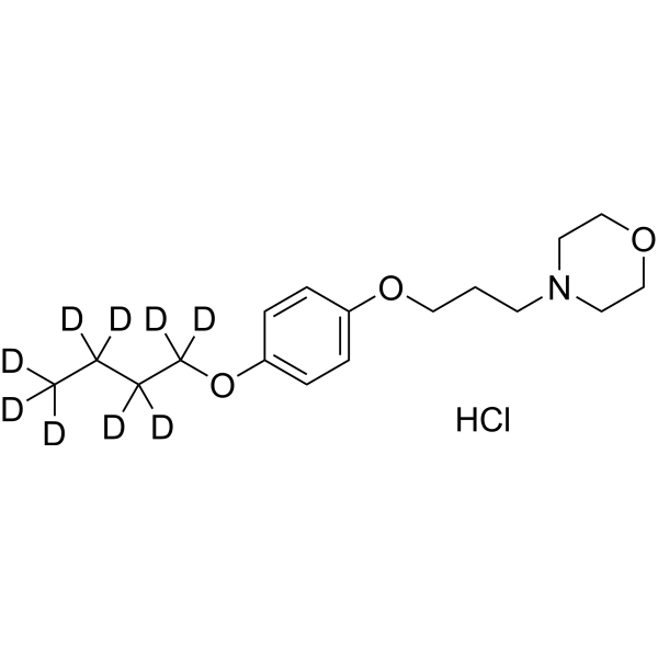
Pramocaine-d9 (Pramoxine-d9) hydrochloride is the d9-labeled Pramocaine hydrochloride (HY-B1319). Pramocaine hydrochloride is a topical surface anesthetic and antipruritic agent. Pramocaine hydrochloride reversibly inhibits voltage-gated sodium channel, reduces transmembrane permeability of sodium ions, stabilizes cell membranes, prevents depolarization, blocks action potential conduction, and inhibits peripheral slow C-fiber pathways associated with pain, pruritus and thermoception. Pramocaine hydrochloride can be used in research related to chronic pruritus, renal pruritus, atopic dermatitis, xerotic pruritus, uremic pruritus, and cutaneous hyperalgesia/pain.
For research use only. We do not sell to patients.
- Formula: C17H19D9ClNO3
- Molecular Weight:338.92
-
Storage:
Please store the product under the recommended conditions in the Certificate of Analysis.

To place orders, for customer services and technical support, please contact: MedChemExpress USA
Tel: 609-228-6898 E-mail: [email protected] [email protected]


-
 Biological Activity
-
Chemical Information
- Protocols
- Purity & Documentation
- References
-
 Help & FAQs




Biological Activity
Description
Application
1. This compound can be used as a tracer
2. This compound can be used as an internal standard for quantitative analysis by NMR, GC-MS, or LC-MS.


Chemical Information
-
Unlabeled CAS 637-58-1
-
Molecular Weight 338.92
-
Formula C17H19D9ClNO3
-
SMILES
[2H]C(C([2H])([2H])C([2H])([2H])C([2H])([2H])[2H])([2H])OC(C=C1)=CC=C1OCCCN2CCOCC2.Cl
-
Synonyms
Pramoxine-d9 hydrochloride
-
Shipping
Room temperature in continental US; may vary elsewhere.
-
Storage
Please store the product under the recommended conditions in the Certificate of Analysis.

Protocols
-
Cardiac voltage-sensitive optical mapping
Cardiac voltage-sensitive optical mapping records changes in transmembrane potential from cardiac tissue by staining the preparation with a voltage-sensitive dye and imaging fluorescence changes during electrical activation; the resulting optical action potentials can be used to map activation time, action potential duration, conduction velocity, wavefront propagation, and arrhythmia dynamics. The optical signal represents a relative fluorescence change from a tissue volume rather than a single-cell intracellular recording, so spatial resolution, sampling rate, voltage resolution, optical magnification, light penetration, and motion control must be considered together when interpreting optical action potentials.
-
Neuronal voltage-sensitive dye imaging
Neuronal voltage-sensitive dye imaging detects membrane-potential-dependent optical changes from dyes associated with neuronal membranes, enabling optical recording of electrical activity from single neurons, dendrites, axons, spines, or neuronal populations in brain slices and cultured neurons. VSD signals are typically reported as fractional fluorescence or absorbance changes over baseline, such as ΔF/F or ΔI/I, and published protocols use high-speed cameras or photodiode arrays because neuronal voltage signals occur on millisecond time scales. Fast VSD imaging can be applied at two common scales: bulk staining of brain slices to measure circuit-level spatiotemporal activity, and single-cell loading or biolistic delivery to record membrane-potential transients from individual neuronal compartments. Optical signals should be interpreted as membrane-potential-related readouts, and validation by simultaneous electrophysiology or pharmacological controls is recommended when the experimen
-
Acute brain-slice whole-cell patch-clamp recording
Acute brain-slice whole-cell patch-clamp recording measures membrane voltage or ionic current from visually targeted cells in living brain slices; after giga-seal formation, the membrane under the pipette is ruptured to provide low-resistance electrical access to the cell interior, enabling current-clamp analysis of excitability and voltage-clamp analysis of synaptic or membrane currents. Acute slices preserve local tissue architecture better than dissociated preparations and allow visually guided recording from defined brain regions or fluorescently labeled cells; however, whole-cell access also permits exchange between pipette solution and cytoplasm, so intracellular dialysis must be considered when interpreting signaling-dependent phenomena.
-
TPA/Croton Oil Ear Edema and Dermatitis
The TPA (12-O-tetradecanoylphorbol-13-acetate) and croton oil-induced mouse ear edema model is a well-established acute cutaneous inflammation system used to evaluate topical anti-inflammatory activity by measuring edema formation, neutrophil infiltration, vascular permeability, and cytokine-mediated skin responses in vivo. The inflammatory response is triggered by topical application of phorbol esters (TPA) or croton oil constituents, leading to rapid activation of protein kinase C signaling, leukocyte recruitment, and increased vascular permeability, which can be quantified by ear thickness, weight, dye extravasation, and biochemical markers such as myeloperoxidase (MPO) activity and pro-inflammatory mediators in ear tissue homogenates. This model is widely used for screening anti-inflammatory agents, where reductions in edema and inflammatory biomarkers reflect suppression of acute dermal inflammation and immune cell infiltration. Histological evaluation typically confirms epidermal
-
Transepithelial/transendothelial electrical resistance assay
TEER measures electrical resistance across epithelial or endothelial monolayers cultured on permeable supports, and the readout reflects ionic conductance through the cell barrier, especially the paracellular pathway regulated by junctional integrity. TEER can be measured without destroying the monolayer and is commonly used before or during transport, permeability, barrier-disruption, and barrier-maturation experiments. TEER values are influenced by biological maturation and technical conditions; reported factors include temperature, medium formulation, passage number, electrode geometry, membrane properties, and junctional length during early monolayer maturation. Therefore, TEER should be interpreted with blank-insert subtraction, area normalization, repeated readings, and, when possible, orthogonal barrier readouts such as FITC-dextran flux or tight-junction staining.


Purity & Documentation
References
[1].
Agarwal A, et al. Topical Pramoxine in Chronic Pruritus: Where do We Stand? Indian J Dermatol. 2021 Sep-Oct;66(5):576.
[Content Brief]
[2]. Chou AK, et al. Intrathecal pramoxine causes long-lasting spinal sensory and motor block in rats. The Journal of pharmacy and pharmacology. 2018 Apr;70(4):543-549. [Content Brief]
[3]. Chou AK, et al. Skin nociceptive block with pramoxine delivery by subcutaneous injection in rats. Pharmacological reports : PR. 2018 Dec;70(6):1180-1184. [Content Brief]

Calculators
Concentration (start) × Volume (start) = Concentration (final) × Volume (final)


Pramocaine-d9 hydrochloride Related Classifications
HY-B1319S1 Related Classifications
 Powered by Bioz