



Pramocaine hydrochloride

Based on 1 Customer Validation
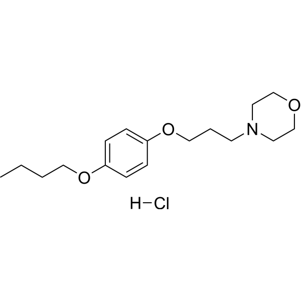
Pramocaine (Pramoxine) hydrochloride is a topical surface anesthetic and antipruritic agent. Pramocaine hydrochloride reversibly inhibits voltage-gated sodium channel, reduces transmembrane permeability of sodium ions, stabilizes cell membranes, prevents depolarization, blocks action potential conduction, and inhibits peripheral slow C-fiber pathways associated with pain, pruritus and thermoception. Pramocaine hydrochloride can be used in research related to chronic pruritus, renal pruritus, atopic dermatitis, xerotic pruritus, uremic pruritus, and cutaneous hyperalgesia/pain.
For research use only. We do not sell to patients.
- Purity : 99.85%
- CAS No.: 637-58-1
- Formula: C17H28ClNO3
- Molecular Weight:329.86
-
Storage:
4°C, sealed storage, away from moisture
* In solvent : -80°C, 6 months; -20°C, 1 month (sealed storage, away from moisture)

To place orders, for customer services and technical support, please contact: MedChemExpress USA
Tel: 609-228-6898 E-mail: [email protected] [email protected]


-
 Biological Activity
-
Chemical Information
-
Solvent & Solubility
- Protocols
- Purity & Documentation
- References
-
 Help & FAQs





Biological Activity
Description
In Vivo
Pramocaine (12-120 μmol; s.c.) hydrochloride elicits dose-related cutaneous antinociception in male Sprague-Dawley rats, with an ED50 of 42.1 μmol[3].
MedChemExpress (MCE) has not independently confirmed the accuracy of these methods. They are for reference only.
-
Animal Model:Sprague-Dawley (male, 298 to 348 g)[2]
-
Dosage:7.74 μmol/kg; 15.48 μmol/kg; 23.22 μmol/kg; 30.96 μmol/kg
-
Administration:intrathecal; single injection
-
Result:Elicited dose-dependent spinal block of nociception, proprioception, and motor function.
Determined ED50 values of 15.47 μmol/kg for nociceptive block, 16.46 μmol/kg for proprioceptive block, and 17.77 μmol/kg for motor block.
Reported mean ED75 and ED25 values across all three block types as 20.83 μmol/kg and 13.17 μmol/kg, respectively.
Produced 100% maximum possible effect (%MPE) for all three block types at 30.96 μmol/kg, with complete blockade times of 36.9 minutes for nociception, 17.8 minutes for proprioception, and 15.4 minutes for motor function.
Recorded full recovery times of 94.2 minutes for nociception, 65.6 minutes for proprioception, and 41.3 minutes for motor function at 30.96 μmol/kg.
Measured AUC values of 6425 (%MPE × min) for nociception, 3709 (%MPE × min) for proprioception, and 2362 (%MPE × min) for motor function at 30.96 μmol/kg.
Showed no significant difference in spinal block duration compared to bupivacaine at equipotent doses (ED75, ED50, ED25).


Chemical Information
-
CAS No. 637-58-1
-
Appearance Solid
-
Molecular Weight 329.86
-
Formula C17H28ClNO3
-
Color White to off-white
-
SMILES
CCCCOC1=CC=C(OCCCN2CCOCC2)C=C1.[H]Cl
-
Synonyms
Pramoxine hydrochloride
-
Shipping
Room temperature in continental US; may vary elsewhere.
-
Storage
4°C, sealed storage, away from moisture
* In solvent : -80°C, 6 months; -20°C, 1 month (sealed storage, away from moisture)


Solvent & Solubility
In Vitro:
DMSO : 100 mg/mL (303.16 mM; Need ultrasonic; Hygroscopic DMSO has a significant impact on the solubility of product, please use newly opened DMSO)
H2O : 100 mg/mL (303.16 mM; Need ultrasonic)
Please refer to the solubility information to select the appropriate solvent. Once prepared, please aliquot and store the solution to prevent product inactivation from repeated freeze-thaw cycles.
Storage method and period of stock solution: -80°C, 6 months; -20°C, 1 month (sealed storage, away from moisture). When stored at -80°C, please use it within 6 months. When stored at -20°C, please use it within 1 month.
* Note: If you choose water as the stock solution, please dilute it to the working solution, then filter and sterilize it with a 0.22 μm filter before use.
Please refer to the solubility information to select the appropriate solvent. Once prepared, please aliquot and store the solution to prevent product inactivation from repeated freeze-thaw cycles.
Storage method and period of stock solution: -80°C, 6 months; -20°C, 1 month (sealed storage, away from moisture). When stored at -80°C, please use it within 6 months. When stored at -20°C, please use it within 1 month.
* Note: If you choose water as the stock solution, please dilute it to the working solution, then filter and sterilize it with a 0.22 μm filter before use.
Concentration (start) × Volume (start) = Concentration (final) × Volume (final)
In Vivo:
Select the appropriate dissolution method based on your experimental animal and administration route.
- For the following dissolution methods, please ensure to first prepare a clear stock solution using an In Vitro approach and then sequentially add co-solvents:
- To ensure reliable experimental results, the clarified stock solution can be appropriately stored based on storage conditions. As for the working solution for In Vivo experiments, it is recommended to prepare freshly and use it on the same day.
- The percentages shown for the solvents indicate their volumetric ratio in the final prepared solution. If precipitation or phase separation occurs during preparation, heat and/or sonication can be used to aid dissolution.
Add each solvent one by one: 10% DMSO 40% PEG300 5% Tween-80 45% Saline
Solubility: ≥ 2.5 mg/mL (7.58 mM); Clear solution
This protocol yields a clear solution of ≥ 2.5 mg/mL (saturation unknown).
Taking 1 mL working solution as an example, add 100 μL DMSO stock solution (25.0 mg/mL) to 400 μL PEG300, and mix evenly; then add 50 μL Tween-80 and mix evenly; then add 450 μL Saline to adjust the volume to 1 mL.
Preparation of Saline: Dissolve 0.9 g sodium chloride in ddH₂O and dilute to 100 mL to obtain a clear Saline solution.
Add each solvent one by one: 10% DMSO 90% (20% SBE-β-CD in Saline)
Solubility: ≥ 2.5 mg/mL (7.58 mM); Clear solution
This protocol yields a clear solution of ≥ 2.5 mg/mL (saturation unknown).
Taking 1 mL working solution as an example, add 100 μL DMSO stock solution (25.0 mg/mL) to 900 μL 20% SBE-β-CD in Saline, and mix evenly.
Preparation of 20% SBE-β-CD in Saline (4°C, storage for one week): 2 g SBE-β-CD powder is dissolved in 10 mL Saline, completely dissolve until clear.
For the following dissolution methods, please prepare the working solution directly:
It is recommended to prepare fresh solutions and use them promptly within a short period of time.
The percentages shown for the solvents indicate their volumetric ratio in the final prepared solution. If precipitation or phase separation occurs during preparation, heat and/or sonication can be used to aid dissolution.
Add each solvent one by one: PBS
Solubility: 100 mg/mL (303.16 mM); Clear solution; Need ultrasonic
In Vivo Dissolution Calculator
Please enter the basic information of animal experiments:
-
-
-
-
Recommended: Prepare an additional quantity of animals to account for potential losses during experiments.
Working solution concentration: 0.22 mg/mL
This product has good water solubility, please refer to the measured solubility data in water/PBS/Saline for details.

Protocols
-
Cardiac voltage-sensitive optical mapping
Cardiac voltage-sensitive optical mapping records changes in transmembrane potential from cardiac tissue by staining the preparation with a voltage-sensitive dye and imaging fluorescence changes during electrical activation; the resulting optical action potentials can be used to map activation time, action potential duration, conduction velocity, wavefront propagation, and arrhythmia dynamics. The optical signal represents a relative fluorescence change from a tissue volume rather than a single-cell intracellular recording, so spatial resolution, sampling rate, voltage resolution, optical magnification, light penetration, and motion control must be considered together when interpreting optical action potentials.
-
Neuronal voltage-sensitive dye imaging
Neuronal voltage-sensitive dye imaging detects membrane-potential-dependent optical changes from dyes associated with neuronal membranes, enabling optical recording of electrical activity from single neurons, dendrites, axons, spines, or neuronal populations in brain slices and cultured neurons. VSD signals are typically reported as fractional fluorescence or absorbance changes over baseline, such as ΔF/F or ΔI/I, and published protocols use high-speed cameras or photodiode arrays because neuronal voltage signals occur on millisecond time scales. Fast VSD imaging can be applied at two common scales: bulk staining of brain slices to measure circuit-level spatiotemporal activity, and single-cell loading or biolistic delivery to record membrane-potential transients from individual neuronal compartments. Optical signals should be interpreted as membrane-potential-related readouts, and validation by simultaneous electrophysiology or pharmacological controls is recommended when the experimen
-
Acute brain-slice whole-cell patch-clamp recording
Acute brain-slice whole-cell patch-clamp recording measures membrane voltage or ionic current from visually targeted cells in living brain slices; after giga-seal formation, the membrane under the pipette is ruptured to provide low-resistance electrical access to the cell interior, enabling current-clamp analysis of excitability and voltage-clamp analysis of synaptic or membrane currents. Acute slices preserve local tissue architecture better than dissociated preparations and allow visually guided recording from defined brain regions or fluorescently labeled cells; however, whole-cell access also permits exchange between pipette solution and cytoplasm, so intracellular dialysis must be considered when interpreting signaling-dependent phenomena.
-
TPA/Croton Oil Ear Edema and Dermatitis
The TPA (12-O-tetradecanoylphorbol-13-acetate) and croton oil-induced mouse ear edema model is a well-established acute cutaneous inflammation system used to evaluate topical anti-inflammatory activity by measuring edema formation, neutrophil infiltration, vascular permeability, and cytokine-mediated skin responses in vivo. The inflammatory response is triggered by topical application of phorbol esters (TPA) or croton oil constituents, leading to rapid activation of protein kinase C signaling, leukocyte recruitment, and increased vascular permeability, which can be quantified by ear thickness, weight, dye extravasation, and biochemical markers such as myeloperoxidase (MPO) activity and pro-inflammatory mediators in ear tissue homogenates. This model is widely used for screening anti-inflammatory agents, where reductions in edema and inflammatory biomarkers reflect suppression of acute dermal inflammation and immune cell infiltration. Histological evaluation typically confirms epidermal
-
Transepithelial/transendothelial electrical resistance assay
TEER measures electrical resistance across epithelial or endothelial monolayers cultured on permeable supports, and the readout reflects ionic conductance through the cell barrier, especially the paracellular pathway regulated by junctional integrity. TEER can be measured without destroying the monolayer and is commonly used before or during transport, permeability, barrier-disruption, and barrier-maturation experiments. TEER values are influenced by biological maturation and technical conditions; reported factors include temperature, medium formulation, passage number, electrode geometry, membrane properties, and junctional length during early monolayer maturation. Therefore, TEER should be interpreted with blank-insert subtraction, area normalization, repeated readings, and, when possible, orthogonal barrier readouts such as FITC-dextran flux or tight-junction staining.


Purity & Documentation
-
Data Sheet (277 KB)
-
SDS (394 KB)
- English - EN (394 KB)
- Français - FR (394 KB)
- Deutsch - DE (394 KB)
- Norwegian - NO (394 KB)
- Español - ES (394 KB)
- Swedish - SV (394 KB)
- Italian - IT (394 KB)
- Korean - KR (394 KB)
- Portuguese - PT (394 KB)
-
Handling Instructions (2659 KB)
References
[1].
Agarwal A, et al. Topical Pramoxine in Chronic Pruritus: Where do We Stand? Indian J Dermatol. 2021 Sep-Oct;66(5):576.
[Content Brief]
[2]. Chou AK, et al. Intrathecal pramoxine causes long-lasting spinal sensory and motor block in rats. The Journal of pharmacy and pharmacology. 2018 Apr;70(4):543-549. [Content Brief]
[3]. Chou AK, et al. Skin nociceptive block with pramoxine delivery by subcutaneous injection in rats. Pharmacological reports : PR. 2018 Dec;70(6):1180-1184. [Content Brief]


Complete Stock Solution Preparation Table
Please refer to the solubility information to select the appropriate solvent. Once prepared, please aliquot and store the solution to prevent product inactivation from repeated freeze-thaw cycles.
Storage method and period of stock solution: -80°C, 6 months; -20°C, 1 month (sealed storage, away from moisture). When stored at -80°C, please use it within 6 months. When stored at -20°C, please use it within 1 month.
| Optional Solvent | Concentration Solvent Mass | 1 mg | 5 mg | 10 mg | 25 mg |
|---|---|---|---|---|---|
| DMSO / H2O | 1 mM | 3.0316 mL | 15.1579 mL | 30.3159 mL | 75.7897 mL |
| 5 mM | 0.6063 mL | 3.0316 mL | 6.0632 mL | 15.1579 mL | |
| 10 mM | 0.3032 mL | 1.5158 mL | 3.0316 mL | 7.5790 mL | |
| 15 mM | 0.2021 mL | 1.0105 mL | 2.0211 mL | 5.0526 mL | |
| 20 mM | 0.1516 mL | 0.7579 mL | 1.5158 mL | 3.7895 mL | |
| 25 mM | 0.1213 mL | 0.6063 mL | 1.2126 mL | 3.0316 mL | |
| 30 mM | 0.1011 mL | 0.5053 mL | 1.0105 mL | 2.5263 mL | |
| 40 mM | 0.0758 mL | 0.3789 mL | 0.7579 mL | 1.8947 mL | |
| 50 mM | 0.0606 mL | 0.3032 mL | 0.6063 mL | 1.5158 mL | |
| 60 mM | 0.0505 mL | 0.2526 mL | 0.5053 mL | 1.2632 mL | |
| 80 mM | 0.0379 mL | 0.1895 mL | 0.3789 mL | 0.9474 mL | |
| 100 mM | 0.0303 mL | 0.1516 mL | 0.3032 mL | 0.7579 mL |
* Note: If you choose water as the stock solution, please dilute it to the working solution, then filter and sterilize it with a 0.22 μm filter before use.


Pramocaine hydrochloride Related Classifications
HY-B1319 Related Classifications
 Powered by Bioz