



Propacetamol hydrochloride

Based on 1 Customer Validation
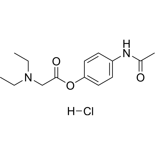
Propacetamol hydrochloride is an orally active prodrug of paracetamol and an inducer of acute liver injury models, with multiple properties including antinociception, antioxidation and gastroprotection. Propacetamol hydrochloride potentiates Tramadol and attenuates Aspirin (HY-14654)-induced gastric mucosal damage and lipid peroxidation. Under specific conditions, Propacetamol hydrochloride also acts as a hepatotoxic inducer, triggering acute liver injury, oxidative stress and apoptosis, with strain differences in toxicity sensitivity. Propacetamol hydrochloride can be used in the research of acute liver injury, drug-induced hepatotoxicity and gastric mucosal damage.
For research use only. We do not sell to patients.
- Purity : 99.74%
- CAS No.: 66532-86-3
- Formula: C14H21ClN2O3
- Molecular Weight:300.79
-
Storage:
4°C, stored under nitrogen, away from moisture
* In solvent : -80°C, 6 months; -20°C, 1 month (stored under nitrogen, away from moisture)

To place orders, for customer services and technical support, please contact: MedChemExpress USA
Tel: 609-228-6898 E-mail: [email protected] [email protected]


-
 Biological Activity
-
Chemical Information
-
Solvent & Solubility
- Protocols
- Purity & Documentation
- References
-
 Help & FAQs




Biological Activity
Description
In Vitro
Propacetamol induces acute liver injury through the following mechanisms: it is metabolized by CYP2E1 to form the toxic intermediate N-acetyl-p-benzoquinone imine (NAPQI), which depletes hepatic GSH and triggers oxidative stress, inflammatory responses and apoptosis; it also inhibits UGT1A1 expression and activates the JNK/ERK pathway. Its core activity mimics the toxicological effects of acetaminophen overdose[1].
MedChemExpress (MCE) has not independently confirmed the accuracy of these methods. They are for reference only. Further protocols information, click here.
In Vivo
Propacetamol (1200-1400 mg/kg; i.p.; single dose) induces acute lethal liver injury in BALB/c mice, with 1400 mg/kg causing 100% mortality by 168 h and 1200 mg/kg reducing survival to 10% over the same period[3].
Propacetamol (1200-1800 mg/kg; i.p.; single dose) induces acute liver injury in C57BL/6 mice with strain-specific tolerance, with 1600 and 1800 mg/kg causing 100% mortality by 168 h and 1200 mg/kg reducing survival to 50% over the same period[3].
Propacetamol (200 mg/kg; i.p.; single dose) produces significant antinociception in mice with acetic acid-induced persistent visceral pain, as measured by reduced abdominal writhing responses[4].
Propacetamol (270-540 mg/kg; i.p.; single dose) does not produce significant antinociception in female mice tested with the hot plate acute thermal pain assay[4].
Propacetamol (125-500 mg/kg; i.g.; single dose; 30 min before ASA) exerts a dose-dependent gastroprotective effect against ASA-induced gastric ulceration in male Wistar rats, with the 500 mg/kg dose reducing ulcer area by 52.9% and gastric mucosal MDA levels by 66.9%, and supporting adaptive antioxidant responses via maintenance of elevated gastric UA and glutathione levels[5].
MedChemExpress (MCE) has not independently confirmed the accuracy of these methods. They are for reference only.
-
Animal Model:BALB/cByJNarl (six-week-old male, 20-22 g, acute liver injury induced by propacetamol injection)[2]
-
Dosage:600 mg/kg (acute liver injury induction); 1200 mg/kg (lethal model)
-
Administration:i.p.; single injection
-
Result:Induced significant acute liver injury, characterized by dark red discoloration of liver tissue, marked elevations in serum alanine aminotransferase (ALT) and aspartate aminotransferase (AST) activities, severe histopathological liver damage (cell vacuolization, membrane loss, nuclear disappearance, bleeding, and elevated histopathological scores), reduced hepatic glutathione (GSH) levels, increased thiobarbituric acid reactive substances (TBARS) and 3-nitrotyrosine (3-NT) levels, suppressed superoxide dismutase (SOD), catalase (CAT), and glutathione peroxidase (GPx) activities, increased serum tumor necrosis factor-alpha (TNF-α) and interleukin-6 (IL-6) levels, increased hepatic Bax expression and Bax/Bcl-2 ratio at 6 h post-injection, increased cleaved caspase-3 expression at 12 h post-injection, 83.00 ± 2.89% TUNEL-positive liver area at 18 h post-injection, increased hepatic cytochrome P450 2E1 (CYP2E1) expression, reduced UDP-glucuronosyltransferase family 1 member A1 (UGT1A1) expression, increased nuclear nuclear factor erythroid 2-related factor 2 (Nrf2) levels at 6 h post-injection, reduced Nrf2 and glutamate cysteine ligase catalytic subunit (GCLC) levels at 18 h post-injection, increased heme oxygenase 1 (HO-1) levels at 18 h post-injection, increased phosphorylated c-jun-N-terminal kinase (p-JNK)/JNK and phosphorylated extracellular signal-regulated kinase (p-ERK)/ERK ratios at 6 h post-injection, and increased hepatic receptor-interacting serine/threonine-protein kinase 1 (RIP1) expression at 6 h post-injection.\nCaused 100% mortality of all tested mice within 14 h post-injection without post-treatment.
-
Animal Model:BALB/cByJNarl (male, 5 weeks old, 20-22 g, acute liver injury induced by intraperitoneal propacetamol injection)[3]
-
Dosage:1200 mg/kg; 1400 mg/kg
-
Administration:i.p.; single dose
-
Result:Resulted in 10% survival at 168 h post-injection (1200 mg/kg).\nResulted in 0% survival at 168 h post-injection (1400 mg/kg).
-
Animal Model:C57BL/6JNarl (male, 5 weeks old, 20-22 g, acute liver injury induced by intraperitoneal propacetamol injection)[3]
-
Dosage:1200 mg/kg; 1400 mg/kg; 1600 mg/kg; 1800 mg/kg
-
Administration:i.p.; single dose
-
Result:Resulted in 50% survival at 168 h post-injection (1200 mg/kg).\nResulted in 40% survival at 168 h post-injection (1400 mg/kg).\nResulted in 0% survival at 168 h post-injection (1600 mg/kg).\nResulted in 0% survival at 168 h post-injection (1800 mg/kg).
-
Animal Model:Wistar (male, 240-270 g, ASA-induced gastric ulceration)[5]
-
Dosage:125 mg/kg; 250 mg/kg; 500 mg/kg
-
Administration:i.g.; single dose; 30 min before ASA
-
Result:Reduced ASA-induced gastric ulcer area by 22.0% (125 mg/kg), 38.5% (250 mg/kg), and 52.9% (500 mg/kg).\nReduced ASA-elevated gastric mucosal MDA levels by 48.4% (125 mg/kg), 56.3% (250 mg/kg), and 66.9% (500 mg/kg).\nElevated gastric mucosal UA levels above control values and significantly higher than the ASA-only group at 250 mg/kg and 500 mg/kg.\nDecreased gastric mucosal GSH and GSSG levels compared to the ASA-only group but remained significantly higher than control values.\nCaused gastric mucosa to appear almost normal, with only occasional bending of superficial epithelium and focal desquamation observed.\nReduced plasma MDA levels by a slight, non-significant 1.5% at 500 mg/kg compared to the ASA-only group.\nElevated plasma UA levels above control values, increasing by 19.8% (250 mg/kg) and 26.7% (500 mg/kg) compared to the ASA-only group.


Chemical Information
-
CAS No. 66532-86-3
-
Appearance Solid
-
Molecular Weight 300.79
-
Formula C14H21ClN2O3
-
Color White to light yellow
-
SMILES
O=C(OC1=CC=C(NC(C)=O)C=C1)CN(CC)CC.[H]Cl
-
Shipping
Room temperature in continental US; may vary elsewhere.
-
Storage
4°C, stored under nitrogen, away from moisture
* In solvent : -80°C, 6 months; -20°C, 1 month (stored under nitrogen, away from moisture)


Solvent & Solubility
In Vitro:
H2O : 200 mg/mL (664.92 mM; Need ultrasonic)
DMSO : 25 mg/mL (83.11 mM; ultrasonic and warming and heat to 80°C; Hygroscopic DMSO has a significant impact on the solubility of product, please use newly opened DMSO)
Please refer to the solubility information to select the appropriate solvent. Once prepared, please aliquot and store the solution to prevent product inactivation from repeated freeze-thaw cycles.
Storage method and period of stock solution: -80°C, 6 months; -20°C, 1 month (stored under nitrogen, away from moisture). When stored at -80°C, please use it within 6 months. When stored at -20°C, please use it within 1 month.
* Note: If you choose water as the stock solution, please dilute it to the working solution, then filter and sterilize it with a 0.22 μm filter before use.
Please refer to the solubility information to select the appropriate solvent. Once prepared, please aliquot and store the solution to prevent product inactivation from repeated freeze-thaw cycles.
Storage method and period of stock solution: -80°C, 6 months; -20°C, 1 month (stored under nitrogen, away from moisture). When stored at -80°C, please use it within 6 months. When stored at -20°C, please use it within 1 month.
* Note: If you choose water as the stock solution, please dilute it to the working solution, then filter and sterilize it with a 0.22 μm filter before use.
Concentration (start) × Volume (start) = Concentration (final) × Volume (final)

Protocols
-
Apoptosis
Apoptosis, also called programmed cell death, is generally characterized by distinct morphological characteristics.
-
TUNEL staining for apoptotic DNA fragmentation
TUNEL staining detects DNA strand breaks by using terminal deoxynucleotidyl transferase to add labeled nucleotides to exposed 3′-OH DNA termini, generating either microscopic staining in fixed cells or tissue sections, or fluorescence/cytometric signal in cell suspensions. TUNEL positivity reflects DNA fragmentation but should not be interpreted alone as definitive apoptosis, because TUNEL can also label necrotic, autolytic, mechanically damaged, or DNA-repair-associated DNA breaks.
-
Annexin V plus membrane-impermeant dye apoptosis staining
Annexin V-based apoptosis assays rely on the detection of phosphatidylserine (PS) externalization from the inner leaflet of the plasma membrane to the outer leaflet, an early biochemical hallmark of apoptosis. Fluorescently labeled Annexin V binds PS in a calcium-dependent manner, enabling identification of early apoptotic cells by flow cytometry or fluorescence microscopy. When combined with a membrane-impermeant DNA-binding dye (e. g. , propidium iodide), this approach allows discrimination between viable (Annexin V−/dye−), early apoptotic (Annexin V+/dye−), and late apoptotic or necrotic (Annexin V+/dye+) cell populations by assessing membrane integrity and PS exposure.
-
ROS/oxidative-stress fluorescent staining
ROS/oxidative-stress fluorescent staining uses cell-permeant fluorogenic probes that become fluorescent after oxidation inside cells or tissues; commonly used examples include DCFH-DA/DCFDA for broad cellular oxidant detection, DHE for superoxide-related signal detection, MitoSOX for mitochondrial superoxide-related signal detection, and CellROX probes for oxidative-stress-associated fluorescence readouts. The assay detects probe oxidation rather than a single ROS species unless the probe and analysis method have been chemically validated for that species. DCFH-DA enters cells, is deacetylated by intracellular esterases to DCFH, and produces fluorescent DCF after oxidation, so the readout is used as an operational measure of total cellular oxidative stress rather than a species-specific ROS measurement. DHE and MitoSOX can report superoxide-related oxidation, but red fluorescence alone can include non-specific ethidium-like oxidation products; HPLC or optimized spectral approaches are
-
Apoptosis Solutions
Apoptosis is a regulated, generally non-lytic cell-death pathway that removes unwanted, damaged, infected, or abnormal cells through coordinated morphological changes, caspase activation, DNA fragmentation, and membrane remodeling. The intrinsic apoptosis pathway is controlled mainly by mitochondrial outer membrane permeabilization, BCL-2 family proteins, cytochrome c release, apoptosome formation, caspase-9 activation, and downstream executioner caspase-3/7 activation. The extrinsic apoptosis pathway is initiated by death receptors such as Fas, TNFR, and TRAIL receptors, which recruit adaptor proteins and activate caspase-8 before engaging executioner caspases or mitochondrial amplification through BID cleavage. Apoptosis is linked to many phenotypes, including cancer cell killing, tissue homeostasis, immune regulation, neurodegeneration, infection response, and treatment-induced cytotoxicity; unresolved questions include how apoptosis interacts with necroptosis, pyroptosis, ferroptos
-
Formalin-Induced Paw Inflammation/Nociceptive Inflammation
The formalin-induced paw inflammation/nociceptive test is a chemical persistent pain model in rodents in which subcutaneous injection of formalin into the hind paw produces spontaneous nocifensive behaviors such as flinching and licking. The response is classically biphasic, consisting of an early acute phase (Phase I) reflecting direct activation of peripheral nociceptors (particularly C-fiber afferents), followed by a later prolonged phase (Phase II) associated with central sensitization in the spinal dorsal horn driven by sustained afferent input and inflammatory signaling. This model is widely used to evaluate analgesic and anti-inflammatory interventions because it captures both peripheral nociception and central sensitization processes within a single assay system.
-
Ferroptosis Solutions
Ferroptosis is an iron-dependent, non-apoptotic form of regulated cell death characterized by lethal lipid peroxidation and sensitivity to suppression by iron chelators or lipophilic radical-trapping antioxidants. The core pathway links cystine uptake through system Xc−, glutathione availability, GPX4-dependent detoxification of phospholipid hydroperoxides, iron-dependent oxidative reactions, and polyunsaturated-phospholipid metabolism into a cell-death program that is biochemically and morphologically distinct from apoptosis, necrosis, and autophagy. The ferroptosis pathway is experimentally linked to phenotype through chemical and genetic perturbation. Erastin induces ferroptosis by inhibiting cystine uptake through system Xc− and weakening antioxidant defenses, while GPX4 inhibition or depletion causes lipid peroxide accumulation and ferroptotic cancer-cell death. ACSL4 and oxidizable arachidonoyl- or adrenoyl-containing phosphatidylethanolamines shape ferroptosis sensitivity by con
-
Hepatotoxicity Study
This protocol evaluates hepatotoxicity using complementary in vivo mouse APAP acute liver injury and in vitro hepatocyte-based cytotoxicity readouts. In vivo APAP injury is assessed by serum ALT/AST, liver histology, hepatic glutathione, protein adducts, necrosis, inflammation, and regeneration-related endpoints. In vitro hepatotoxicity is assessed by loss of viability, leakage of ALT/AST/LDH, oxidative-stress markers, mitochondrial function, nuclear morphology, intracellular calcium, and high-content imaging endpoints.


Purity & Documentation
-
Data Sheet (277 KB)
-
SDS (394 KB)
- English - EN (394 KB)
- Français - FR (394 KB)
- Deutsch - DE (394 KB)
- Norwegian - NO (394 KB)
- Español - ES (394 KB)
- Swedish - SV (394 KB)
- Italian - IT (394 KB)
- Korean - KR (394 KB)
- Portuguese - PT (394 KB)
-
Handling Instructions (2659 KB)
References
[1]. Tsai MS, et al. Kaempferol protects against propacetamol-induced acute liver injury through CYP2E1 inactivation, UGT1A1 activation, and attenuation of oxidative stress, inflammation and apoptosis in mice. Toxicol Lett. 2018 Jun 15;290:97-109. [Content Brief]
[2]. Tsai MS, et al. Kaempferol protects against propacetamol-induced acute liver injury through CYP2E1 inactivation, UGT1A1 activation, and attenuation of oxidative stress, inflammation and apoptosis in mice. Toxicol Lett. 2018;290:97-109. [Content Brief]
[3]. Liou GG, et al. N-Acetyl Cysteine Overdose Inducing Hepatic Steatosis and Systemic Inflammation in Both Propacetamol-Induced Hepatotoxic and Normal Mice. Antioxidants (Basel). 2021;10(3):442. Published 2021 Mar 12. [Content Brief]
[4]. Zhang Y, et al. Enhanced analgesic effects of propacetamol and tramadol combination in rats and mice. Biol Pharm Bull. 2011;34(3):349-353. [Content Brief]
[5]. Galunska B, et al. Effects of paracetamol and propacetamol on gastric mucosal damage and gastric lipid peroxidation caused by acetylsalicylic acid (ASA) in rats. Pharmacol Res. 2002;46(2):141-147. [Content Brief]


Complete Stock Solution Preparation Table
Please refer to the solubility information to select the appropriate solvent. Once prepared, please aliquot and store the solution to prevent product inactivation from repeated freeze-thaw cycles.
Storage method and period of stock solution: -80°C, 6 months; -20°C, 1 month (stored under nitrogen, away from moisture). When stored at -80°C, please use it within 6 months. When stored at -20°C, please use it within 1 month.
| Optional Solvent | Concentration Solvent Mass | 1 mg | 5 mg | 10 mg | 25 mg |
|---|---|---|---|---|---|
| DMSO / H2O | 1 mM | 3.3246 mL | 16.6229 mL | 33.2458 mL | 83.1145 mL |
| 5 mM | 0.6649 mL | 3.3246 mL | 6.6492 mL | 16.6229 mL | |
| 10 mM | 0.3325 mL | 1.6623 mL | 3.3246 mL | 8.3114 mL | |
| 15 mM | 0.2216 mL | 1.1082 mL | 2.2164 mL | 5.5410 mL | |
| 20 mM | 0.1662 mL | 0.8311 mL | 1.6623 mL | 4.1557 mL | |
| 25 mM | 0.1330 mL | 0.6649 mL | 1.3298 mL | 3.3246 mL | |
| 30 mM | 0.1108 mL | 0.5541 mL | 1.1082 mL | 2.7705 mL | |
| 40 mM | 0.0831 mL | 0.4156 mL | 0.8311 mL | 2.0779 mL | |
| 50 mM | 0.0665 mL | 0.3325 mL | 0.6649 mL | 1.6623 mL | |
| 60 mM | 0.0554 mL | 0.2770 mL | 0.5541 mL | 1.3852 mL | |
| 80 mM | 0.0416 mL | 0.2078 mL | 0.4156 mL | 1.0389 mL | |
| H2O | 100 mM | 0.0332 mL | 0.1662 mL | 0.3325 mL | 0.8311 mL |
* Note: If you choose water as the stock solution, please dilute it to the working solution, then filter and sterilize it with a 0.22 μm filter before use.


Propacetamol hydrochloride Related Classifications
HY-108292 Related Classifications
 Powered by Bioz