



ALK-7 Protein, Human (HEK293, Fc)

Based on 1 Customer Validation
ALK-7 is a type I receptor serine-threonine kinase mediate inhibitory as well as stimulatory signals for growth and differentiation by binding to members of the TGF-β superfamily. ALK-7 combined with specific ligands, such as Nodal, activin B and growth differentiation factor (GDF), can activate Smads and other signaling pathways, thereby regulating cell proliferation, differentiation and apoptosis in various cells. ALK-7 Protein, Human (HEK293, Fc) is produced in HEK293 cells with a C-Terminal Fc-tag. It consists of 113 amino acids (M1-E113).
- Species: Human
- Source: HEK293
-
Storage:Stored at -20°C for 2 years from date of receipt. After reconstitution, it is stable at 4°C for 1 week or -20°C for longer (with carrier protein). It is recommended to freeze aliquots at -20°C or -80°C for extended storage.


To place orders, for customer services and technical support, please contact: MedChemExpress USA
Tel: 609-228-6898 E-mail: [email protected] [email protected]


-
 Biological Activity
-
Technical Parameters
-
Product Properties
- Documentation
- References
- Help & FAQs




Biological Activity
Description
ALK-7 is a type I receptor serine-threonine kinase mediate inhibitory as well as stimulatory signals for growth and differentiation by binding to members of the TGF-β superfamily. ALK-7 combined with specific ligands, such as Nodal, activin B and growth differentiation factor (GDF), can activate Smads and other signaling pathways, thereby regulating cell proliferation, differentiation and apoptosis in various cells[1][2][3]. ALK-7 Protein, Human (HEK293, Fc) is produced in HEK293 cells with a C-Terminal Fc-tag. It consists of 113 amino acids (M1-E113).
Background
ALK-7, also known as ACVR1C, is a serine/threonine kinase consistent with the characteristics of a type-I receptor. ALK-7 is predominantly expressed in central nervous system. ALK-7 can form complexes with type II receptor serine-threonine kinases for TGF-β and activin in a ligand-dependent manner[1].
The ALK-7 gene encodes a 55-kDa cell-surface protein that exhibits up to 78% amino acid sequence identity in the kinase domain to previously isolated type I receptors for TGF-β and activin. In the extracellular domain, however, ALK-7 is more divergent, displaying comparable similarities with all members of the ALK subfamily. Originally identified and cloned from rat brain, ALK-7 mRNA is present throughout the digestive and central nervous system of rats. The function of ALK-7 as a type I receptor was confirmed with a constitutively activemutant form that activated a TGF-β/activin response reporter. ALK-7 has also been found to activate some components of the Smad pathway, such as Smad2 and Smad3, in fetal and adult rat pancreas. In the rat pheochromocytoma PC12 cell line, ALK-7 not only activated both Smad2, Smad3, and the MAPK of extracellular signal-regulated kinase and JNK, but it inhibits cell proliferation as well. The human gene for ALK-7 has been mapped to the genetic location of 2q24.1-q3, with most of the mRNA located in the brain, pancreas, and colon. ALK-7 mediates high-ambient glucose-induced cardiomyoblasts apoptosis through the activation of Smad2/3[1][2][3].
ALK-7 combined with specific ligands, such as Nodal, activin B and growth differentiation factor (GDF), can activate Smads and other signaling pathways, thereby regulating cell proliferation, differentiation and apoptosis in various cells. Besides that, ALK-7, along with ALK-5 and ALK-6, participated in renal interstitial fibrosis[1][2][3].
In Vitro
Activin AB binds ActRIIA-Fc with a high affinity, whereas activin AB shows little binding to ALK7-Fc or ALK4-Fc (.5-5 μg). An estimated Kd of activin AB to ActRIIA-Fc is 333 pM. The affinity of activin AB for ActRIIA-Fc/ALK7-Fc is greater than that for ActRIIA-Fc and has an estimated Kd of 125 pM. This indicates the enhanced activin AB binding to the activin receptors in the presence of ALK7[5].
Verified Bioactivity
1.This product does not contain protein kinase domain.
2.Measured by its binding ability in a functional ELISA. When Recombinant Human ALK-7 is immobilized at 2 µg/mL (100 µL/well) can bind Biotinylated Recombinant Human Nodal. The ED50 for this effect is 243.4 ng/mL.
MCE Validation Data
-
Purity - SDS-PAGE
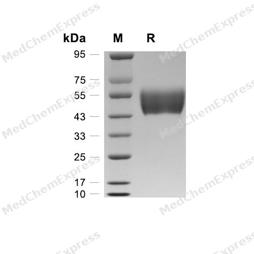
Purity - SDS-PAGE
-
Bioactivity - ELISA
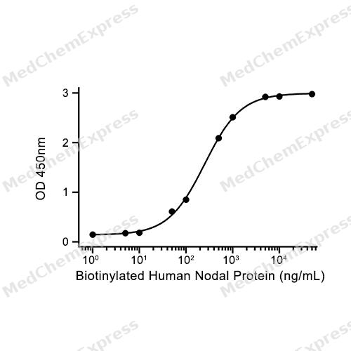
Bioactivity - ELISA

Assay Procedure
Materials
Biotin
Pre-chilled sterile water
Wash buffer: 1X PBS-T (PBS with 0.05% Tween-20)
Blocking buffer: 1% BSA in 1X PBS-T
TMB substrate solution: Mix substrate solution A and B at a 1:1 ratio immediately before use
Stop solution: 2 M H2SO4
ALK-7 Protein, Human (HEK293, Fc) (HY-P74417)
Procedure
Biotinylation
1. Dissolution of Biotin Reagent: Remove Biotin from -10 to -30°C and immediately dissolve 1 tube of Biotin in 180 μL of cold purified water. Gently pipette to mix and obtain a 10 mM Biotin solution.
2. Calculation of EZ-LinkTM Sulfo-NHS-LC-LC-Biotin, No-WeighTM Format Volume: Calculate the volume to be added.
3. Incubation: Incubate at room temperature in the dark for 45 minutes.
4. Quantification of Biotinylated Protein: Use the BCA assay to quantify the protein concentration. The concentration of biotinylated Nodal is 900 μg/mL.
ELISA Detection
1. Coating: Dilute ALK-7 protein to 2 μg/mL in 1X PBS, add 100 μL/well, and incubate overnight at 4°C.
2. Washing: Wash the plate with wash buffer (260 μL/well) , once, and pat dry.
3. Blocking: Add 150 μL/well of blocking buffer (1% BSA in 1X PBS-T) , incubate at 25°C with shaking at 450 rpm for 4 hours.
4. Dilution of Biotinylated Nodal: Prepare dilutions at concentrations of 0, 0.1, 0.5, 1, 5, 10, 50, 100, 500, 1000, 5000, 10000, 500000 ng/mL.
5. Add Biotinylated Nodal Protein: Add 100 μL/well, incubate at 25°C with shaking at 450 rpm for 2 hours.
6. Washing: Wash the plate with Wash Buffer (260 μL/well) , five times, with each wash lasting 1 minute, then pat dry.
7. Add Secondary Antibody: Dilute the secondary antibody at a 1:2000 ratio, add 100 μL/well, incubate at 25°C with shaking at 450 rpm for 1 hour.
8. Washing: Wash the plate with wash buffer (260 μL/well) , three times, with each wash lasting 1 minute, then pat dry.
9. Add TMB Substrate Solution: Add 100 μL/well, incubate at room temperature in the dark for 15 minutes.
10. Stop reaction: Add 100 μL H2SO4 2 M/well.
11. Read plate: Measure absorbance at 450 nm.
12. Using GraphPad Prism software, plot a curve with the OD value as the vertical axis and the Biotinylated Human Nodal protein concentration as the horizontal axis, and calculate the ED50 value.


Technical Parameters
-
Species Human
-
Source HEK293
-
Tag C-hFc
-
Accession
Q8NER5 (L22-E113)
-
Molecular Construction
-
N-term
-
ALK-7 (L22-E113)
Accession # Q8NER5 -
hFc
-
C-term
-
-
Protein Length
Extracellular Domain
-
Synonyms
ACVR1C; Activin Receptor Type-1C; Activin A Receptor Type 1C; Activin Receptor Type IC; ALK7; ACTR-IC; ACVRLK7; ALK-7; Activin Receptor-Like Kinase 7; Uncharacterized Protein ACVR1C; Activin A Receptor, Type IC
-
AA Sequence
LSPGLKCVCLLCDSSNFTCQTEGACWASVMLTNGKEQVIKSCVSLPELNAQVFCHSSNNVTKTECCFTDFCNNITLHLPTASPNAPKLGPME
-
Molecular Weight
Approximately 43-56 kDa, based on SDS-PAGE under reducing conditions.
-
Purity
≥ 95%, as determined by reducing SDS-PAGE.


Product Properties
Lyophilized powder.
Lyophilized from a 0.22 μm filtered solution of PBS, pH 7.4.
<1 EU/μg, determined by LAL method.
It is not recommended to reconstitute to a concentration less than 100 μg/mL in ddH2O. For long term storage it is recommended to add a carrier protein (0.1% BSA, 5% HSA, 10% FBS or 5% Trehalose).
Stored at -20°C for 2 years from date of receipt. After reconstitution, it is stable at 4°C for 1 week or -20°C for longer (with carrier protein). It is recommended to freeze aliquots at -20°C or -80°C for extended storage.
Room temperature in continental US; may vary elsewhere.


Documentation
-
Data Sheet (264 KB)
-
SDS (252 KB)
- English - EN (252 KB)
- Français - FR (252 KB)
- Deutsch - DE (252 KB)
- Norwegian - NO (252 KB)
- Español - ES (252 KB)
- Swedish - SV (252 KB)
- Italian - IT (252 KB)
- Korean - KR (252 KB)
- Portuguese - PT (252 KB)
-
Handling Instructions (2659 KB)


References
[1]. M Rydén, et al. A novel type I receptor serine-threonine kinase predominantly expressed in the adult central nervous system. J Biol Chem. 1996 Nov 29;271(48):30603-9. [Content Brief]
[2]. Byung-Chul Kim, et al. Activin receptor-like kinase-7 induces apoptosis through activation of MAPKs in a Smad3-dependent mechanism in hepatoma cells. J Biol Chem. 2004 Jul 2;279(27):28458-65. [Content Brief]
[3]. Wen-bo Li, et al. Silencing of activin receptor-like kinase 7 alleviates aortic stiffness in type 2 diabetic rats. Acta Diabetol. 2015 Aug;52(4):717-26. [Content Brief]
[4]. Kunihiro Tsuchida, et al. Activin isoforms signal through type I receptor serine/threonine kinase ALK7. Mol Cell Endocrinol. 2004 May 31;220(1-2):59-65. [Content Brief]

Calculators
Concentration (start) × Volume (start) = Concentration (final) × Volume (final)


 Powered by Bioz

