



Ro 41-0960

Based on 1 Customer Validation
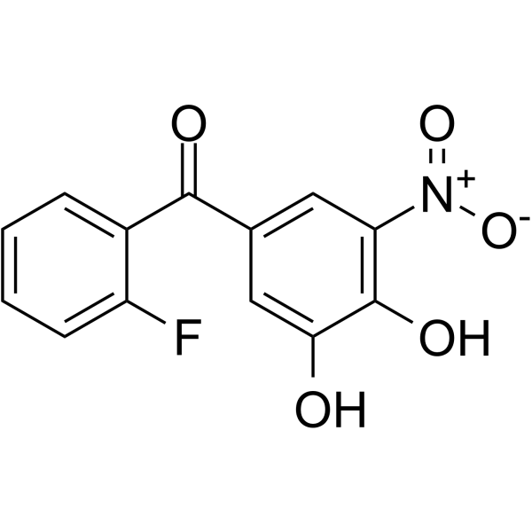
Ro 41-0960 is a CNS-penetrant, orally active catechol-O-methyl transferase (COMT) inhibitor. Ro 41-0960 reduces dopamine catabolism, increases striatal dopamine and DOPAC levels, decreases striatal HVA levels, induces apoptosis, inhibits proliferation and extracellular matrix formation in uterine fibroid cells. Ro 41-0960 arrests or shrinks uterine fibroid lesions in rats. Ro 41-0960 can be used for the research of Parkinson’s disease, uterine leiomyomas, and breast cancer.
For research use only. We do not sell to patients.
- Purity : 98.22%
- CAS No.: 125628-97-9
- Formula: C13H8FNO5
- Molecular Weight:277.20
-
Storage:Powder -20°C, 3 years ; In solvent -80°C, 6 months , -20°C, 1 month

To place orders, for customer services and technical support, please contact: MedChemExpress USA
Tel: 609-228-6898 E-mail: [email protected] [email protected]


-
 Biological Activity
-
Chemical Information
-
Solvent & Solubility
- Protocols
- Purity & Documentation
- References
-
 Help & FAQs


-
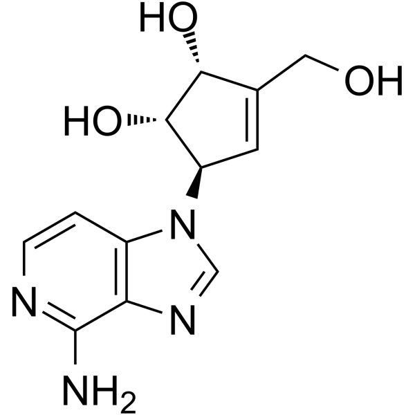
HY-10442
3-Deazaneplanocin AEZH2 Inhibitor -
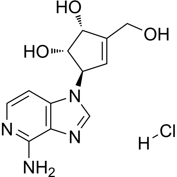
HY-12186
3-Deazaneplanocin A hydrochlorideEZH2 Inhibitor -
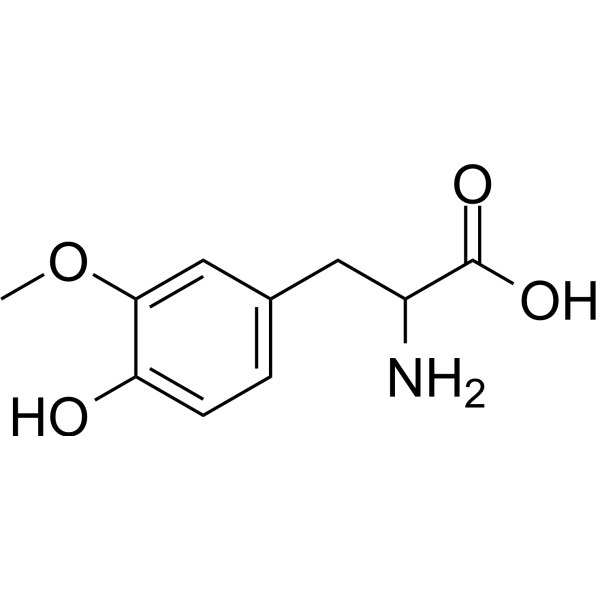
HY-113468
3-O-Methyl-DL-DOPAEndogenous Metabolite -

HY-113468R
3-O-Methyl-DL-DOPA (Standard)Endogenous Metabolite -

HY-139182
MAGL-IN-3 -
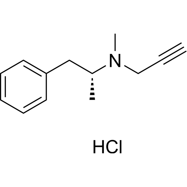
HY-14199
Selegiline hydrochlorideMAO-B Inhibitor -
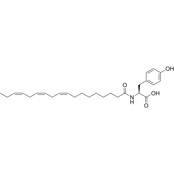
HY-145512
N-(α-Linolenoyl) tyrosineDopaminergic Agonist -

HY-10442R
3-Deazaneplanocin A (Standard)Reference Standard -
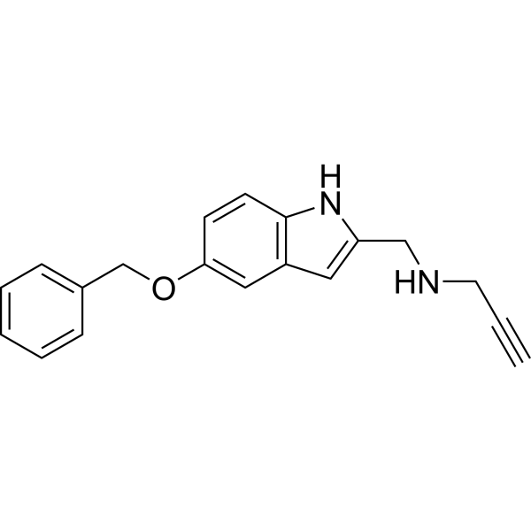
HY-120419
PF9601NMAO-B Inhibitor -
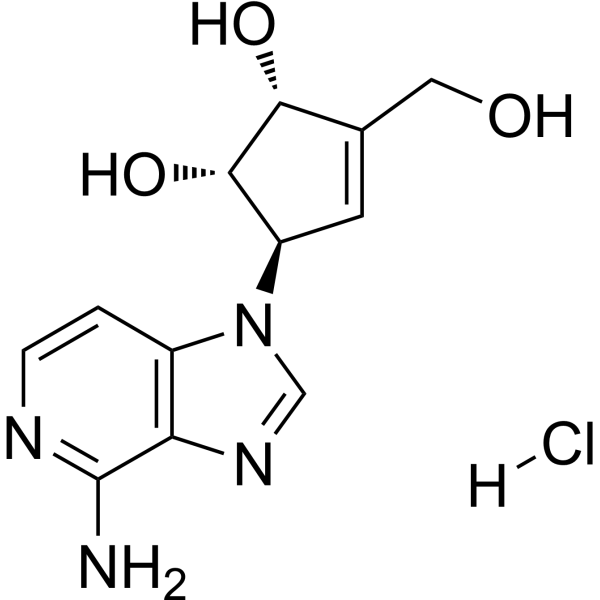
HY-12186G
3-Deazaneplanocin A hydrochloride (GMP)EZH2 Inhibitor (GMP) -

HY-139008
RC363GPx1 Activator -

HY-21197
Perfluoroenanthic acidPollutant


Biological Activity
Description
Cellular Effect
|
Cell Line

|
Type | Value | Description | References |
|---|---|---|---|---|
| HT-29 | EC50 |
8.66 μM
Compound: Ro41-0960
|
Agonist activity at GPR35 in human HT-29 cells assessed as induction of whole cell dynamic mass redistribution after 50 mins by resonant waveguide grating biosensor analysis
Agonist activity at GPR35 in human HT-29 cells assessed as induction of whole cell dynamic mass redistribution after 50 mins by resonant waveguide grating biosensor analysis
|
10.1039/C2MD20210G |
| U2OS | EC50 |
54.4 μM
Compound: Ro41-0960
|
Agonist activity at GPR35 in human U2OS cells assessed as induction of beta-arrestin translocation after 5 hrs by beta-lactamase reporter gene assay
Agonist activity at GPR35 in human U2OS cells assessed as induction of beta-arrestin translocation after 5 hrs by beta-lactamase reporter gene assay
|
10.1039/C2MD20210G |
In Vitro
Ro 41-0960 (0.05-50 µM; 20 min) directly activates SERCA2a in pig cardiac sarcoplasmic reticulum microsomes with an EC50 of 27 µM and increases ATPase turnover by 38%[3].
Ro 41-0960 (0.5-100 µM; 30 min) exerts concentration-dependent, contrasting marginal effects on Ca2+ transient amplitude and CaTD50, and minimal effects on beat rate, in human iPSC-derived cardiomyocytes[3].
Ro 41-0960 inhibits rat brain COMT with an IC50 of 16 nmol/L and rat liver COMT with an IC50 of 42 nmol/L[4].
Ro 41-0960 (30 min) potently inhibits COMT activity in cytosolic fractions from healthy human mammary tissues, with IC50 values ranging from 5.1 nM to 42.1 nM across 7 individual tissue samples, and fully blocks activity at 0.3 μM[5].
Ro 41-0960 (10 μM; 5-6 h) inhibits COMT activity in MCF-7 human mammary tumor cells by ~98%, reducing catechol estrogen inactivation and increasing catechol estrogen-induced DNA damage by ~200% without causing cytotoxicity[5].
MedChemExpress (MCE) has not independently confirmed the accuracy of these methods. They are for reference only. Further protocols information, click here.
In Vivo
Ro 41-0960 (150 mg/kg; s.c.; twice daily; 28 days) arrests growth or shrinks uterine fibroids in Eker rats[2].
Ro 41-0960 (30 mg/kg; i.p.; single dose) attenuates or completely prevents L-Dopa (HY-N0304)-induced changes in sulfur amino acid metabolite concentrations in rat brain regions, peripheral tissues, and plasma, while having minimal effects on these metabolites when administered alone[4].
MedChemExpress (MCE) has not independently confirmed the accuracy of these methods. They are for reference only.
-
Animal Model:Wistar rats (male, 270-320 g)[1]
-
Dosage:20 mg/kg
-
Administration:i.p.; single dose (acute); daily; 5 days (chronic)
-
Result:Produced a statistically significant 123.6% increase in striatal DA content, a 234.6% increase in striatal DOPAC content, and a 58.0% reduction in striatal HVA content relative to saline controls (acute treatment).
Produced a 122.5% increase in striatal DA content, a 225.5% increase in striatal DOPAC content, and a 13.1% reduction in striatal HVA content relative to saline controls (chronic treatment).
Caused a significantly greater reduction in HVA content compared to the acute treatment.
-
Animal Model:Eker rats (female, 14-16 months old, germline mutation in tuberous sclerosis-2 tumor suppressor gene)[2]
-
Dosage:150 mg/kg
-
Administration:s.c.; twice daily; 28 days
-
Result:Exhibited fibroid volumes of 86% and 105% of initial burden at 2 and 4 weeks post-treatment, respectively.
Increased the urinary 2-hydroxy E2/16-hydroxy E2 ratio.
Increased p53 mRNA levels.
Decreased PARP1-positive cells.
Decreased PCNA-positive cells.
Decreased cyclin D1-positive cells.
Decreased TGFb3 mRNA levels.
Did not alter normal tissue histology, serum liver enzyme levels (AST, ALT, total bilirubin), or urinary DPD levels.
-
Animal Model:Sprague-Dawley rats (male, ~200 g)[4]
-
Dosage:30 mg/kg
-
Administration:i.p.; single dose
-
Result:Showed no significant difference in mean SAM and SAH concentrations in cortex, hippocampus, cerebellum, spleen, kidney, and liver.
Increased mean striatal SAM concentration.
Decreased mean striatal SAH concentration.
Showed no significant difference in mean total plasma homocysteine concentration.
Increased mean SAM concentrations significantly in cortex, hippocampus, cerebellum, striatum, spleen, and kidney.


Chemical Information
-
CAS No. 125628-97-9
-
Appearance Solid
-
Molecular Weight 277.20
-
Formula C13H8FNO5
-
Color Light yellow to yellow
-
SMILES
O=C(C1=CC([N+]([O-])=O)=C(C(O)=C1)O)C2=CC=CC=C2F
-
Shipping
Room temperature in continental US; may vary elsewhere.
-
Storage
Powder -20°C 3 years In solvent -80°C 6 months -20°C 1 month


Solvent & Solubility
In Vitro:
DMSO : ≥ 100 mg/mL (360.75 mM; Hygroscopic DMSO has a significant impact on the solubility of product, please use newly opened DMSO)
* "≥" means soluble, but saturation unknown.
Please refer to the solubility information to select the appropriate solvent. Once prepared, please aliquot and store the solution to prevent product inactivation from repeated freeze-thaw cycles.
Storage method and period of stock solution: -80°C, 6 months; -20°C, 1 month. When stored at -80°C, please use it within 6 months. When stored at -20°C, please use it within 1 month.
Please refer to the solubility information to select the appropriate solvent. Once prepared, please aliquot and store the solution to prevent product inactivation from repeated freeze-thaw cycles.
Storage method and period of stock solution: -80°C, 6 months; -20°C, 1 month. When stored at -80°C, please use it within 6 months. When stored at -20°C, please use it within 1 month.
Concentration (start) × Volume (start) = Concentration (final) × Volume (final)
In Vivo:
Select the appropriate dissolution method based on your experimental animal and administration route.
- For the following dissolution methods, please ensure to first prepare a clear stock solution using an In Vitro approach and then sequentially add co-solvents:
- To ensure reliable experimental results, the clarified stock solution can be appropriately stored based on storage conditions. As for the working solution for In Vivo experiments, it is recommended to prepare freshly and use it on the same day.
- The percentages shown for the solvents indicate their volumetric ratio in the final prepared solution. If precipitation or phase separation occurs during preparation, heat and/or sonication can be used to aid dissolution.
Add each solvent one by one: 10% DMSO 40% PEG300 5% Tween-80 45% Saline
Solubility: ≥ 2.5 mg/mL (9.02 mM); Clear solution
This protocol yields a clear solution of ≥ 2.5 mg/mL (saturation unknown).
Taking 1 mL working solution as an example, add 100 μL DMSO stock solution (25.0 mg/mL) to 400 μL PEG300, and mix evenly; then add 50 μL Tween-80 and mix evenly; then add 450 μL Saline to adjust the volume to 1 mL.
Preparation of Saline: Dissolve 0.9 g sodium chloride in ddH₂O and dilute to 100 mL to obtain a clear Saline solution.
Add each solvent one by one: 10% DMSO 90% (20% SBE-β-CD in Saline)
Solubility: ≥ 2.5 mg/mL (9.02 mM); Clear solution
This protocol yields a clear solution of ≥ 2.5 mg/mL (saturation unknown).
Taking 1 mL working solution as an example, add 100 μL DMSO stock solution (25.0 mg/mL) to 900 μL 20% SBE-β-CD in Saline, and mix evenly.
Preparation of 20% SBE-β-CD in Saline (4°C, storage for one week): 2 g SBE-β-CD powder is dissolved in 10 mL Saline, completely dissolve until clear.
In Vivo Dissolution Calculator
Please enter the basic information of animal experiments:
-
-
-
-
Recommended: Prepare an additional quantity of animals to account for potential losses during experiments.
Please enter your animal formula composition:
-
%DMSO +
Recommended: Keep the proportion of DMSO in working solution below 2% if your animal is weak.
-
%+
-
+%Tween-80 + +
-
%Saline +
The co-solvents required include: DMSO, . All of co-solvents are available by MedChemExpress (MCE). , Tween 80. All of co-solvents are available by MedChemExpress (MCE).
Working solution concentration: 0.22 mg/mL
Method for preparing stock solution: mg drug dissolved in μL DMSO. Stock solution concentration: mg/mL.
1. Take μL DMSO stock solution;
2. Add μL .
μL , mix evenly;
3. Then add μL Tween 80, mix evenly;
4. Then add μL
Please ensure that the stock solution in the first step is dissolved to a clear state, and add co-solvents in sequence. You can use ultrasonic heating (ultrasonic cleaner, recommended frequency 20-40 kHz), vortexing, etc. to assist dissolution.

Protocols
-
Fibrosis/Collagen Morphometry
Fibrosis and collagen morphometry is based on the quantitative visualization of fibrillar collagen deposition in tissue sections using histochemical stains such as Sirius Red (Picrosirius Red) or Masson's trichrome, followed by image-based or polarization-enhanced analysis to estimate collagen proportional area as a surrogate of extracellular matrix accumulation during fibrotic remodeling. Sirius Red combined with polarized light microscopy enhances detection of collagen fibers due to birefringence properties, enabling more specific visualization of collagen type I and III fibrils compared to conventional bright-field histology, while whole-section or region-restricted digital morphometry reduces field-selection bias in fibrosis assessment. Alternative quantitative approaches include second harmonic generation (SHG) and two-photon excited fluorescence microscopy, which enable label-free detection of fibrillar collagen and have been validated against histological staining and biochemica
-
Collagen: Sirius Red Staining
Sirius Red or picrosirius red staining is a histochemical method for visualizing collagen-rich extracellular matrix in tissue sections, and collagen fibers are detected as red-stained structures under bright-field microscopy with enhanced birefringence under polarized light. Picrosirius red is useful for assessing total collagen organization, distribution, and fibrosis burden, but polarized color should not be interpreted as a definitive collagen type I versus type III readout because color is affected by fiber orientation, thickness, and packing.
-
Apoptosis
Apoptosis, also called programmed cell death, is generally characterized by distinct morphological characteristics.
-
TUNEL staining for apoptotic DNA fragmentation
TUNEL staining detects DNA strand breaks by using terminal deoxynucleotidyl transferase to add labeled nucleotides to exposed 3′-OH DNA termini, generating either microscopic staining in fixed cells or tissue sections, or fluorescence/cytometric signal in cell suspensions. TUNEL positivity reflects DNA fragmentation but should not be interpreted alone as definitive apoptosis, because TUNEL can also label necrotic, autolytic, mechanically damaged, or DNA-repair-associated DNA breaks.
-
Annexin V plus membrane-impermeant dye apoptosis staining
Annexin V-based apoptosis assays rely on the detection of phosphatidylserine (PS) externalization from the inner leaflet of the plasma membrane to the outer leaflet, an early biochemical hallmark of apoptosis. Fluorescently labeled Annexin V binds PS in a calcium-dependent manner, enabling identification of early apoptotic cells by flow cytometry or fluorescence microscopy. When combined with a membrane-impermeant DNA-binding dye (e. g. , propidium iodide), this approach allows discrimination between viable (Annexin V−/dye−), early apoptotic (Annexin V+/dye−), and late apoptotic or necrotic (Annexin V+/dye+) cell populations by assessing membrane integrity and PS exposure.
-
ECM-Embedded Organoid (Matrigel/Dome) Culture
ECM-embedded organoid dome culture embeds epithelial stem cells, crypts, organoid fragments, or tumor-derived epithelial cells in a basement-membrane-like hydrogel such as Matrigel, allowing 3D growth, self-organization, lumen formation, budding or cystic morphogenesis, and lineage maintenance under defined niche-factor-containing medium. The primary readouts are organoid establishment efficiency, growth, morphology, passaging capacity, lineage-marker expression, and, when fluorescently labeled lines are used, microscopy- or flow-cytometry-based quantification of population behavior in 3D culture.
-
Apoptosis Solutions
Apoptosis is a regulated, generally non-lytic cell-death pathway that removes unwanted, damaged, infected, or abnormal cells through coordinated morphological changes, caspase activation, DNA fragmentation, and membrane remodeling. The intrinsic apoptosis pathway is controlled mainly by mitochondrial outer membrane permeabilization, BCL-2 family proteins, cytochrome c release, apoptosome formation, caspase-9 activation, and downstream executioner caspase-3/7 activation. The extrinsic apoptosis pathway is initiated by death receptors such as Fas, TNFR, and TRAIL receptors, which recruit adaptor proteins and activate caspase-8 before engaging executioner caspases or mitochondrial amplification through BID cleavage. Apoptosis is linked to many phenotypes, including cancer cell killing, tissue homeostasis, immune regulation, neurodegeneration, infection response, and treatment-induced cytotoxicity; unresolved questions include how apoptosis interacts with necroptosis, pyroptosis, ferroptos
-
Matrigel/ECM Transwell Invasion Assay
The Matrigel/ECM Transwell invasion assay measures the ability of cells to move toward a chemoattractant while crossing an extracellular-matrix barrier placed on a porous membrane; therefore, the readout reflects both chemotactic motility and matrix invasion rather than migration alone. Matrigel is a basement-membrane-rich matrix derived from Engelbreth-Holm-Swarm mouse sarcoma and has been used as a reconstituted basement membrane barrier in chemoinvasion assays. The assay readout is generated by quantifying cells that reach the underside of the insert membrane or lower compartment after incubation, commonly by staining and counting invaded cells or by fluorescence-based quantification.
-
Breast Cancer Modeling
Breast cancer is a heterogeneous cancer, and it has been distinguished into four subtypes: luminal A, luminal B, HER2-positive and basal-like. Molecular mutations, epigenetic alterations, hormone exposure and immune microenvironment are related to the progression of breast cancer.
-
Cell Viability Determination by MTT Colorimetric Assay
The following protocol uses the MTT colorimetric assay as a classic literature-established method for assessing cell viability/metabolic activity in cultured mammalian cells. MTT[3-(4,5-dimethylthiazol-2-yl)-2,5-diphenyltetrazolium bromide] is reduced by metabolically active cells to a colored formazan product; the amount of formazan is quantified spectrophotometrically and provides an indirect measure of metabolically active viable cells. Importantly, MTT reduction reflects cellular oxidoreductase/metabolic activity rather than an absolute direct count of living cells, so changes in cellular metabolism can alter the signal independently of cell number.
-
Matrigel Transwell/Boyden Chamber Invasion Assay
Matrigel Transwell/Boyden chamber invasion assay measures the ability of cells to degrade or traverse an extracellular matrix-coated porous membrane and move from an upper chamber toward a chemoattractant in a lower chamber. Invasion is distinguished from migration by coating the membrane with Matrigel or basement membrane matrix; uncoated inserts measure migration, while coated inserts require cells to cross an ECM barrier before reaching the underside of the membrane.


Purity & Documentation
-
Data Sheet (282 KB)
-
SDS (251 KB)
- English - EN (251 KB)
- Français - FR (251 KB)
- Deutsch - DE (251 KB)
- Norwegian - NO (251 KB)
- Español - ES (251 KB)
- Swedish - SV (251 KB)
- Italian - IT (251 KB)
- Korean - KR (251 KB)
- Portuguese - PT (251 KB)
-
Handling Instructions (2659 KB)
References
[2]. Hassan MH, et al. Towards non-surgical therapy for uterine fibroids: catechol-O-methyl transferase inhibitor shrinks uterine fibroid lesions in the Eker rat model. Hum Reprod. 2011;26(11):3008-3018. [Content Brief]
[3]. Aguayo-Ortiz R, et al. A multiscale approach for bridging the gap between potency, efficacy, and safety of small molecules directed at membrane proteins. Sci Rep. 2021 Aug 16;11(1):16580. [Content Brief]
[4]. Miller JW, et al. Effect of L-Dopa and the catechol-O-methyltransferase inhibitor Ro 41-0960 on sulfur amino acid metabolites in rats. Clin Neuropharmacol. 1997 Feb;20(1):55-66. [Content Brief]
[5]. van Duursen MB, et al. Phytochemicals inhibit catechol-O-methyltransferase activity in cytosolic fractions from healthy human mammary tissues: implications for catechol estrogen-induced DNA damage. Toxicol Sci. 2004;81(2):316-324. [Content Brief]


Complete Stock Solution Preparation Table
Please refer to the solubility information to select the appropriate solvent. Once prepared, please aliquot and store the solution to prevent product inactivation from repeated freeze-thaw cycles.
Storage method and period of stock solution: -80°C, 6 months; -20°C, 1 month. When stored at -80°C, please use it within 6 months. When stored at -20°C, please use it within 1 month.
| Optional Solvent | Concentration Solvent Mass | 1 mg | 5 mg | 10 mg | 25 mg |
|---|---|---|---|---|---|
| DMSO | 1 mM | 3.6075 mL | 18.0375 mL | 36.0750 mL | 90.1876 mL |
| 5 mM | 0.7215 mL | 3.6075 mL | 7.2150 mL | 18.0375 mL | |
| 10 mM | 0.3608 mL | 1.8038 mL | 3.6075 mL | 9.0188 mL | |
| 15 mM | 0.2405 mL | 1.2025 mL | 2.4050 mL | 6.0125 mL | |
| 20 mM | 0.1804 mL | 0.9019 mL | 1.8038 mL | 4.5094 mL | |
| 25 mM | 0.1443 mL | 0.7215 mL | 1.4430 mL | 3.6075 mL | |
| 30 mM | 0.1203 mL | 0.6013 mL | 1.2025 mL | 3.0063 mL | |
| 40 mM | 0.0902 mL | 0.4509 mL | 0.9019 mL | 2.2547 mL | |
| 50 mM | 0.0722 mL | 0.3608 mL | 0.7215 mL | 1.8038 mL | |
| 60 mM | 0.0601 mL | 0.3006 mL | 0.6013 mL | 1.5031 mL | |
| 80 mM | 0.0451 mL | 0.2255 mL | 0.4509 mL | 1.1273 mL | |
| 100 mM | 0.0361 mL | 0.1804 mL | 0.3608 mL | 0.9019 mL |


Ro 41-0960 Related Classifications
HY-125339 Related Classifications
 Powered by Bioz